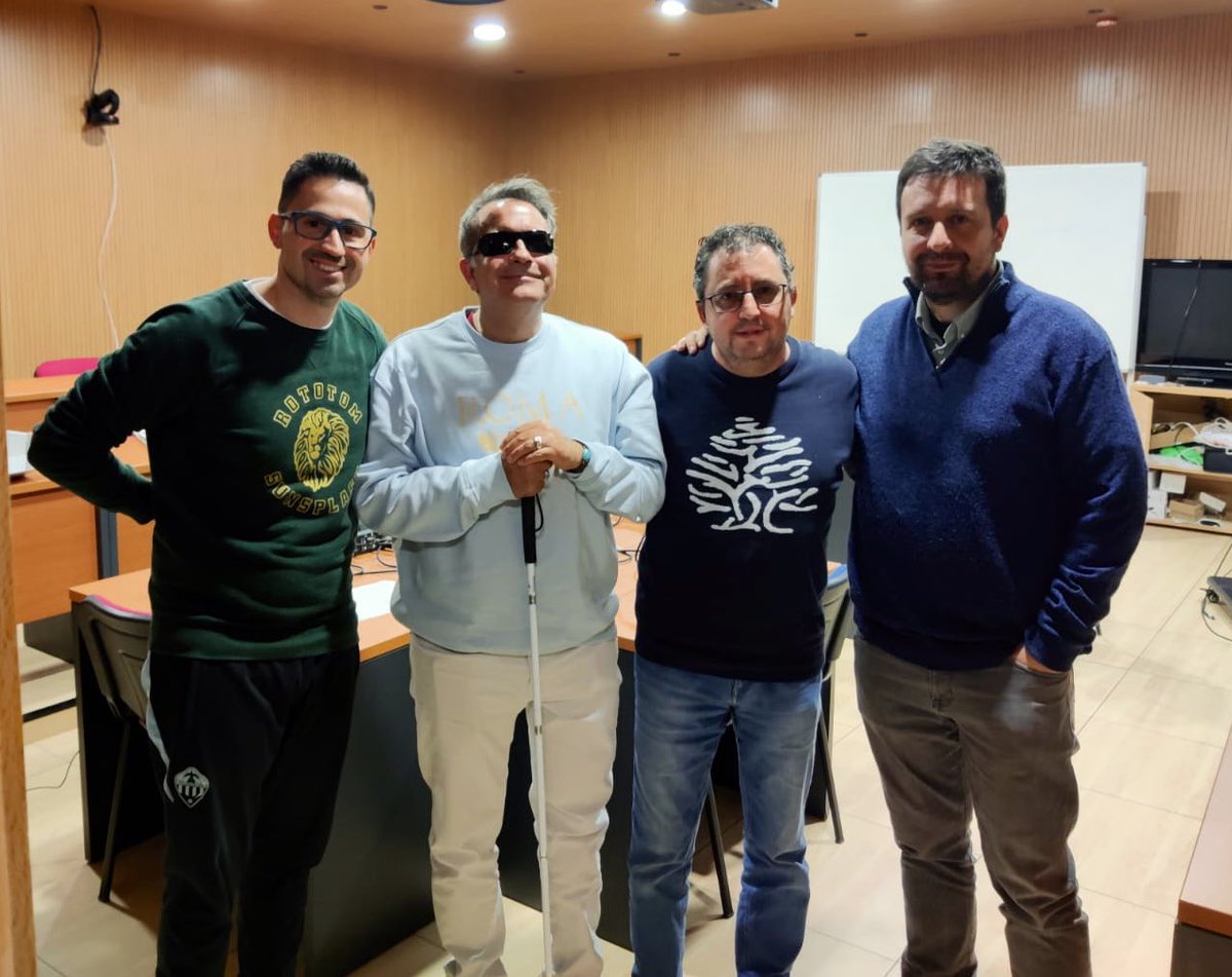
YA tenéis disponible un NUEVO PROGRAMA: “Evolución y Complejidad”
Con Fernando Ballesteros premio Cozzarelli de la Academia de Ciencias de Estados Unidos.
Desde el @IFICorpuscular
go.ivoox.com/rf/172419077

Español
Oscilador Armónico
562 posts

@OA_Podcast
Podcast de Física. Desde el @IFICorpuscular


X-ray images are among the most commonly used tools in medicine. They are ordered daily in emergency rooms, clinics and wards, often regarded as routine, almost trivial investigations. Yet few clinicians — and even fewer patients — stop to consider the physical principles that make these images possible, or that determine what we can (and cannot) see in them. Medical X-ray imaging is not simply applied anatomy; it is applied physics. *⃣ An X-ray image is not a photograph in the optical sense. It does not rely on reflection or colour, but on the interaction between high-energy photons and biological tissues. In the X-ray tube, electrons accelerated at high voltage strike a metal target, producing a spectrum of photons with different energies. This dispersion of energies is essential: tissues attenuate low- and high-energy photons differently, and image contrast emerges from this differential interaction. 1⃣ As the beam passes through the patient, photons are absorbed or transmitted according to tissue thickness, density and atomic composition. Bone, rich in calcium and therefore higher atomic number elements, strongly absorbs X-rays and appears white. Soft tissues absorb less and generate intermediate greys. Air absorbs almost nothing and appears black. *⃣ The detector does not “see” anatomy directly; it records the spatial distribution of photons that survive the journey through the body. The radiographic image is, in essence, a quantitative map of photon attenuation. In this sense, the body behaves like a biological filter. Much like a prism disperses light according to wavelength, human tissues selectively modify the X-ray beam according to physical properties. The emerging photon pattern contains encoded information about internal structures, which digital detectors translate into greyscale values. Clinicians then learn to read these values as ribs, lungs, consolidations or fractures. What appears intuitive at the bedside is, in reality, the result of probabilistic physical events. 2⃣ However, absorption is only part of the story. A second phenomenon, the Compton effect, plays a central role in defining image quality. When an X-ray photon collides with a loosely bound outer electron, it may not be absorbed but scattered. The photon loses energy, changes direction, and continues travelling through the body. Many of these scattered photons still reach the detector. *⃣ From a diagnostic perspective, this is problematic. Scattered photons no longer carry accurate spatial information about their point of origin. Instead of contributing to useful contrast, they add a background signal that reduces image sharpness and masks subtle differences between tissues. This is why X-ray images, particularly of the chest and abdomen, are never perfectly crisp. *⃣ Compton scattering depends mainly on electron density and beam energy, not on atomic number. As a result, it predominates in soft tissues and increases with patient thickness and higher X-ray energies. This explains why low-contrast lesions can be difficult to detect and why radiographic technique — beam energy, collimation and patient positioning — matters so much. *⃣ Modern radiology actively manages this unavoidable physics. Anti-scatter grids absorb obliquely scattered photons, collimation limits the irradiated volume, and digital post-processing improves contrast. Yet none of these strategies can eliminate scatter entirely. Every medical X-ray image is a compromise between penetration, contrast, noise and dose. *⃣ Understanding X-ray imaging therefore requires more than anatomical knowledge. It requires recognising that every radiograph is a controlled physical experiment performed in a living human. Medicine asks the clinical question, but physics sets the rules — and ultimately defines the limits — of what the image can reveal.

YA tenéis disponible un NUEVO PROGRAMA: “Computación en Física Experimental de Partículas: de las Emulsiones Nucleares a la IA” Con José Salt profesor de investigación experto en computación para la física de partículas. Desde el @IFICorpuscular go.ivoox.com/rf/170777785


YA tenéis un NUEVO PROGRAMA: “Mecánica Cuántica X: Interpretaciones de la Mecánica Cuántica” ¿Qué nos dice la mecánica cuántica sobre cómo es la realidad? Con @sientoquinse Desde el @IFICorpuscular #podcast #ciencia go.ivoox.com/rf/169971392

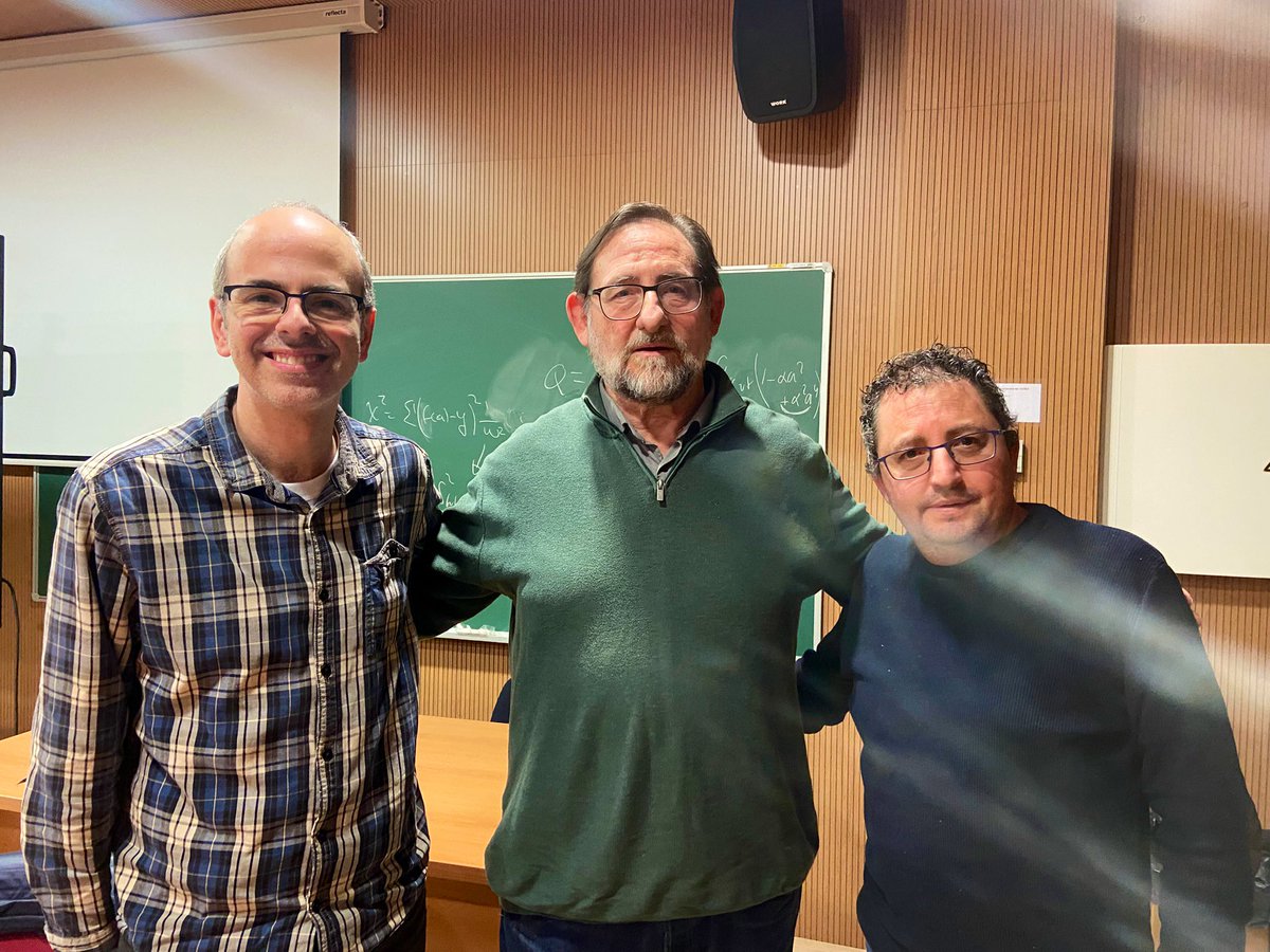
YA tenéis disponible un NUEVO PROGRAMA: “La Influencia del Clima en la Historia” con Francisco Jiménez Espejo @IACT_CSIC geoquímico experto en Paleoclimatología y José Soto Chica, doctor en Historia Medieval @DespertaFerro Desde el @IFICorpuscular go.ivoox.com/rf/169525365

YA tenéis disponible un NUEVO PROGRAMA: “La Influencia del Clima en la Historia” con Francisco Jiménez Espejo @IACT_CSIC geoquímico experto en Paleoclimatología y José Soto Chica, doctor en Historia Medieval @DespertaFerro Desde el @IFICorpuscular go.ivoox.com/rf/169525365