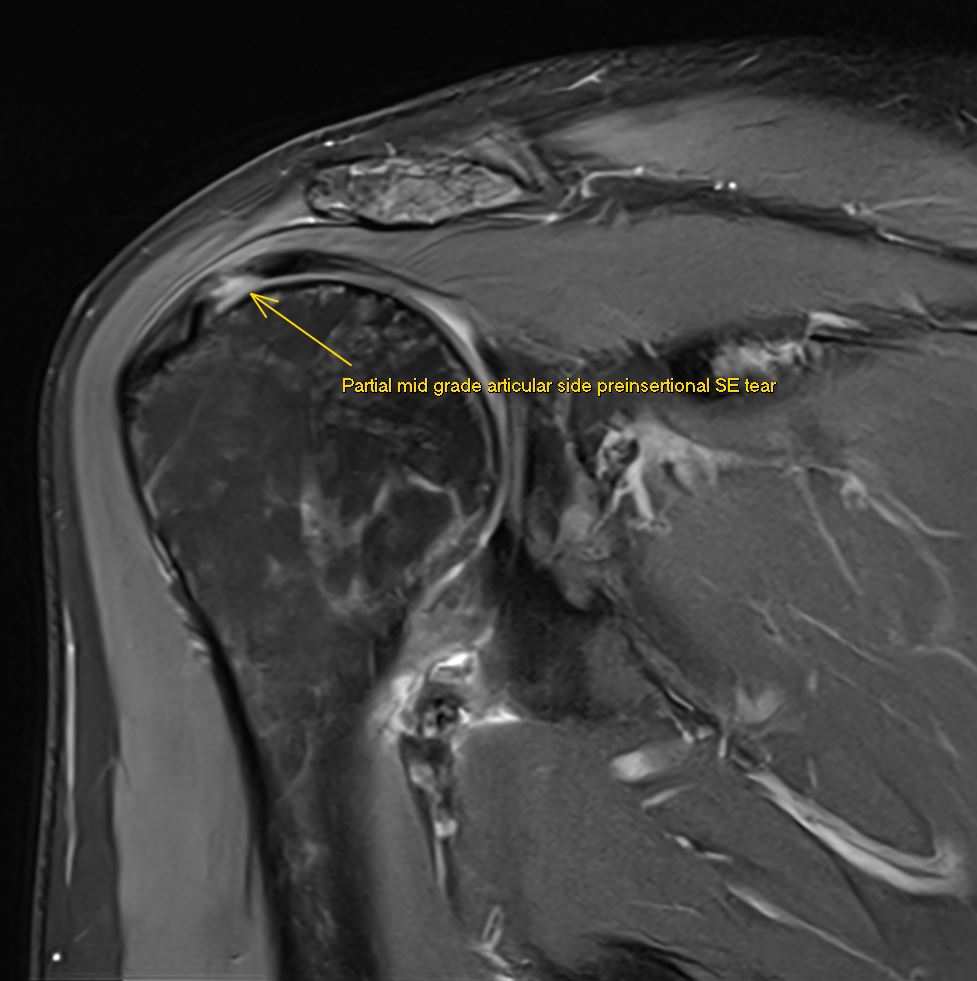
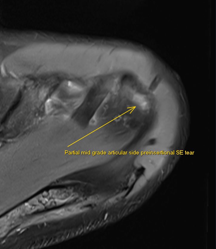
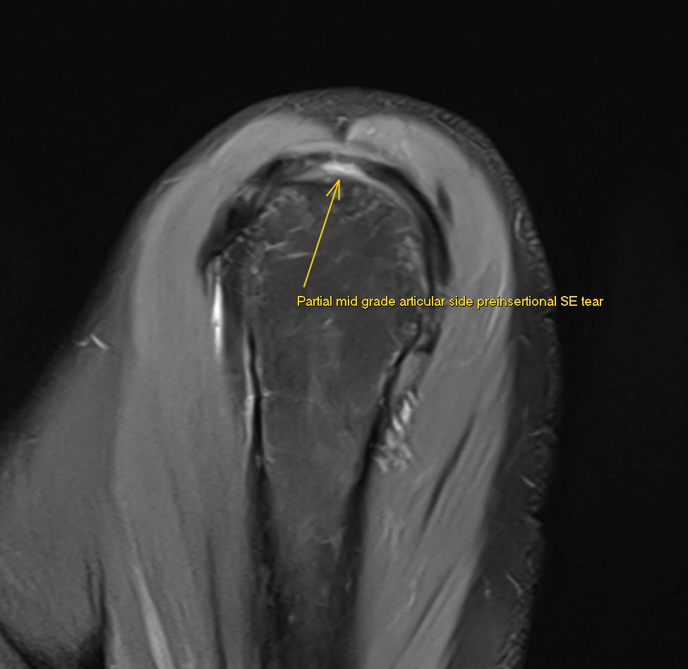
JuanMiranda

1.3K posts

JuanMiranda

JuanMiranda

@themskarchive

Musculoskeletal Radiologist. Husband. Obsessive reader. Investor. Python. Grateful to share my daily MSK and Tanzanian activities #radiology #radtwitter #MSKRad

Madrid, Comunidad de Madrid Beigetreten Aralık 2022
115 Folgt2.9K Follower
JuanMiranda
JuanMiranda@themskarchive·
16/12/2025🏃‍♂️💥Málaga Marathon (14 Dec 2025) → 1 day later I wanted to see the DOMS, not just feel it. 🧲🦵 
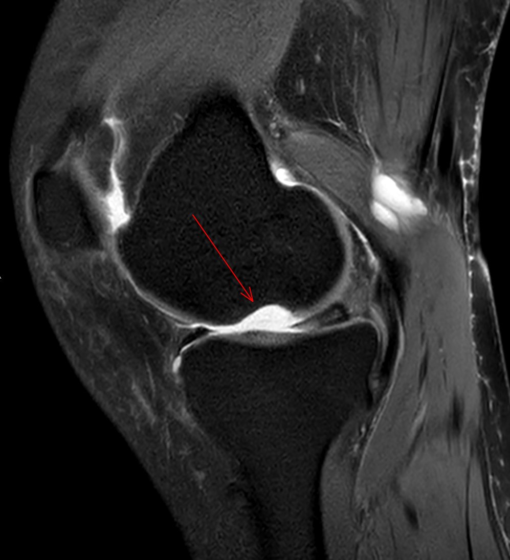
Axial FSDP (R thigh): patchy exercise-related edema in VL/VI+adductor magnus+semitendinosus — classic post-endurance micro-trauma, not a tear. #Marathon #MalagaMarathon
JuanMiranda tweet mediaJuanMiranda tweet media
English
0
5
26
3.4K
JuanMiranda
JuanMiranda@themskarchive·
26/11/2025.🖐️37M with central dorsal wrist pain. US-guided arthrography showed immediate contrast passage from RC into the MC joint → intrinsic ligament tear. 📸 CT & MR arthrograms confirmed complete lunotriquetral ligament rupture #MSK #Radiology #SLIL #WristPain #MRI #CT🩻
JuanMiranda tweet mediaJuanMiranda tweet mediaJuanMiranda tweet media
English
0
5
17
1.3K
JuanMiranda
JuanMiranda@themskarchive·
19/11/2025. 🦵 55 y/o woman with knee pain. FSPD #MRI, 1 year apart: 📍 Baseline: large osteochondral defect in the lateral aspect of the medial femoral condyle 🔧 Arthroscopic microperforations (microfracture) ✨ Follow-up: defect filled with fibrocartilage-like tissue👀 #MSK
JuanMiranda tweet mediaJuanMiranda tweet mediaJuanMiranda tweet mediaJuanMiranda tweet media
English
2
10
50
3.7K
JuanMiranda
JuanMiranda@themskarchive·
17/11/2025 – 53 y/o woman with heel pain. Sagittal & axial ankle FSPD MRI: ✅ Insertional plantar fasciopathy 💥 Plantar fat pad edema 💫 Edema deep to FDB + inferior calcaneal process ⚠️ Deep central band fibers disrupted → medium-grade partial tear #Radiology #MSKR #Ankle
JuanMiranda tweet mediaJuanMiranda tweet media
English
1
14
58
4.3K
JuanMiranda
JuanMiranda@themskarchive·
24/10/2025🧗‍♂️ 21-year-old climber with sudden pain + “pop” at the PIP joint of the right middle finger. US (volar, sagittal) shows widening between the volar cortex of P1 and the flexor tendons, with fluid occupying the gap → acute A2 pulley injury—classic in climbers. #MSK🖐️🔍
JuanMiranda tweet media
English
0
1
4
877
Manigoldo
Manigoldo@cristoateo69·
@themskarchive @pepelermarx @JM_Zulueta_O @drwillyjara ecografía de hombro. Me gustaría saber si usted me puede colaborar respondiendo una pequeña pregunta técnica que me permita poder implementar un método de validación de este nuevo protocolo. Agradezco inmensamente por leerme. Espero me pueda ayudar. Feliz día.
Español
1
0
0
36
JuanMiranda
JuanMiranda@themskarchive·
5/6/2024. 👌 Thrilled to have improved MRI protocols with my teammate @pepelermarx! This #shoulder MRI clearly shows a preinsertional partial articular-sided mid-grade supraspinatus tear in three planes 💪. #MRI
JuanMiranda tweet mediaJuanMiranda tweet mediaJuanMiranda tweet media
English
7
15
86
6.8K
JuanMiranda
JuanMiranda@themskarchive·
21/10/2025.🦵 46F, knee MRI. Coronal & sagittal DP (± fat sat): AVM in Hoffa’s fat pad. 🩸 Serpiginous tubular flow-voids = anomalous arteries 🔎 Perivascular fat signal change → nidus #Radiology #MSK #KneeMRI #AVM #HoffasFatPad #FlowVoid
JuanMiranda tweet media
English
0
1
37
2.3K
JuanMiranda
JuanMiranda@themskarchive·
9/10/2025. 🟥🦴 What do you see here? (AP pelvis X-ray) Notice the altered trabecular pattern in the right femoral head — with a subtle articular collapse and a well-defined subchondral lytic lesion with sclerotic margins. Any guesses? 👀 #Radiology #MedTwitter
JuanMiranda tweet media
English
7
3
41
4.7K
JuanMiranda
JuanMiranda@themskarchive·
Axial view.
JuanMiranda tweet media
Français
0
0
5
454
JuanMiranda
JuanMiranda@themskarchive·
3/10/2025.🦴⚠️69yo male, h/o bilateral TKA, c/o R knee pain. XR (Right knee): 🟥 Loss of polyethylene + metal-on-metal contact 🟨 Prosthesis loosening 🟩 Capsular distension w/ metallic debris ✅ Left knee: normal ❌ Right knee: advanced metallosis #Metallosis #Radiology #TKA
JuanMiranda tweet media
English
2
9
58
4.1K
JuanMiranda
JuanMiranda@themskarchive·
29/9/2025.🟥 14y boy, h/o distal tibia epiphysiolysis II (7mo ago). MRI shows: Altered physeal/periphyseal signal Edema epi/metaphysis Oval lesion disrupting trilaminar pattern ➡️ Suspicious for physeal bar → risk of growth arrest, deformity, LLD. CT pending for confirmation.
JuanMiranda tweet mediaJuanMiranda tweet media
English
0
6
29
2.6K