Bilu Huang@BiluHuang
The damage accumulation theory of aging is clearly incorrect because a valid theory cannot allow for contradictory evidence or loopholes. The DNA damage accumulation theory of aging is riddled with loopholes and can be determined to be wrong based on the following 22 evidences:
1. All human cells accumulate waste molecules like lipofuscin. In 1973, Tappel et al. added the free radical scavenger vitamin E to the feed of adult mice for one year and found that neuronal lipofuscin indeed decreased, but there was no reduction in mortality [Tappel A, Fletcher B, Deamer D. Effect of antioxidants and nutrients on lipid peroxidation fluorescent products and aging parameters in the mouse. J Gerontol. 1973 Oct;28(4):415-24. doi: 10.1093/geronj/28.4.415.]. Feeding mice a diet with 2% protein for 9-15 weeks led to the formation of large amounts of lipofuscin in the nervous system. Subsequently, feeding them a diet with 25% protein reduced lipofuscin [Monocha SL. Acta Histochen 1977;58:219.]. However, a high-protein diet actually shortens lifespan, indicating that cellular and individual aging are not caused by the accumulation of cross-linked, denatured waste molecules inside or outside cells.
2. In 2007, scientists at the University of Washington demonstrated that mitochondrial DNA mutations do not cause premature aging in mice. To observe whether point mutations (a single DNA base) in mtDNA directly affect aging, a research team designed "mitochondrial mutator" mice. Comparing the mtDNA mutation frequency between mutator mice and normal mice, they found that mutator mice had 50 times more mtDNA mutations but showed no signs of premature aging such as osteoporosis, hair loss, or decreased fertility [Vermulst M, Bielas JH, Kujoth GC, et al. Mitochondrial point mutations do not limit the natural lifespan of mice. Nat Genet. 2007 Apr;39(4):540-3. doi: 10.1038/ng1988.].
3. In plants and animals without a specific immune system, cells with DNA damage mutations in the nucleus cannot be selectively cleared. For example, both planarian and tree somatic cells generate DNA damage, but planarians achieve immortality through fission reproduction and trees through branch cutting propagation, indicating that the DNA damage accumulation theory of aging is incorrect. The immortality of Turritopsis dohrnii is also unaffected by DNA damage.
4. In 1975, Wright and Hayflick replaced the nucleus of an aged cell with that of a young cell, resulting in the aged cell regaining youth and continuing to divide for the same number of divisions as the young cell [Wright WE, Hayflick L. Nuclear control of cellular aging demonstrated by hybridization of anucleate and whole cultured normal human fibroblasts. Exp Cell Res. 1975 Nov;96(1):113-21. doi: 10.1016/s0014-4827(75)80043-7.]. This shows that the determinant of cellular aging lies in the nucleus, not in the mutated mitochondria, lipofuscin, or various denatured and cross-linked waste macromolecules in the cytoplasm.
5. Compared to humans, cockroaches and tardigrades have superstrong DNA repair capabilities, but under suitable conditions, their lifespans are only a few months.
6. Radiation can cause DNA mutations. However, irradiating fruit flies with 45 Gy反而 resulted in a longer lifespan compared to the control group. If irradiation does not cause cancer, irradiating mice can also extend their lifespan.
7. At the individual level, mutated nuclear DNA (1) can be repaired; (2) if repair fails, apoptosis is initiated; (3) if neither repair nor apoptosis occurs, the cell is ultimately cleared by the immune system. Therefore, the increase in DNA-mutated cells with age is solely due to the aging of the immune system.
8. HeLa cells also rapidly accumulate non-telomeric DNA damage. However, HeLa cells still have an unlimited number of divisions.
9. Nuclear DNA mutations do not cause aging [Robinson, P.S., Coorens, T.H.H., Palles, C. et al. Increased somatic mutation burdens in normal human cells due to defective DNA polymerases. Nat Genet 53, 1434–1442 (2021).].
10. The number of mutations in aged yeast cells is quite low. Some genetically engineered mouse strains with high levels of free radicals or mutation rates do not seem to age prematurely nor have shorter lifespans than wild-type mice. This seems to indicate that mutation load (accumulation) may not influence aging as strongly as once thought.
11. The damage accumulation theory cannot explain the vast difference in lifespan between *C. elegans*, which lives only about ten days, and the Arctic clam, which lives 400–500 years. Why is the wear rate so different in the same environment? Why didn't clearing lipofuscin extend the lifespan of mice? Why is there such a huge difference in lifespan between neurons (which can last a hundred years) and white blood cells (which last only hours or days) within the same human body? Why does a variety of African killifish with a lifespan of only 3 months accumulate liver lipofuscin faster than a variety with a lifespan of 16 months, despite having similar body structures?
12. Long-lived cells like neurons and cardiomyocytes also demonstrate that damage accumulation can be overcome.
13. Cellular reprogramming phenomena, where aged cells can be reversed into young cells, also show that damage accumulation can be overcome.
14. No signs of aging are observed during growth and development. Since organisms can remain free from damage accumulation for extended periods during growth, continuously becoming stronger, if there were no mechanisms to overcome damage accumulation, there would be no reason for this trend to continue.
15. During mouse aging, the increased plasma proteins are mostly detrimental, while the decreased ones are mostly beneficial. If the damage accumulation theory holds, the compensatory overexpression of proteins triggered by damage accumulation should all be beneficial repair proteins. Furthermore, damage is non-directional, so the composition of plasma proteins should not change in a regular pattern, which contradicts the facts.
16. Autophagy in cells can clear damaged mitochondria and cross-linked, denatured macromolecules. But why do resveratrol and curcumin, which enhance autophagy, fail to extend mouse lifespan according to tests by the National Institute on Aging? Moreover, enhancing autophagy can accelerate ovarian aging in mice, and enhancing autophagy in the intestine or brain of *C. elegans* can shorten its lifespan.
17. The turquoise killifish, living in Africa and South America where waters can dry up at any time, has evolved a lifespan of less than 6 months, making it the shortest-lived vertebrate [Dance A. (2016). Live fast, die young. Nature, 535(7612), 453–455. doi.org/10.1038/535453a]. Within its less than 6-month lifespan, it recapitulates various hallmarks of human aging: genomic instability appears, harmful mutations accumulate widely [Cui, R., Medeiros, T., Willemsen, D., Iasi, L. N. M., Collier, G. E., Graef, M., Reichard, M., & Valenzano, D. R. (2019). Relaxed Selection Limits Lifespan by Increasing Mutation Load. Cell, 178(2), 385–399.e20. doi.org/10.1016/j.cell…]; the thymus rapidly shrinks [Morabito G, Donertas HM, Seidel J, Poursadegh A, Poeschla M, Valenzano DR. (2023). Spontaneous onset of cellular markers of inflammation and genome instability during aging in the immune niche of the naturally short-lived turquoise killifish (Nothobranchius furzeri). bioRxiv 2023.02.06.527346; doi: doi.org/10.1101/2023.0…]; telomeres shorten, cancer develops, regenerative capacity is lost, motor activity decreases, cognitive ability declines, etc. [de Bakker, D. E. M., & Valenzano, D. R. (2023). Turquoise killifish: A natural model of age-dependent brain degeneration. Ageing research reviews, 90, 102019. Advance online publication. doi.org/10.1016/j.arr.…]. Changes in gut microbiota during aging are similar to those in humans! Older fish have much lower bacterial diversity, closely correlated with their aging process [elifesciences.org/for-the-press/…].
Since turquoise killifish rapidly accumulate gene-mutated cells and develop cancer within less than 6 months, along with rapid changes in gut microbiota, it suggests that the accumulation of gene-mutated cells and cancer development are due to the aging of the immune system. Changes in gut microbiota are also caused by changes in intestinal secretions or, in other words, the aging of the intestine itself.
18. Taking killifish as another example, in Zimbabwe, where there are only brief rainy seasons followed by rapid drying of ponds, the killifish lifespan is only 3 months, matching the length of the rainy season. In Mozambique, where the rainy season is 4 times longer, the killifish can live for 9 months. Another killifish species living in an area with two rainy seasons can live up to 16 months. When these three types of killifish are raised under identical artificial conditions, their lifespan differences persist. This suggests that aging is program-controlled, not the result of random damage accumulation, because random damage accumulation cannot explain why the lifespans of these three congeneric fish (with extremely similar body structures) differ so greatly and "coincidentally" match the length of the rainy season.
19. Free radicals can increase DNA mutations and protein cross-linking/denaturation. However, in August 2015, a aging research center in California stated that free radicals actually play a crucial role in skin healing and healthy regeneration in people under 50. Scientists injected mice with excessive free radicals, expecting to see rapidly aged, wrinkled skin, but the opposite happened – the mice's skin actually improved. This indicates that DNA damage and protein cross-linking/denaturation are not the fundamental causes of aging.
20. Heterozygous mutation of mitochondrial superoxide dismutase (SOD2) in mice, while leading to increased oxidative damage, did not shorten the animals' lifespan [Van Remmen H, Ikeno Y, Hamilton M, et al. Life-long reduction in MnSOD activity results in increased DNA damage and higher incidence of cancer but does not accelerate aging. Physiol Genomics. 2003 Dec 16;16(1):29-37. doi: 10.1152/physiolgenomics.00122.2003.].
21. An article titled "Why Do We Age? DNA Damage A Likely Cause" [forbes.com/sites/williamh…] suggests that DNA damage might be a cause of cellular aging because the gene expression patterns in cells exposed to DNA damaging agents are very similar to those in normal aging. Progeroid syndromes like Cockayne syndrome are also characterized by DNA damage.
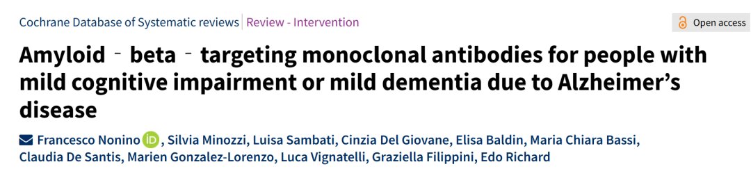
22. Regarding the neurodegenerative diseases you mentioned, the mainstream approach to treating Alzheimer's disease is to eliminate Aβ and tau proteins through various methods. However, according to a 2018 report released by the Association for the Study of Pharmaceutical Manufacturers and Research in the United States, from 2000 to 2017, global pharmaceutical companies invested over $600 billion in Alzheimer's disease research and development, with over 300 clinically approved drugs failing, a failure rate exceeding 99%. This is also a major piece of evidence refuting the theory that aging is caused by damage accumulation.