
Toby Schmitt, MD

263 posts

Toby Schmitt, MD banner
Toby Schmitt, MD

Toby Schmitt, MD

@TobyASchmitt

Dermpath fellow | Developer of https://t.co/mXLTq4CIbQ, https://t.co/S3gFkr0ifl

Vancouver, Canada Katılım Ekim 2022
364 Takip Edilen1.3K Takipçiler
Sabitlenmiş Tweet
Toby Schmitt, MD
Toby Schmitt, MD@TobyASchmitt·
Couldn't be more excited to announce the launch of pathlibrary.com, the virtual slide site I wish had existed during my pathology residency. You can: - Order special stains to work up cases - See annotated histologic features - Prepare for exams with board-style questions
Toby Schmitt, MD tweet media
English
19
99
387
195.3K
Toby Schmitt, MD
Toby Schmitt, MD@TobyASchmitt·
@dgbrackett True... Sebaceous glands don't really sway me as they can be present in both.
English
0
0
1
15
Diane Brackett, MD
Diane Brackett, MD@dgbrackett·
@TobyASchmitt I'm glad they are both benign😅 Do the little foci of sebaceous glands help point one way or another?
Diane Brackett, MD tweet media
English
1
0
3
129
Toby Schmitt, MD
Toby Schmitt, MD@TobyASchmitt·
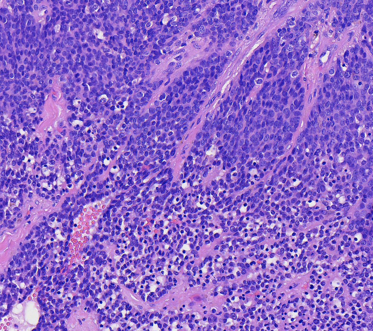
Mesenchymal chondrosarcoma shows a histologic triad of: * Mature cartilage * Atypical small round blue cells * Thin-walled, branching vessels See the labelled whole slide at: pathlibrary.com/slidebox/?case…
Toby Schmitt, MD tweet mediaToby Schmitt, MD tweet mediaToby Schmitt, MD tweet media
English
2
15
39
2K
Toby Schmitt, MD
Toby Schmitt, MD@TobyASchmitt·
@ReyPathology Are you referring to the whole slide image (on the left) or the static images from the differential diagnoses (on the right)?
English
0
0
0
200
Severino Rey MD, PhD
Severino Rey MD, PhD@ReyPathology·
@TobyASchmitt Congratulations on this important initiative. I am asking for your help to be able to open the cases. I hover over the image to enlarge it, but it does not move. I have tried both on my iPhone and on a desktop computer without success. Could you guide me on how to achieve this
English
1
0
0
230
Toby Schmitt, MD
Toby Schmitt, MD@TobyASchmitt·
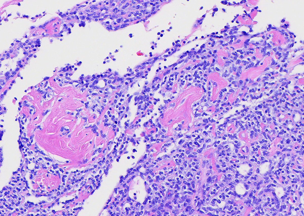
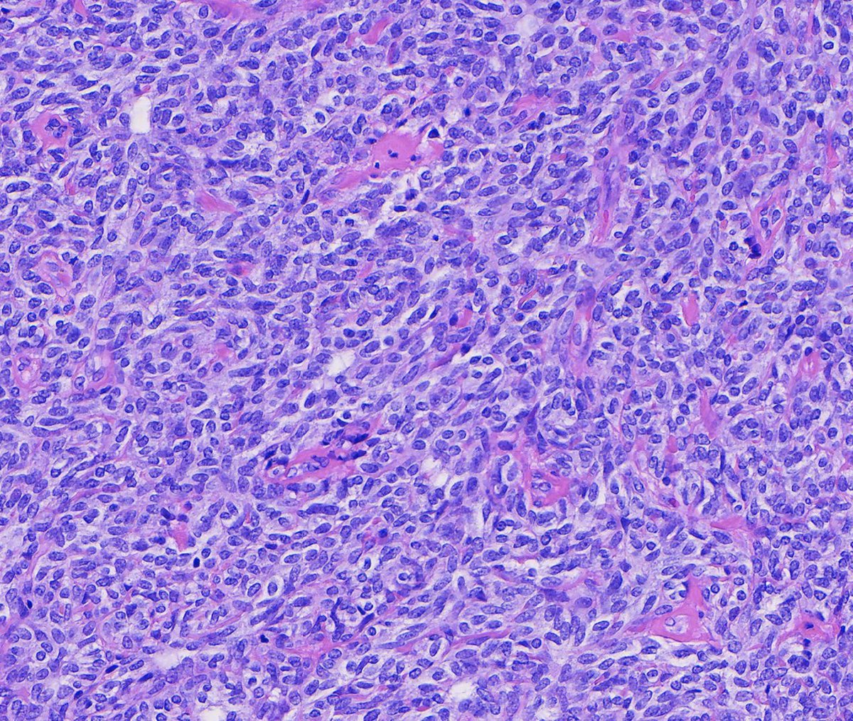
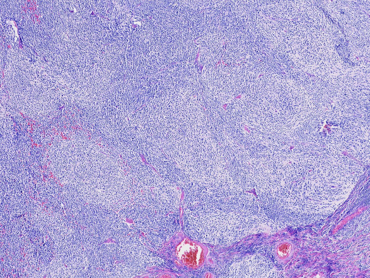
New pathlibrary.com cases dropped: We're past the 1450 cases mark. Try your hand at working up this challenging soft tissue tumor (case #1450). (Hint: FOSB IHC+)
Toby Schmitt, MD tweet media
English
8
13
62
5.1K
Toby Schmitt, MD
Toby Schmitt, MD@TobyASchmitt·
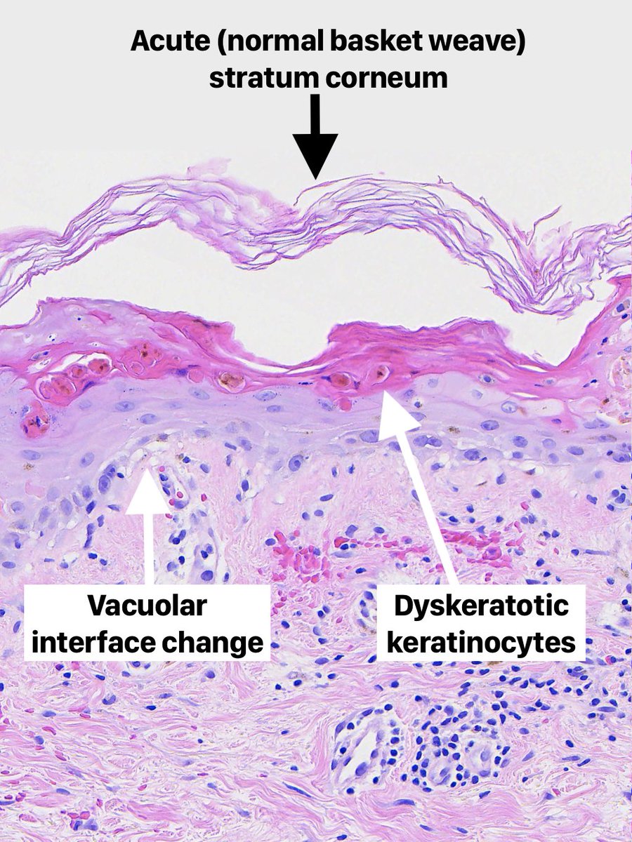
Acute vacuolar interface change with dyskeratosis: A pattern with a narrow differential: * Erythema multiforme, TEN/SJS * Fixed drug eruption (usually has dermal melanin) * Acute GvHD * Reactive infectious mucocutaneous eruption (RIME) WSI & discussion: pathlibrary.com/slidebox/?case…
Toby Schmitt, MD tweet mediaToby Schmitt, MD tweet media
English
1
10
30
1.1K
Toby Schmitt, MD
Toby Schmitt, MD@TobyASchmitt·
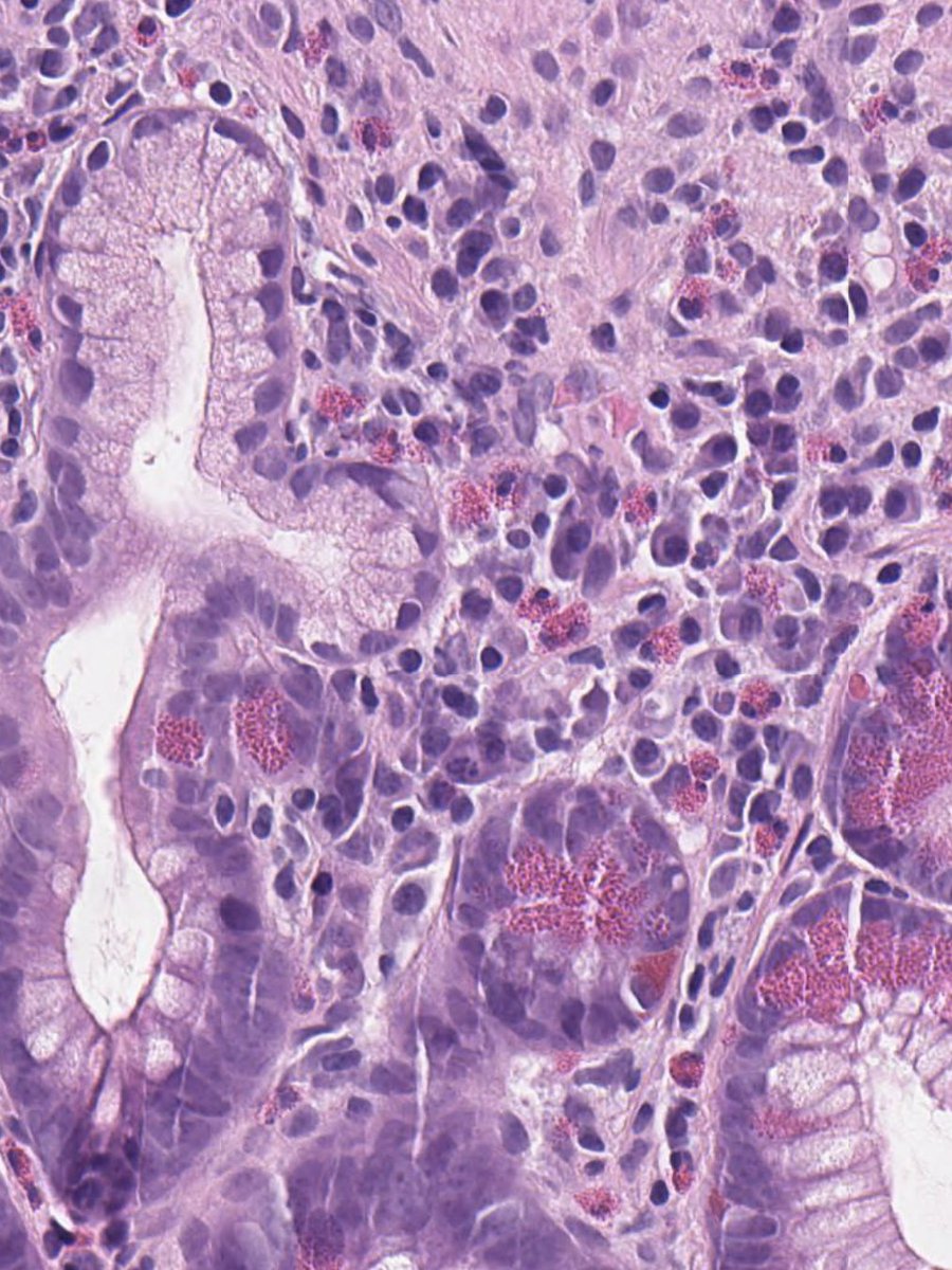
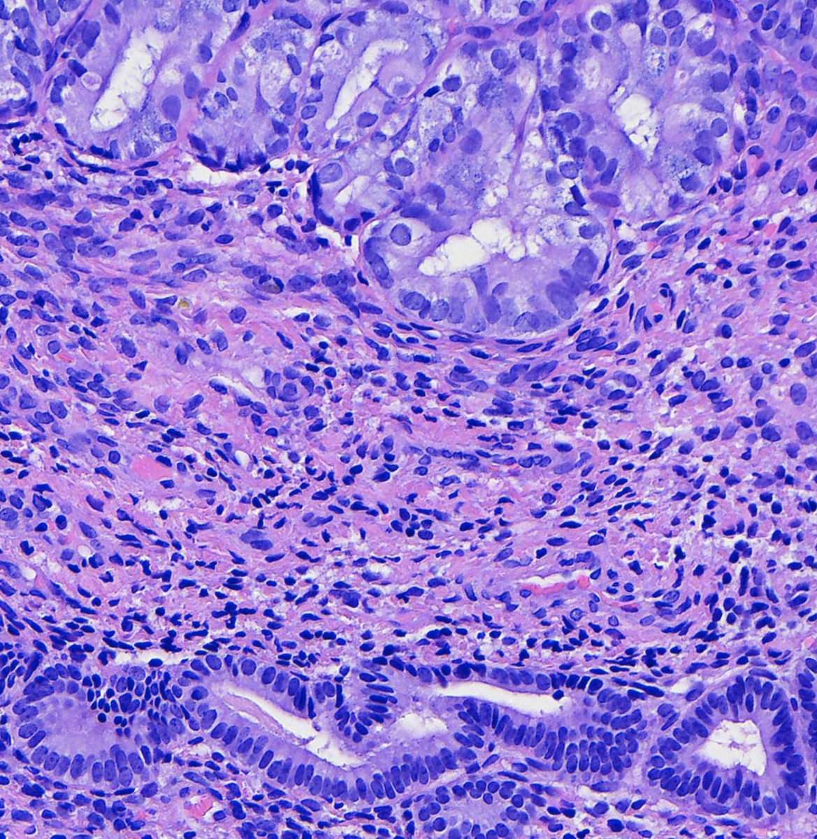
Chronic colitis with pyloric gland metaplasia (top left) and Paneth cell metaplasia (bottom right - their granules look similar to eosinophil cytoplasm). Paneth cells are normal in the right side of the colon but signify colitis in the left side. WSI: pathlibrary.com/slidebox/?case…
Toby Schmitt, MD tweet media
English
1
10
39
1.2K
Toby Schmitt, MD
Toby Schmitt, MD@TobyASchmitt·
A case of pityriasis lichenoides showing its classic features: Mounds of parakeratosis, red cell extravasation, lichenoid inflammation, and lymphocytic exocytosis. Labelled whole slide: pathlibrary.com/slidebox/?case…
Toby Schmitt, MD tweet mediaToby Schmitt, MD tweet mediaToby Schmitt, MD tweet media
English
1
4
27
1.3K
Toby Schmitt, MD
Toby Schmitt, MD@TobyASchmitt·
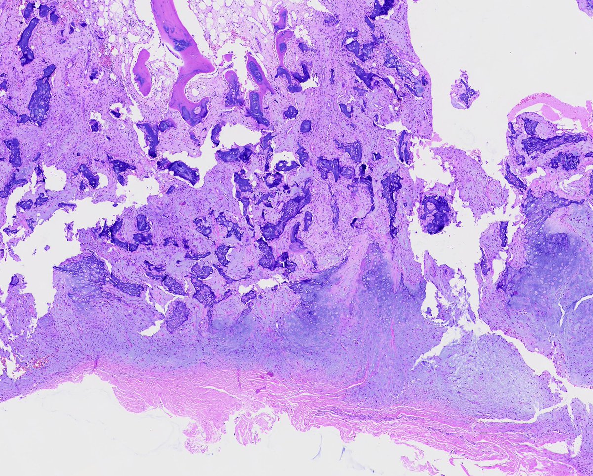
A bizarre parosteal osteochondromatous proliferation (BPOP) with disorganized cartilage, disorganized bone, and bland fibrous tissue. BPOP is benign but locally destructive and has a propensity for the small bones of the hands & feet. Labelled WSI: pathlibrary.com/slidebox/?case…
Toby Schmitt, MD tweet mediaToby Schmitt, MD tweet mediaToby Schmitt, MD tweet mediaToby Schmitt, MD tweet media
English
2
24
57
2K
Toby Schmitt, MD
Toby Schmitt, MD@TobyASchmitt·
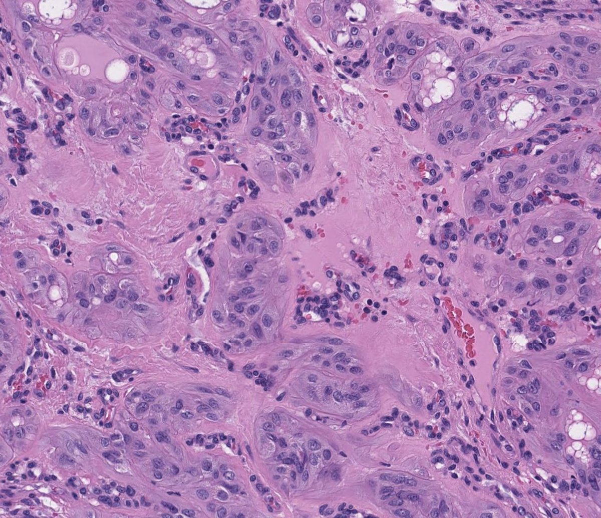
@BaiBai31141 This would be a tough one to get on frozen, although the monomorphic cytology would point away from clear cell carcinoma.
English
0
0
0
30
bai cherry
bai cherry@BaiBai31141·
@TobyASchmitt A very good case.I am curious about how to differentiated from clear cell carcinoma in frozen section ?
English
1
0
0
43
Toby Schmitt, MD
Toby Schmitt, MD@TobyASchmitt·
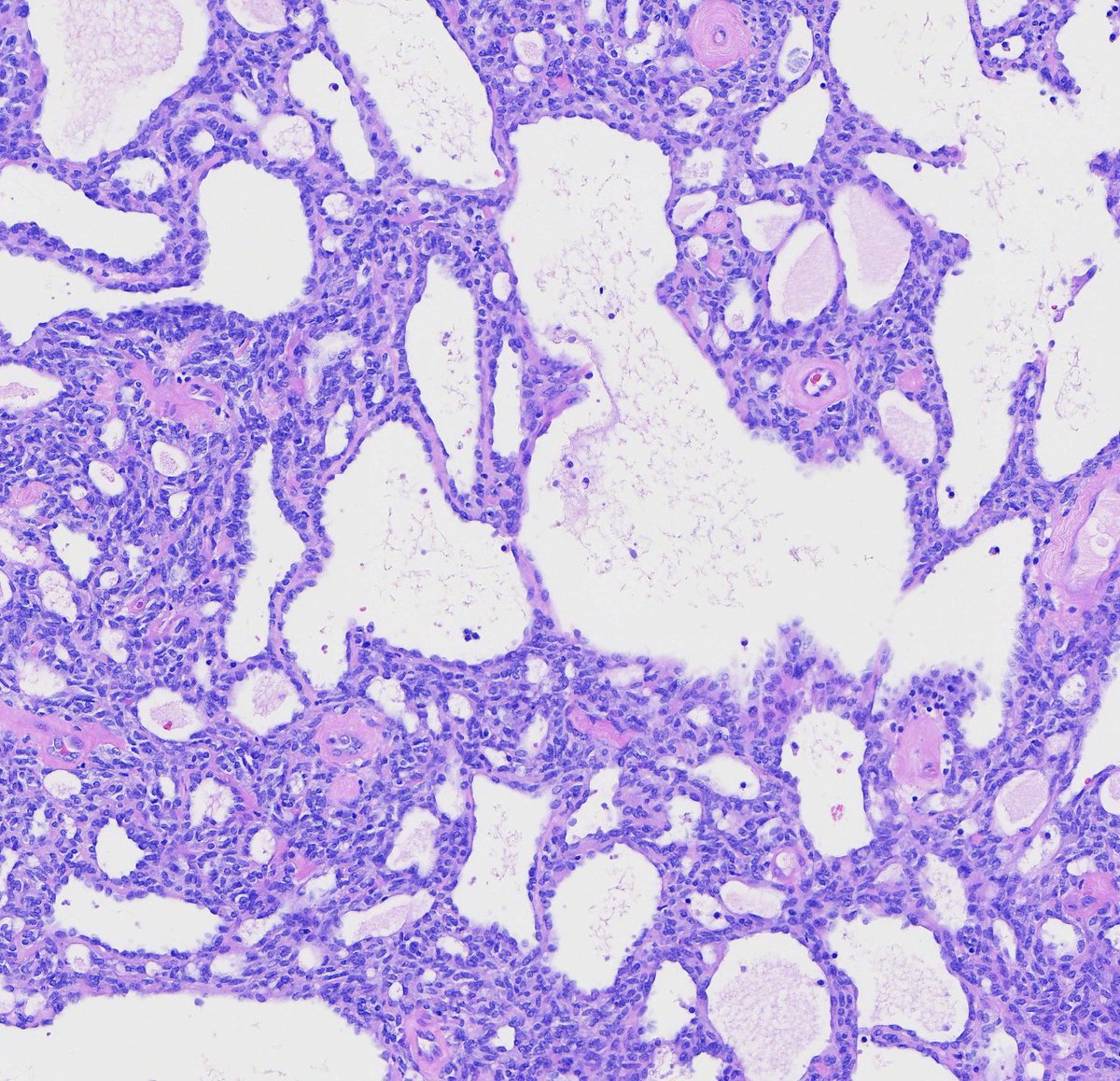
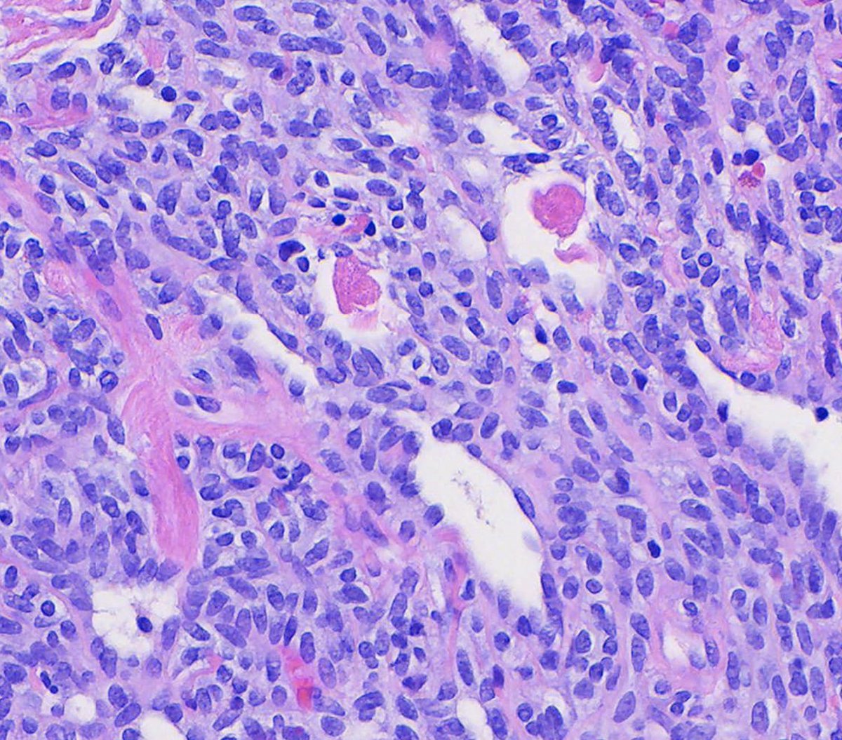
A case of a Wolffian tumor (previously female adnexal tumor of probable Wolffian origin/FATWO). These lesions typically show a variety of architectural patterns including tubular, cystic, and solid. Tubules filled with eosinophilic material are common. WSI: pathlibrary.com/slidebox/?case…
Toby Schmitt, MD tweet mediaToby Schmitt, MD tweet mediaToby Schmitt, MD tweet mediaToby Schmitt, MD tweet media
English
4
14
62
1.9K
Toby Schmitt, MD
Toby Schmitt, MD@TobyASchmitt·
Atypical endometrial hyperplasia (top) and background benign endometrial glands (bottom). Note the contrast between small, dark benign nuclei and enlarged, pale, vesicular nuclei of atypical hyperplasia. Labelled whole slide: pathlibrary.com/slidebox/?case…
Toby Schmitt, MD tweet mediaToby Schmitt, MD tweet media
English
0
15
57
2K
Toby Schmitt, MD
Toby Schmitt, MD@TobyASchmitt·
A desmoplastic nodular medulloblastoma showing characteristic paler areas among more densely-packed small round blue cells. Molecularly, this subtype of medulloblastoma tends to be SHH (sonic hedgehod pathway)-activated. See the whole slide at: pathlibrary.com/slidebox/?case…
Toby Schmitt, MD tweet mediaToby Schmitt, MD tweet media
English
2
7
26
1.1K
Toby Schmitt, MD
Toby Schmitt, MD@TobyASchmitt·
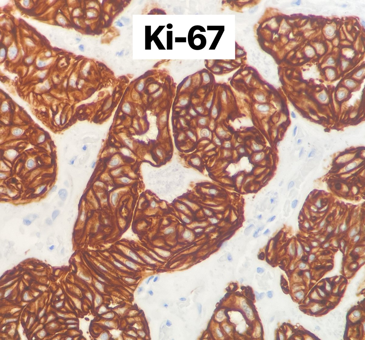
Hyalinizing trabecular tumor is an aptly-named benign thyroid neoplasm. Ki-67 shows an unusual membranous pattern in these lesions. What other lesion characteristically shows this Ki-67 staining pattern? Answer at: pathlibrary.com/slidebox/?case…
Toby Schmitt, MD tweet mediaToby Schmitt, MD tweet mediaToby Schmitt, MD tweet mediaToby Schmitt, MD tweet media
English
4
42
98
4.5K
Toby Schmitt, MD
Toby Schmitt, MD@TobyASchmitt·
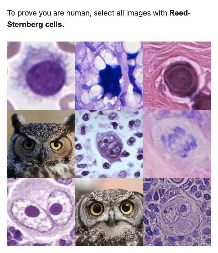
Trying to combat the number of bot accounts signing up for PathLibrary...
Toby Schmitt, MD tweet media
English
1
7
44
1.3K
Toby Schmitt, MD
Toby Schmitt, MD@TobyASchmitt·
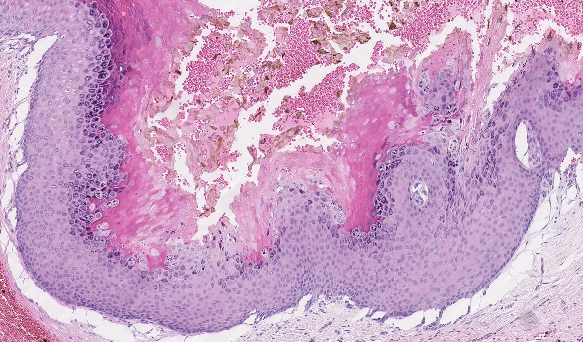
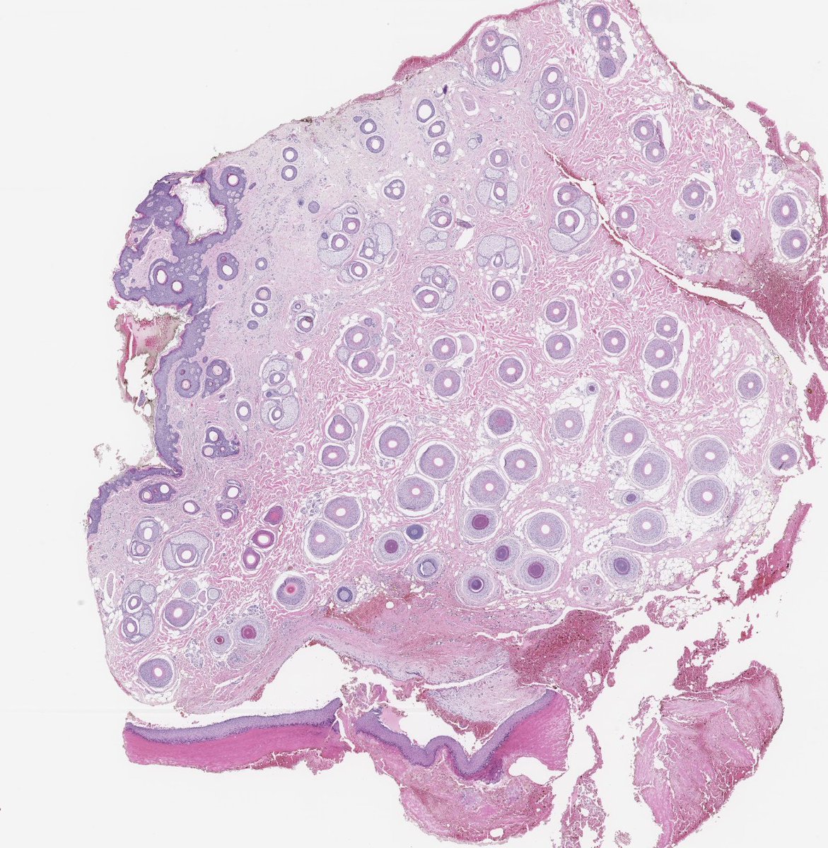
A verrucous cyst showing the same papillomatosis, hypergranulosis, and coarse keratohyalin granules of verruca vulgaris. These lesions may result from low-risk HPV infection of pre-existing epidermoid cysts. Whole slide: pathlibrary.com/slidebox/?case…
Toby Schmitt, MD tweet mediaToby Schmitt, MD tweet mediaToby Schmitt, MD tweet media
English
0
5
21
1.5K
Toby Schmitt, MD
Toby Schmitt, MD@TobyASchmitt·
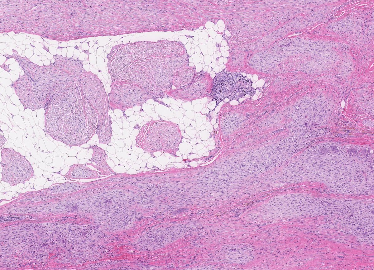
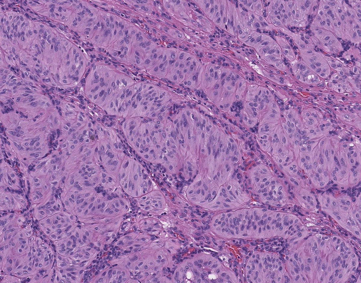
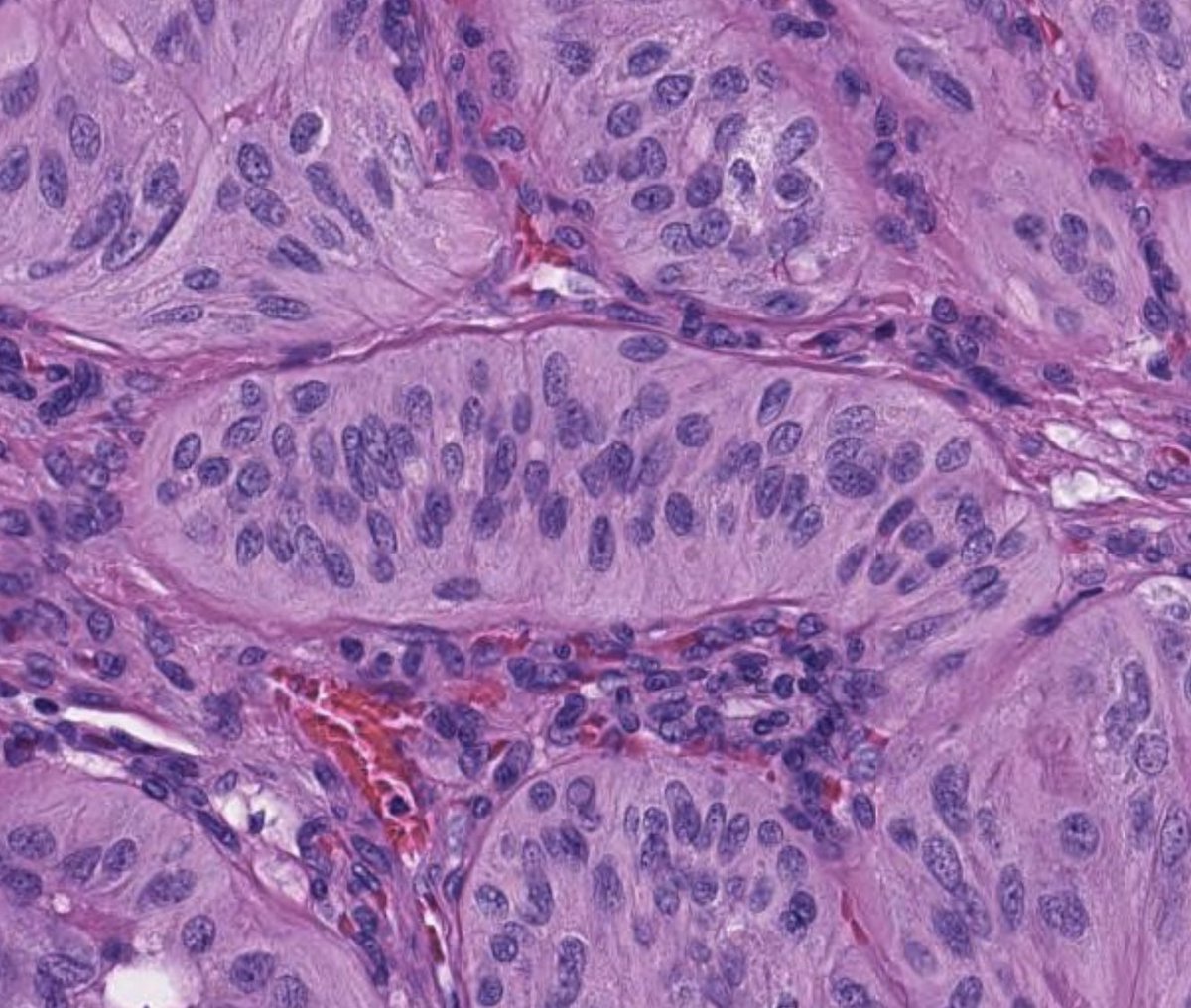
Plexiform fibrohistiocytic tumors are rare soft tissue tumors of intermediate malignant potential. They have a plexiform (woven) architecture with fascicles of histiocytoid cells in a collagenous stroma. See the whole slide: pathlibrary.com/slidebox/?case…
Toby Schmitt, MD tweet mediaToby Schmitt, MD tweet mediaToby Schmitt, MD tweet mediaToby Schmitt, MD tweet media
English
0
19
46
1.4K
Toby Schmitt, MD
Toby Schmitt, MD@TobyASchmitt·
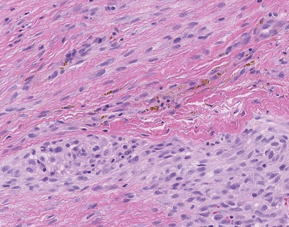
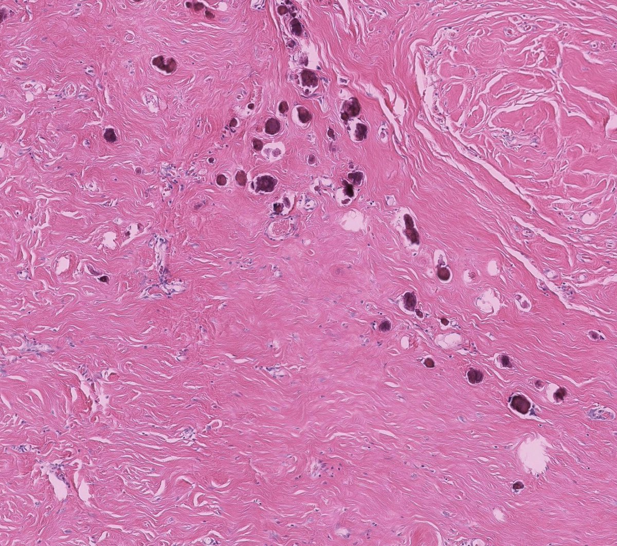
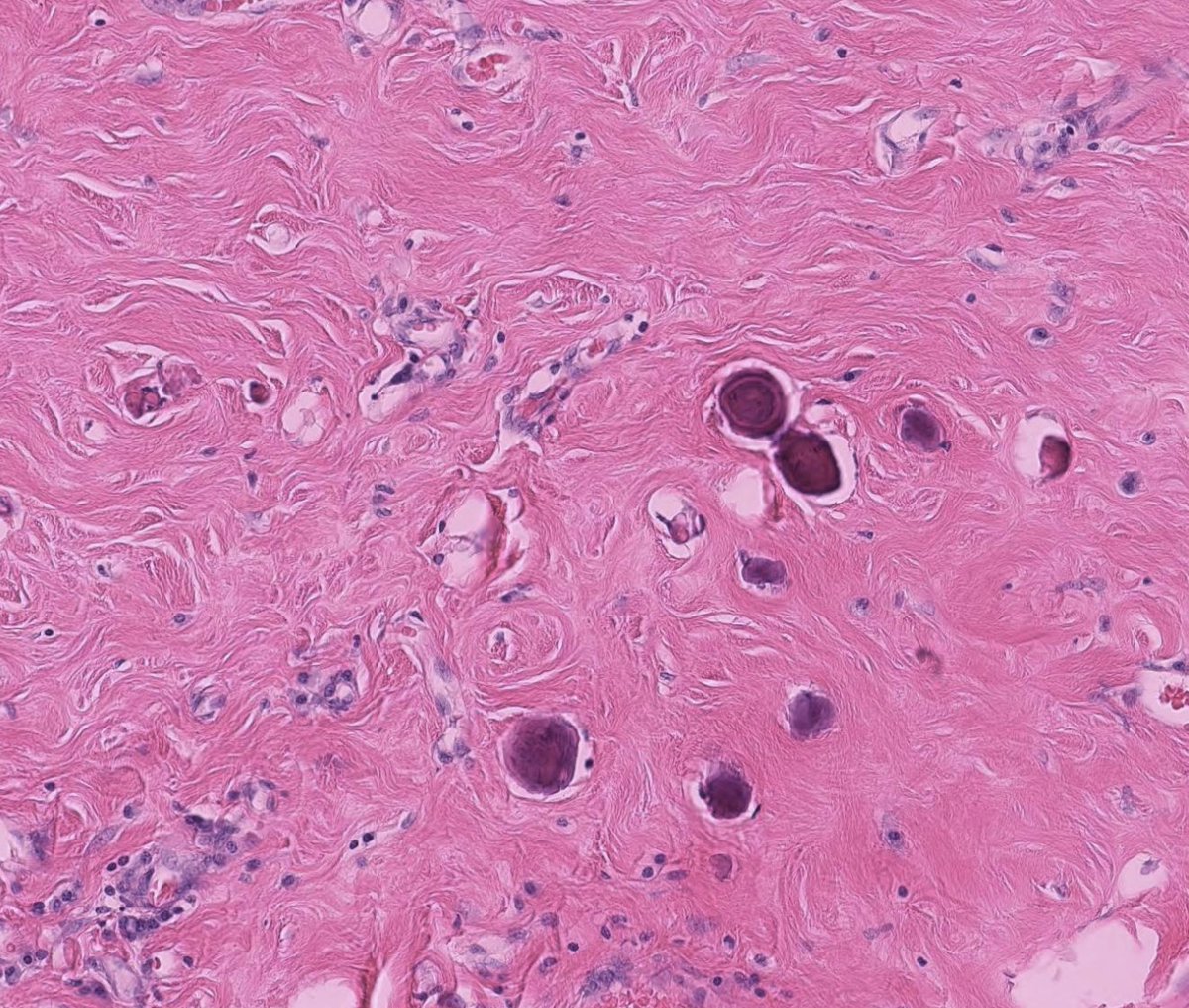
A case of calcifying fibrous tumor, an entity characterized by dense, compact collagen and scattered calcification. These lesions may represent burnt-out IgG4-related disease. Whole slide: pathlibrary.com/slidebox/?case…
Toby Schmitt, MD tweet mediaToby Schmitt, MD tweet mediaToby Schmitt, MD tweet media
English
1
16
55
1.9K