Закреплённый твит
Alex
19.6K posts


Alex ретвитнул

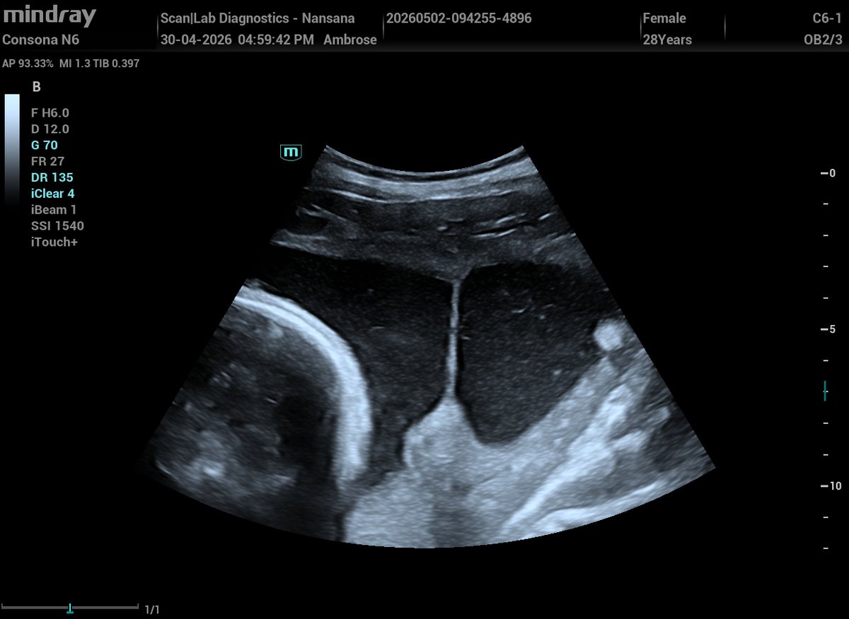
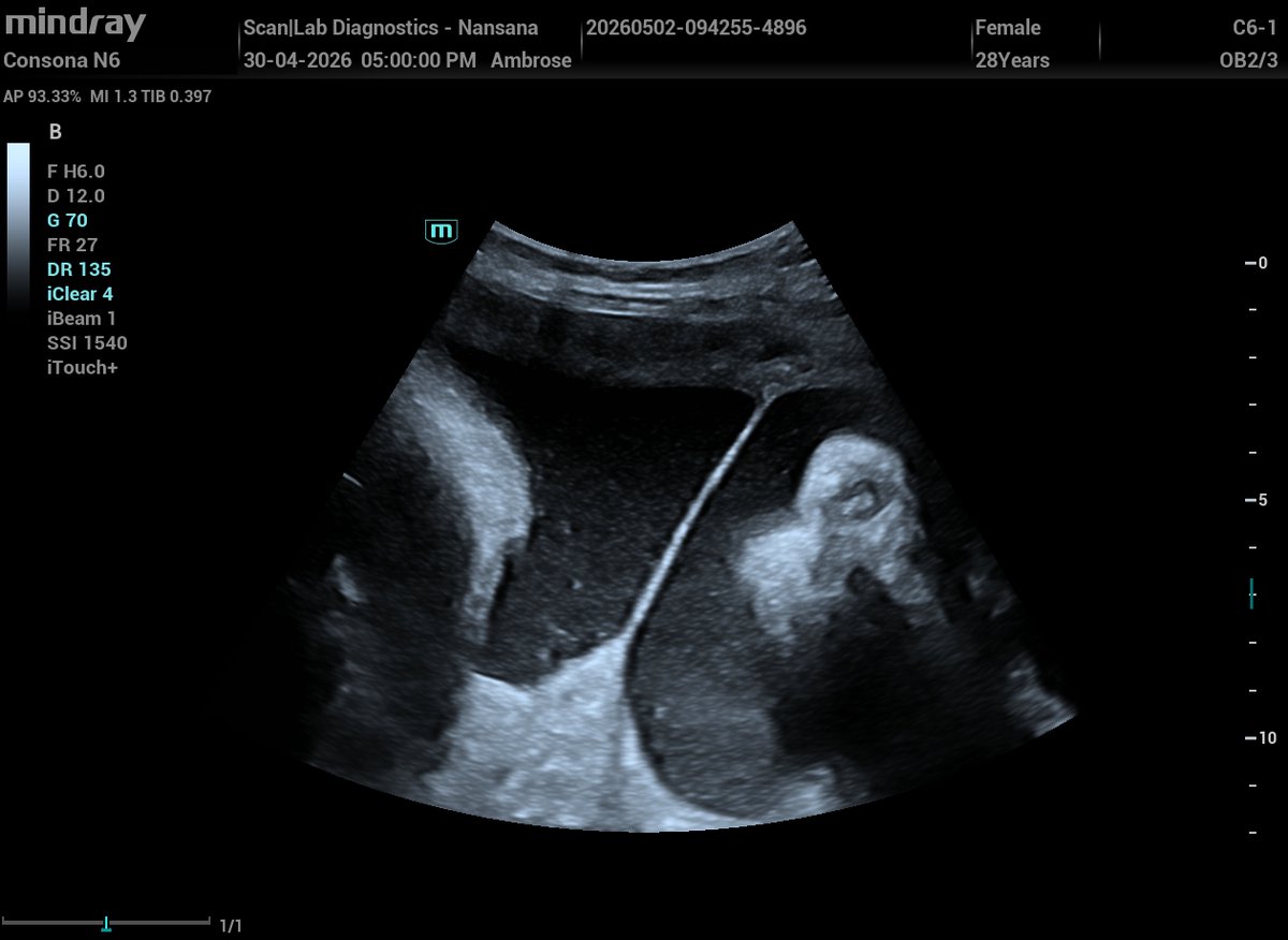
Amniotic Sheet (Synechia) vs Amniotic Band
Amniotic Sheet
Echogenic thick septa just like a separating septum in twin pregnancy,
A broad base attached to uterine wall, free edge but not free‑floating
No attachment to fetal parts,
Often shows Doppler flow along the membrane
Benign and incidental finding, but can influence fetal lie,
Always a single sheet.
Amniotic Band (ABS)
Thin, irregular, free‑floating strands
Attaches to fetal parts restricting movement
Can cause constriction rings, amputations, craniofacial defects
No Doppler flow
It is associated with fetal abnormalities
Case example:
At 34 weeks, a scan shows a thick septum arising from the placental edge and extending across the uterine cavity to the anterior wall.
This is a classic amniotic sheet; It is formed when the amnion drapes over a uterine synechia.
An amniotic sheet is a normal variant with no fetal risk whereas an amniotic band is pathological and can cause serious fetal deformities.
Amniotic sheet 👇👇

English
Alex ретвитнул

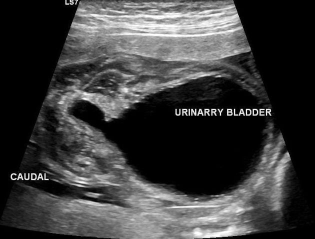
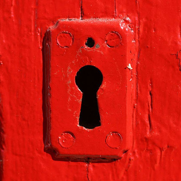
THE “KEYHOLE SIGN” YOU SHOULD NEVER MISS ON FETAL ULTRASOUND 🚨
🔑 THE KEYHOLE SIGN is an important prenatal ultrasound finding in male fetuses suggestive of Posterior Urethral Valves (PUV).
📸 On ultrasound, the dilated posterior/proximal urethra along with a distended urinary bladder create a configuration that resembles a keyhole, hence the name.
📍 Posterior Urethral Valves (PUV), also known as Congenital Obstructing Posterior Urethral Membranes (COPUM) represent the most common cause of congenital urethral obstruction in male infants and can lead to obstructive uropathy, bladder dysfunction, and even long-term renal compromise if not addressed.
#Ultrasound #FetalUltrasound
English
Alex ретвитнул

Alex ретвитнул
The tau sign represents the appearance of a persistent primitive trigeminal artery on the sagittal plane of an angiogram or on sagittal MRI.
It resembles the Greek letter τ, pronounced "tau."
🚨 SIMPLE QUIZ
1. The vertical and anterior horizontal limbs of "tau" are formed by the ____________ artery.
2. The posterior horizontal limb is formed by the ________________ artery.

English
Alex ретвитнул

Alex ретвитнул

Alex ретвитнул


Alex ретвитнул

1/Do radiologists sound like they are speaking a different language when they talk about MRI?
T1 shortening what? T2 prolongation who?
Here’s a translation w/an introductory thread to MRI.

English
Alex ретвитнул

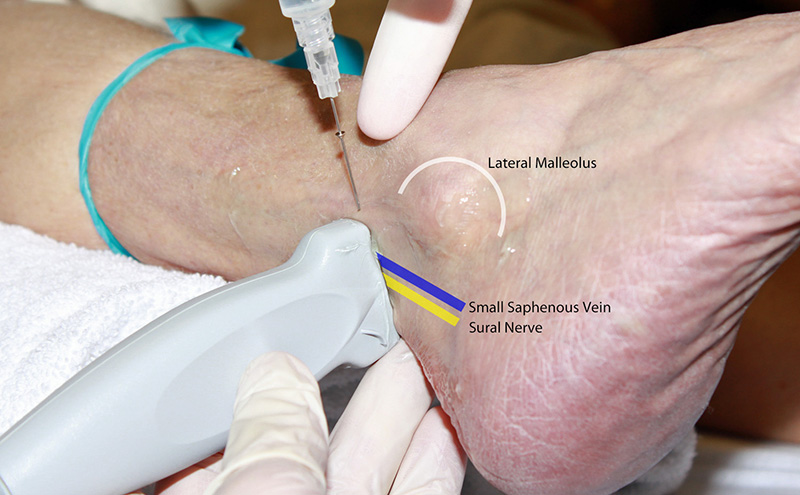
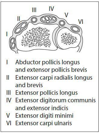
✅حقن المخدر الموضعي تحت الجلد خلف الـ lateral malleolus راح يخدر الـ sural nerve، وهذا العصب أصلاً عصب حسي بحت ما فيه وظيفة حركية، وهو يجي من تفرع الـ tibial nerve والـ common fibular nerve مع بعض، ويمشي من الجزء الخلفي للساق لين يوصل لمنطقة الكعب الخارجي ويطلع على ظهر القدم من الجهة الخارجية وحتى الخنصر، يعني الـ lateral aspect of the foot والـ little toe هي المنطقة اللي راح تتخدر، والفايدة من هالحقنة إنك تقدر تسوي أي إجراء جراحي أو تشخيصي في هالمنطقة بالذات زي عمليات الـ lateral foot، أو تثبيت كسور في منطقة الـ 5th metatarsal، أو حتى أخذ خزعة من الجلد في هالمنطقة، وأحياناً يستخدمونه في حالات الـ chronic pain اللي تكون في توزيع هذا العصب زي الـ sural neuritis، والحقنة نفسها سهلة وآمنة لأن العصب يمشي مع الـ small saphenous vein خلف الـ lateral malleolus مباشرةً، ف الـ landmark واضح وما تحتاج ultrasound في الغالب.
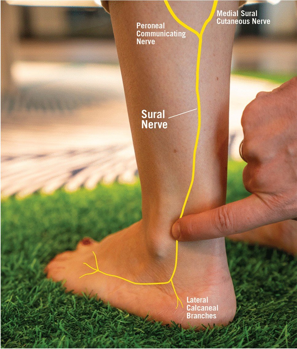
احمد@AB_drmd
🚨🚨🚨حقن مخدر موضعي تحت الجلد خلف الـ lateral malleolus، يخدر اي عصب؟ A) Sural nerve B) Superficial fibular nerve C) Saphenous nerve D) Posterior tibial nerve
العربية
Alex ретвитнул

Excited to share our article on Imaging of Ewing Sarcoma, now available open access in @skeletaljournal rdcu.be/fgmJU link.springer.com/article/10.100…
English
Alex ретвитнул
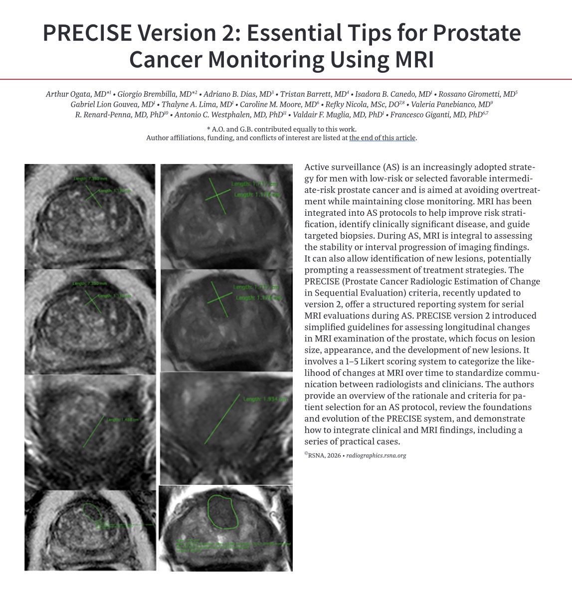
📢 Exciting Read for Radiologists and Urologists🚀
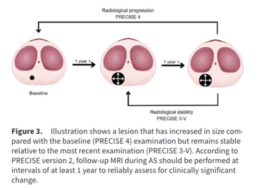
📚Just came across this fantastic educational article in @RadioGraphics “PRECISE Version 2: Essential Tips for Prostate Cancer MRI Reporting”
🔔This paper is a must-read for anyone involved in prostate MRI!
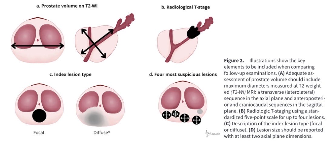
✅It provides clear, practical guidance on using the updated PRECISE v2 system to standardize reporting for patients on active surveillance.
👍Key highlights include refined scoring for assessing disease stability/progression, helpful checklists, case examples, and tips to reduce ambiguity in serial MRI interpretation!
💡Whether you’re a radiologist fine-tuning your reports, a urologist discussing imaging with your patients, or a trainee building your skills — this valuable resource makes complex concepts more approachable and clinically impactful!
🥇Congrats to @giga_fra and the entire team for this nice work!
🔗 Full article: pubs.rsna.org/doi/10.1148/rg…
#Radiology #ProstateCancer #MRI #ActiveSurveillance #RadioGraphics

English
Alex ретвитнул

✅Diffuse leptomeningeal glioneuronal tumor (DL-GNT).
💡Think of it when you see widespread leptomeningeal nodules with cystic-like components in a child. It can mimic infection or metastatic spread, but slow progression and this pattern should raise suspicion.
doi.org/10.3390/diagno…
Doc Navarrow@DocNavarrow
🧒4-year-old child with progressive gait disturbance and cranial nerve symptoms. MRI reveals multiple cystic-appearing nodular intradural lesions with diffuse leptomeningeal enhancement, extending through the posterior fossa and spinal canal. 🤔What is your differential?
English
Alex ретвитнул

Alex ретвитнул

Alex ретвитнул
1/Does your ability to remember temporal lobe anatomy seem, well, temporary?
Or are you feeling temporally challenged when it comes to this complex region?
Here’s a thread to help you remember the structures of the temporal lobe!

English
Alex ретвитнул

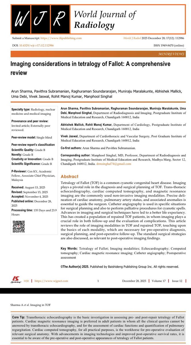
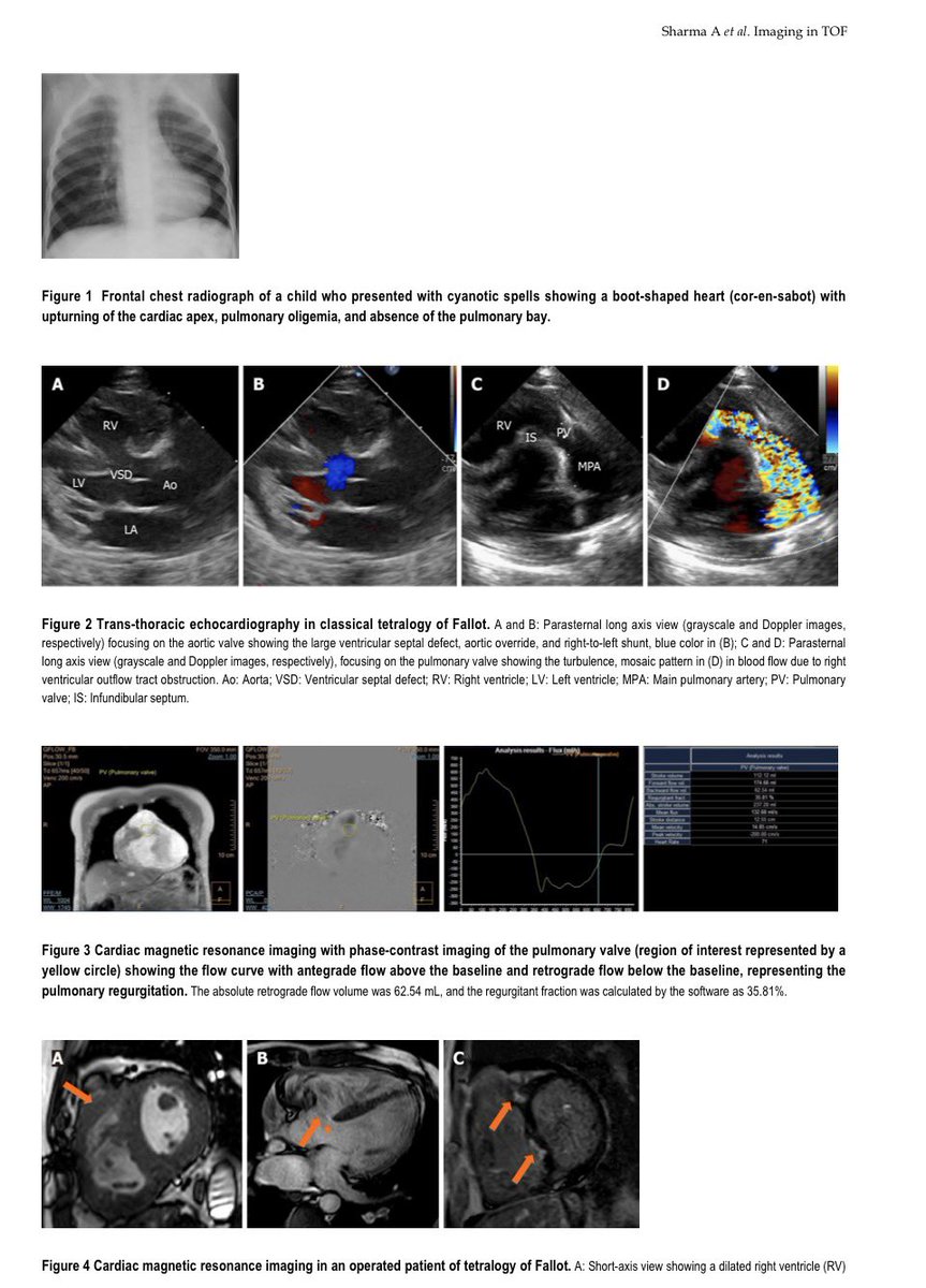
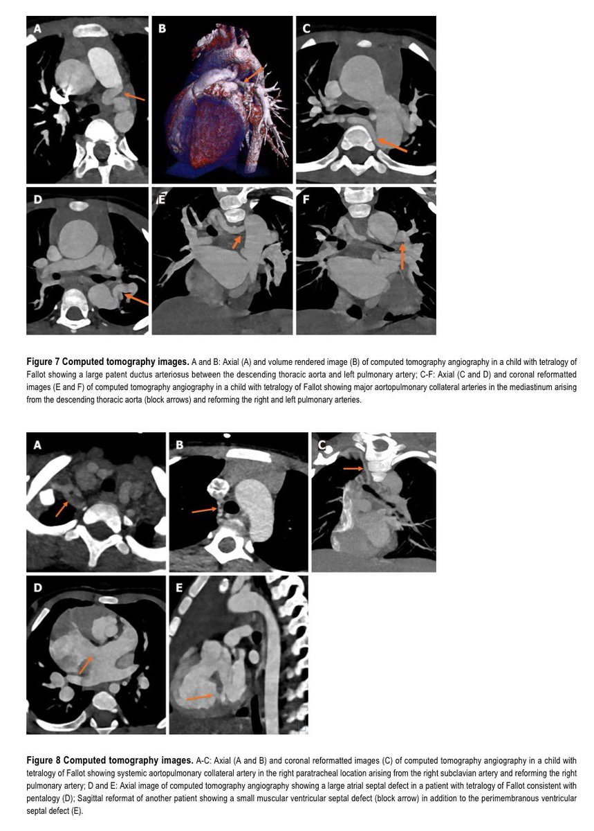
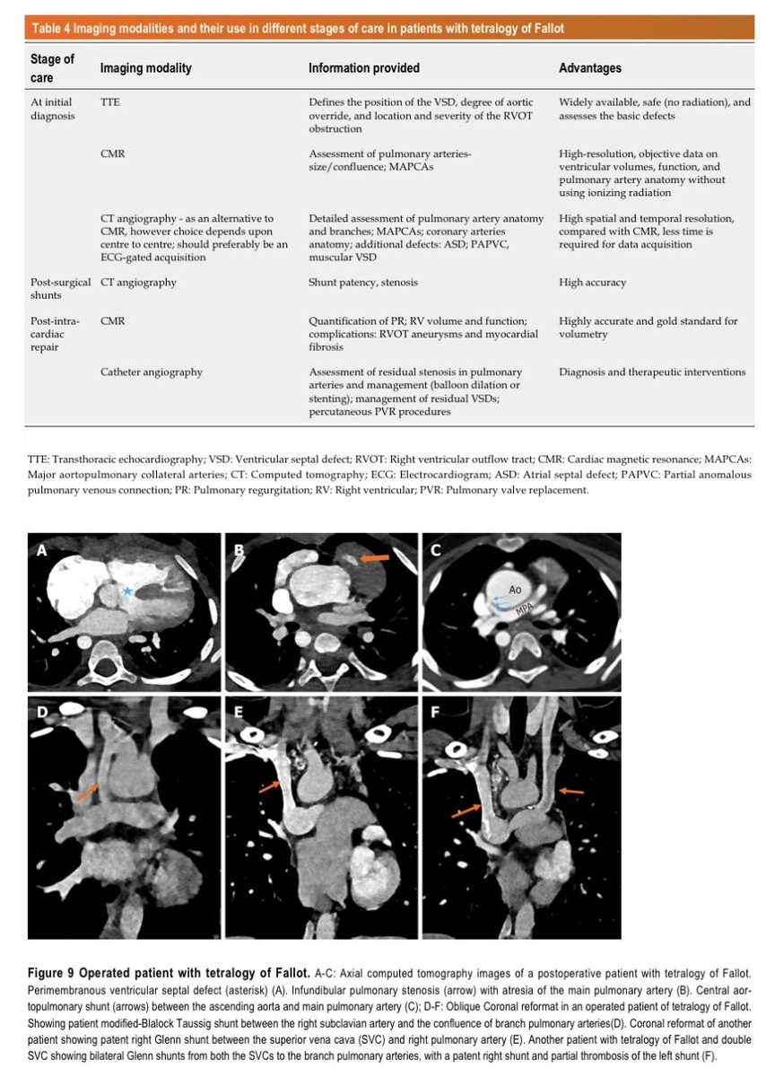
Imaging considerations in #tetralogy_Fallot
a comprehensive approach.
#echofirst #whyCMR #yesCCT
#CHD #cardioped
doi.org/10.4329/wjr.v1…
English
Alex ретвитнул

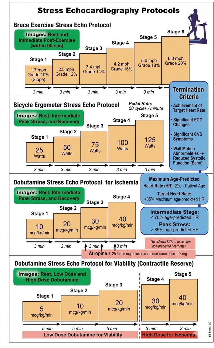
Stress Echocardiography Protocols
Read it. Save it. Share it 😍
English
Alex ретвитнул

Alex ретвитнул

English
Alex ретвитнул

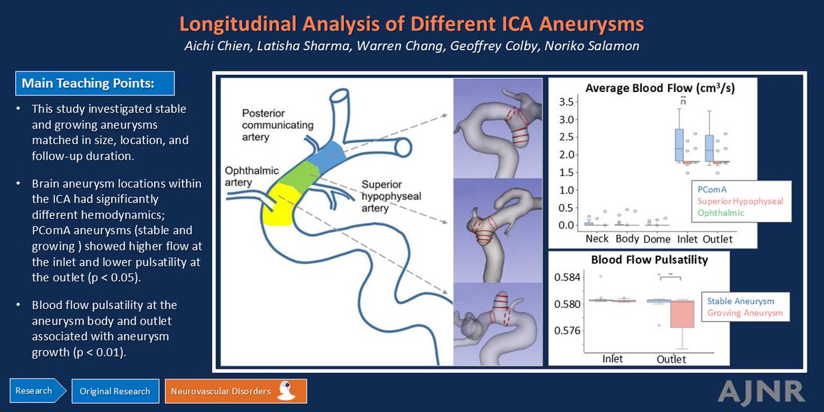
"Longitudinal Analysis of Location- and Growth-Associated Hemodynamics in Matched Paraclinoid Aneurysms"
doi.org/10.3174/ajnr.A…
@Chienlab_UCLA @RadiologyUCLA
English