We are so excited that Jeff was named a 2023 #DoD Vannevar Bush Faculty Fellow, and look forward to participating in this unique and rewarding program!
news.rice.edu/news/2023/rice…
English
Tabor Lab
163 posts
@LabTabor
We are a synthetic biology research group in the Bioengineering Department at Rice University.


I'm happy to share our work on computer-aided synthesis planning of hybrid enzymatic/synthetic reaction pathways with @Geneticdesigner and @cwcoley! nature.com/articles/s4146…
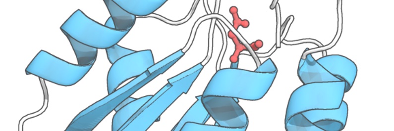
Interested in bacterial peptide sensing? Check out my latest paper! Thanks to all of my amazing co-authors in @LabTabor lab + beyond: @synthetic_queer Andrew Mu @KGroszman @KyVanHoang @kevin_lorch @bhpogostin @DrGunn43 @jeffrey_j_tabor nature.com/articles/s4158…
Here's our new article -- potentially, everything you ever wanted to know about light-regulated gene expression in bacteria (& much more). frontiersin.org/articles/10.33… Welcome opportunity to team up once again with @RobertOhlendorf (this guy knows stuff!). Enjoy


The Department of BioSciences @RiceUniversity is hiring an Assistant Professor in Neuroscience! Beyond the department, there are unique opportunities to engage with Neuroengineers @RiceNeuro and faculty at world class medical institutions @TXMedCenter. apply.interfolio.com/110165 PRT
1/n PAPER ALERT: In collaboration with @davidrliu, we combined eVOLVER + PACE (= ePACE) to facilitate the evolution of compact Nme2Cas9 variants that can target single-nucleotide (previously-inaccessible) PAM sequences for precision genome editing. go.nature.com/3d1A9Y3