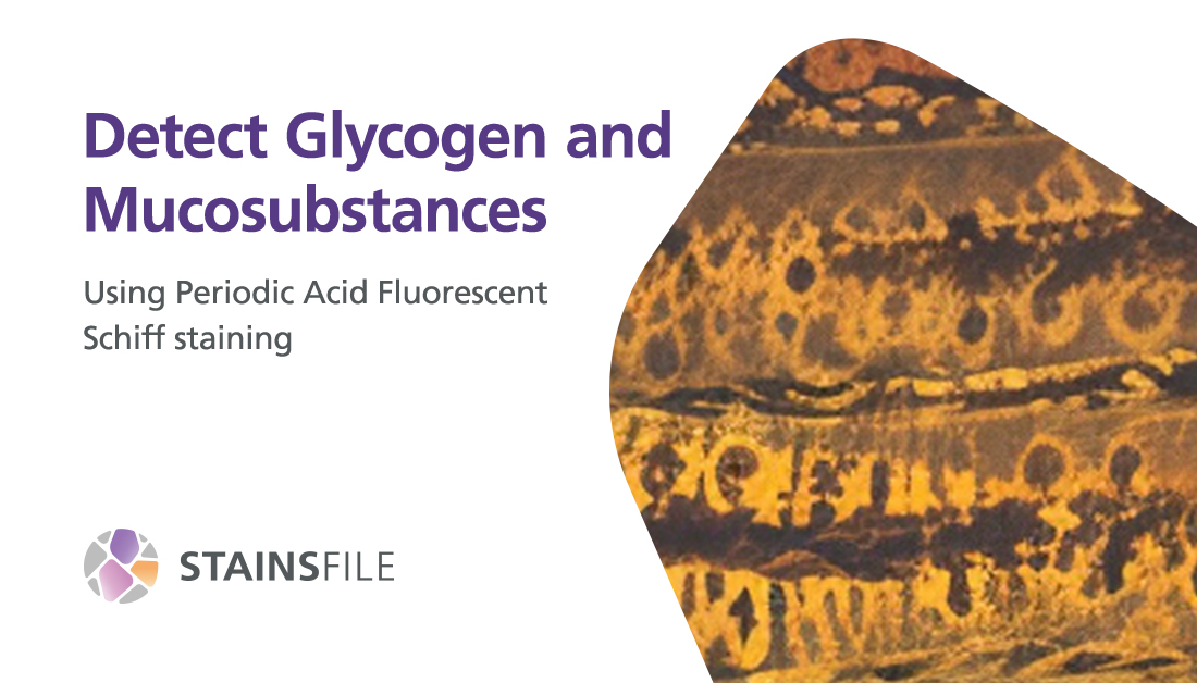
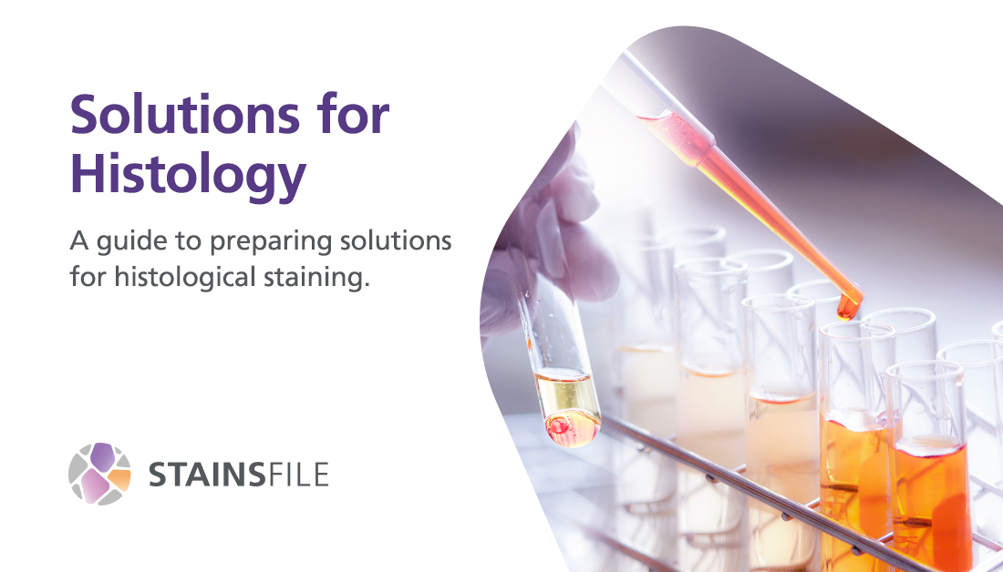
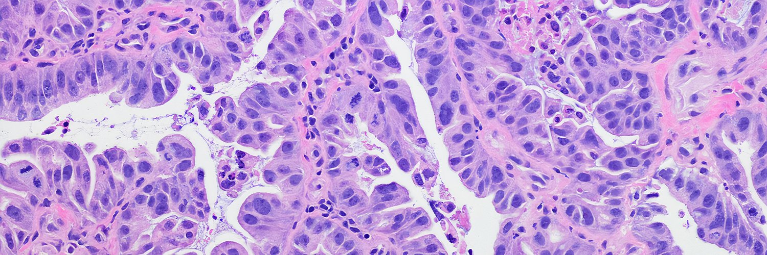
StainsFile

905 posts

StainsFile banner
StainsFile

StainsFile

@StainsFile

Let’s talk about visualizing cells! 👩🏽‍🔬🔬 We’re an online resource for open-access protocols, reagents, theory, and more. Brought to you by @STEMCELLTech.

Bergabung Ekim 2019
542 Mengikuti166 Pengikut
StainsFile
StainsFile@StainsFile·
In a new @SciReports study, researchers performed 3D reconstructive, histological analyses and immunohistochemistry on postmortem human inner ear specimens. This sheds new light on endolymphatic hydrops, paving the way for new therapies: go.nature.com/4kgZ9IS
StainsFile tweet media
English
0
0
0
8
StainsFile
StainsFile@StainsFile·
☕ + 🧫 = 🔬 Researchers at @tugraz have discovered that ordinary espresso can be used as an environmentally friendly alternative to toxic uranyl acetate for staining biological samples in #ElectronMicroscopy! Check out how they did it: bit.ly/4ti2lrQ
StainsFile tweet media
English
0
0
1
14
StainsFile
StainsFile@StainsFile·
🔎 Glycogen and mucosubstances don’t stand a chance. This periodic acid fluorescent Schiff protocol helps you get crisp, high-contrast fluorescence imaging from neutral buffered formalin–fixed tissue samples: bit.ly/4kw7ecT
StainsFile tweet media
English
0
0
0
28
StainsFile
StainsFile@StainsFile·
Passive tissue-clearing methods rely on sodium dodecyl sulfate as a delipidating detergent, which can cause tissue damage. To address this, researchers developed a novel passive tissue clearing technique: OptiMuS-prime. More details here: go.nature.com/3MiRtcM
StainsFile tweet media
English
0
0
0
67
StainsFile me-retweet
WebPathology
WebPathology@WebPathology·
Adult female with history of high-grade uroth. CA. Surveillance cystoscopy showed multiple yellow-tan plaques near trigone. What's your diagnosis? #gupath for #pathresidents Diagnosis & additional images in the comment below.
WebPathology tweet mediaWebPathology tweet media
English
5
11
41
2.2K
StainsFile
StainsFile@StainsFile·
A new dual-light microscope lets researchers observe micro- and nanoscale activity inside living cells without using dyes. 😯 This bidirectional quantitative scattering microscope detects both forward and backward scattered light from cells. bit.ly/4rDcmyg
StainsFile tweet media
English
0
0
0
20
StainsFile me-retweet
Allen Institute
Allen Institute@AllenInstitute·
How do different brain cell types connect with each other? We're mapping the brain to learn more. In this video from our #ElectronMicroscopy team, we see how complex brain cells can be. #BrainAwarenessWeek
English
8
75
312
18.3K
StainsFile
StainsFile@StainsFile·
This one's for all staining experts.🫟 As you advance your knowledge to answer the big questions, take a moment to brush up on the basics of staining. Learn about staining chemistry, methods, and more. bit.ly/45WakRx
StainsFile tweet media
English
0
0
0
8
StainsFile
StainsFile@StainsFile·
📶 Level up your #FluorescenceMicroscopy with hybrid-EDA! Dr. Suliana Manley's team at @EPFL_en have developed an event-driven acquisition (EDA) framework that combines the gentleness and richness of phase-contrast with the specificity of fluorescence: go.nature.com/4by4xFo
StainsFile tweet media
English
0
0
0
12
StainsFile me-retweet
WebPathology
WebPathology@WebPathology·
This bladder tumor had a little bit of everything....adult male with gross hematuria and a large bladder mass. #gupath
WebPathology tweet mediaWebPathology tweet mediaWebPathology tweet mediaWebPathology tweet media
English
3
9
32
2.2K
StainsFile
StainsFile@StainsFile·
🧬 Pathobiology 2026 is coming! Don’t miss the @ASIPath Annual Meeting in Fort Myers, Florida from May 16–19. You'll be able to share discoveries, connect with peers, and explore the latest in disease biology, #pathology, and translational science: bit.ly/3Mt6MiW
StainsFile tweet media
English
0
0
1
38
StainsFile
StainsFile@StainsFile·
Looking for a solution required for histological staining? 🧪 Get simple and reliable recipes for them: bit.ly/46al2UG
StainsFile tweet media
English
0
0
0
17
StainsFile
StainsFile@StainsFile·
Quantitative phase imaging (QPI) is great for non-invasive analysis of transparent specimens, but it can be slow, complex, and have image degradation. The Wetzstein lab's (@Stanford) new QPI platform addresses this by integrating nanophotonic metasurfaces with AI: go.nature.com/4q136Tn
StainsFile tweet media
English
0
0
0
19
StainsFile
StainsFile@StainsFile·
🤖 New research reveals that some AI-powered pathology models may be learning data center “fingerprints” instead of real cancer biology, quietly undermining detection accuracy across datasets. This deep dive unpacks how hidden biases creep into computational pathology and why fixing them is critical for cancer diagnostics: go.nature.com/4a9hoeJ
StainsFile tweet media
English
0
0
0
20
StainsFile me-retweet
Nikon Small World
Nikon Small World@NikonSmallWorld·
We hear 2016 is making a comeback, so let's give this 2016 honorable mention showing Micrasterias rotata cell division some love this #SmallWorldInMotionMonday! bit.ly/3OEnKf3 Credit: Wim van Egmond
English
4
112
628
22.1K