Gu, MD

1.3K posts

Gu, MD banner
Gu, MD

Gu, MD

@gumd_echo

Cardiologist & Echocardiographist . Husband. Father of 2. Cancer survivor.

参加日 Haziran 2018
315 フォロー中185 フォロワー
Gu, MD がリツイート
European Society of Cardiology
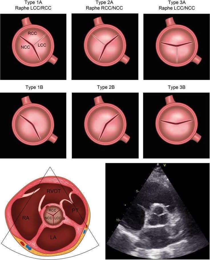
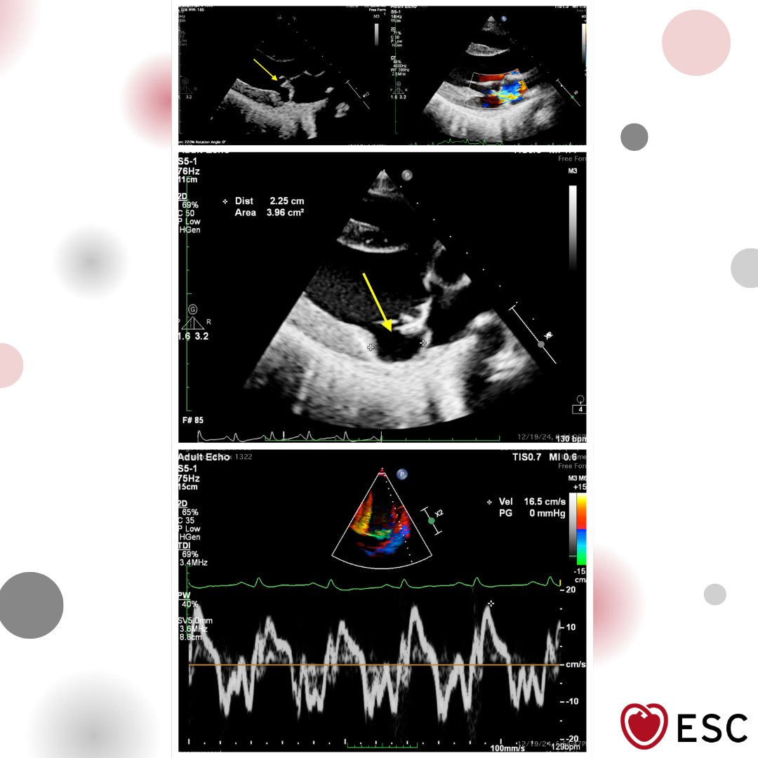
#ImageOfTheWeek by Avishkar Agrawal, Sumit Kumar, and Souvik Sardar from All India Institute of Medical Sciences, New Delhi, India A 28-year-old woman with mitral valve prolapse. What additional high-risk echo finding may be present? ow.ly/V7iS50YJNIU
European Society of Cardiology tweet media
English
2
11
45
5.4K
Gu, MD がリツイート
Alexander Mladenow MD
Alexander Mladenow MD@alex1708ander·
Structural (eccentric) MR Carpentier Type II 📌 3D ECHO → identifies: •Where MR •Why MR (mechanism) 📌 VOLUMETRY → calculates the “how much” •RegVol MR •RegFraction MR ❌ PISA doesn’t work ❌ #3Dfirst #echofirst @NMerke
English
3
15
49
2.5K
Gu, MD がリツイート
American Society of Echocardiography
This video provides a step-by-step roadmap for healthcare providers to teach and troubleshoot the Goal Directed Valsalva technique and addresses common pitfalls. Watch our Goal Directed Valsalva Education Series, featuring free educational resources! bit.ly/4lR9MmF
English
1
8
22
2.8K
Gu, MD がリツイート
Ekaterina Stellbrink
Ekaterina Stellbrink@StellEkaterina·
@echo_batman Really nice work 👏 already using this in my talks, super helpful perspective on TR👍🏻✨
Ekaterina Stellbrink tweet media
English
1
1
14
256
Gu, MD がリツイート
Ahmed Ata
Ahmed Ata@Ahmedata7777·
TAPSE ● Measure of RV longitudinal systolic function (tricuspid annulus motion) ● Method: M-mode in apical 4-chamber at lateral tricuspid annulus. ● Key Cutoff > 17 mm → Normal ≤ 17 mm → RV dysfunction ● Severity 13–17 mm → Mild 10–13 mm → Moderate ≤ 10 mm → Severe 》Clinical pearl ● Always interpret TAPSE with other RV parameters, especially: ▪︎ RV S’ (TDI) ▪︎ FAC (Fractional Area Change) ▪︎ TAPSE/PASP ratio → useful for RV–pulmonary artery coupling
Ahmed Ata tweet media
English
2
62
307
20.8K
Gu, MD がリツイート
Ritika Tuli
Ritika Tuli@RitikaTuliMD·
🫀 #CardioNugget: Predicting SAM after MV repair = GEOMETRY 🎯 📉 AML:PML ↓ (big PML) → anterior coaptation 📏 Coaptation-septum distance <2.5 cm → coaptation near LVOT 📐 Narrow aortomitral angle → flow drags AML 📊 Thick septum → LVOT crowding #EchoBoards #CardioTwitter #MedEd #CardioNuggets
Ritika Tuli tweet media
English
0
39
125
5.3K
Gu, MD がリツイート
Ahmed Ata
Ahmed Ata@Ahmedata7777·
HFpEF RV dysfunction Stages Staging system for right ventricular (RV) dysfunction in Heart Failure with Preserved Ejection Fraction, based on RV–pulmonary artery (RV-PA) coupling using the TAPSE/PASP ratio and the presence of tricuspid regurgitation (TR). 💡 Clinical Pearls ● TAPSE/PASP ratio is a powerful prognostic marker in HFpEF. ● TR is not just a consequence—it worsens RV failure (vicious cycle). ● Transition from Stage 2 → Stage 3 marks a key turning point (loss of RV adaptation). ● Stage 4 = highest mortality risk. 🧠 One-line Summary 👉 HFpEF RV dysfunction progresses from compensated coupling → TR development → RV-PA uncoupling → end-stage RV failure with TR.
Ahmed Ata tweet media
English
2
26
87
5K
Gu, MD がリツイート
Alexander Mladenow MD
Alexander Mladenow MD@alex1708ander·
In this #3D TEE mid-esophageal RV in-out view you can better understand the location of different types of VSD in echo #echofirst supracristal VSD lying more cranial than the infracristal. 👉 for more morphological details check this out doi.org/10.1016/j.atho…
Jorge Faerron@cardiopedhnn

@exp0sse @DrRajeshG1 @swatigar @OungSavly @iamritu @alex1708ander Using the International Society for Nomenclature of Pediatric & #CHD, the type of interventricular communication shown in the images, is an #outlet_muscular_VSD wo malalignment (4.a.i-07.11.06) #cardioped #pedsheart

English
0
45
151
28.3K
Gu, MD がリツイート
WithAScalpel - Fumiya Yoneyama, MD, PhD
Mitral Valve Complex🧐 👉Understanding the mitral complex starts with anatomy: leaflets, annulus, chordae, and papillary muscles function as one unit. 👉Accurate interpretation of mitral valve motion requires integrated assessment of the entire subvalvular and annular apparatus. @AEPCcongenital @iamritu @WGACHDChair @CASivaram1 @alexsfelixecho @echoleolopez @loomba_rohit @alex1708ander @SIwa23288585
WithAScalpel - Fumiya Yoneyama, MD, PhD tweet media
Oung Savly MD FACC FASE FESC FAAP FEACVI 🇰🇭@OungSavly

10y with severe MR,AML appears prolapsed, but it’s false caused by PML tethering👉🏻abnormal coaptation geometry 📌Not all AML prolapse is real! @AEPCcongenital @iamritu @WGACHDChair @CASivaram1 @alexsfelixecho @echoleolopez @loomba_rohit @alex1708ander @SIwa23288585 @WithAScalpel

English
0
31
122
18.7K
Gu, MD がリツイート
Alexander Mladenow MD
Alexander Mladenow MD@alex1708ander·
How 2D images correlate to 3D images aka CLOCKING - take this #echofirst challenge and put the 🅰️ in the right place on the 3D image (surgeon’s view) * patient with paravalvular leakage (PVL) after MV replacement
Nicolas Merke@NMerke

2D #echofirst is the basic diagnostic tool we use in clinical practice As in this case of Pat with SOP and haemolysis But 2D has it’s limits for our understanding

English
4
23
58
7.8K
Gu, MD がリツイート
Edgar Argulian
Edgar Argulian@argulian·
In this patient with HFpEF and high left ventricular filling pressures, shortening of the aortic regurgitation pressure decay time is seen, while aortic regurgitation is mild. Important hemodynamic concept!
Edgar Argulian tweet media
English
2
22
101
7K
Gu, MD がリツイート
EHJ-IMP Editor-in-Chief
EHJ-IMP Editor-in-Chief@EHJIMPEiC·
#EHJIMP 🫀In >24,000 hypertensive participants from the UK Biobank, CMR-defined LVH phenotypes showed distinct imaging profiles and prognostic trajectories. Eccentric LVH carried the highest risk (↑MACE, markedly ↑HF), while concentric LVH was strongly linked to incident heart failure. Read more 👉doi.org/10.1093/ehjimp… Highlighting the value of CMR phenotyping for risk stratification in hypertension. #CardioImaging #CMR #Hypertension #LVH #HeartFailure #MACE #PrecisionCardiology
EHJ-IMP Editor-in-Chief tweet media
English
1
22
74
4K
Gu, MD がリツイート
Ahmed Ata
Ahmed Ata@Ahmedata7777·
Bicuspid aortic valve (BAV) morphology and its echocardiographic appearance. The valve is categorized based on: ▪︎ Number of raphes (fusion lines) ▪︎ Which cusps are fused. ✅ Type A (one raphe) ● Type 1A (Raphe LCC/RCC) Fusion between Left coronary cusp (LCC) and Right coronary cusp (RCC) → most common. ● Type 2A (Raphe RCC/NCC) Fusion between Right coronary cusp (RCC) and Non-coronary cusp (NCC). ● Type 3A (Raphe LCC/NCC) Fusion between Left coronary cusp (LCC) and Non-coronary cusp (NCC). 👉 These appear as asymmetric bicuspid valves with a visible raphe. ✅ Type 1B / 2B / 3B (no raphe) True bicuspid valves with: ▪︎ No raphe ▪︎ Two symmetric cusps Subtypes depend on orientation of commissures. 👉 Typically more symmetrical (“fish-mouth” opening). 🔹 Key Clinical Pearls: ■ Most common BAV type: LCC–RCC fusion BAV predisposes to: ▪︎ Aortic stenosis. ▪︎ Aortic regurgitation. ▪︎ Ascending aortopathy. ■ Echo hallmark: ▪︎ Systolic doming. ▪︎ Eccentric closure line in diastole. #cardiology #medicine
Ahmed Ata tweet media
English
2
33
116
9.9K