WithAScalpel - Fumiya Yoneyama, MD, PhD

1.6K posts

WithAScalpel - Fumiya Yoneyama, MD, PhD banner
WithAScalpel - Fumiya Yoneyama, MD, PhD

WithAScalpel - Fumiya Yoneyama, MD, PhD

@WithAScalpel

Cardiac Surgeon | Assistant Professor | @TexasChildrens | Visual insights in surgery, cardiology & intensive care | Views my own

Katılım Temmuz 2025
102 Takip Edilen7.3K Takipçiler
WithAScalpel - Fumiya Yoneyama, MD, PhD
Surgical Hand Coordination — Building Precision Through Daily Practice! 👍 👉Finger dexterity and bimanual coordination underpin controlled needle handling, tissue manipulation, and knot tying. 👉Short, focused daily drills refine movement economy, precision, and reproducibility for operative performance.
Dr Luis Felipe Cabrera Vargas MD FACS@PipeCabreraV

Knot and counter knot! How to do it! Only in the advanced surgical skills training workshop of @Uniandes @MediUniandes ! Proud of be one of the faculty’s of the marvellous course! @FSFB_Salud ! @SWexner @pferrada1 @TomVargheseJr @juliomayol @CiruAndes2 @BJSAcademy @AmCollSurgeons

English
0
0
0
60
WithAScalpel - Fumiya Yoneyama, MD, PhD
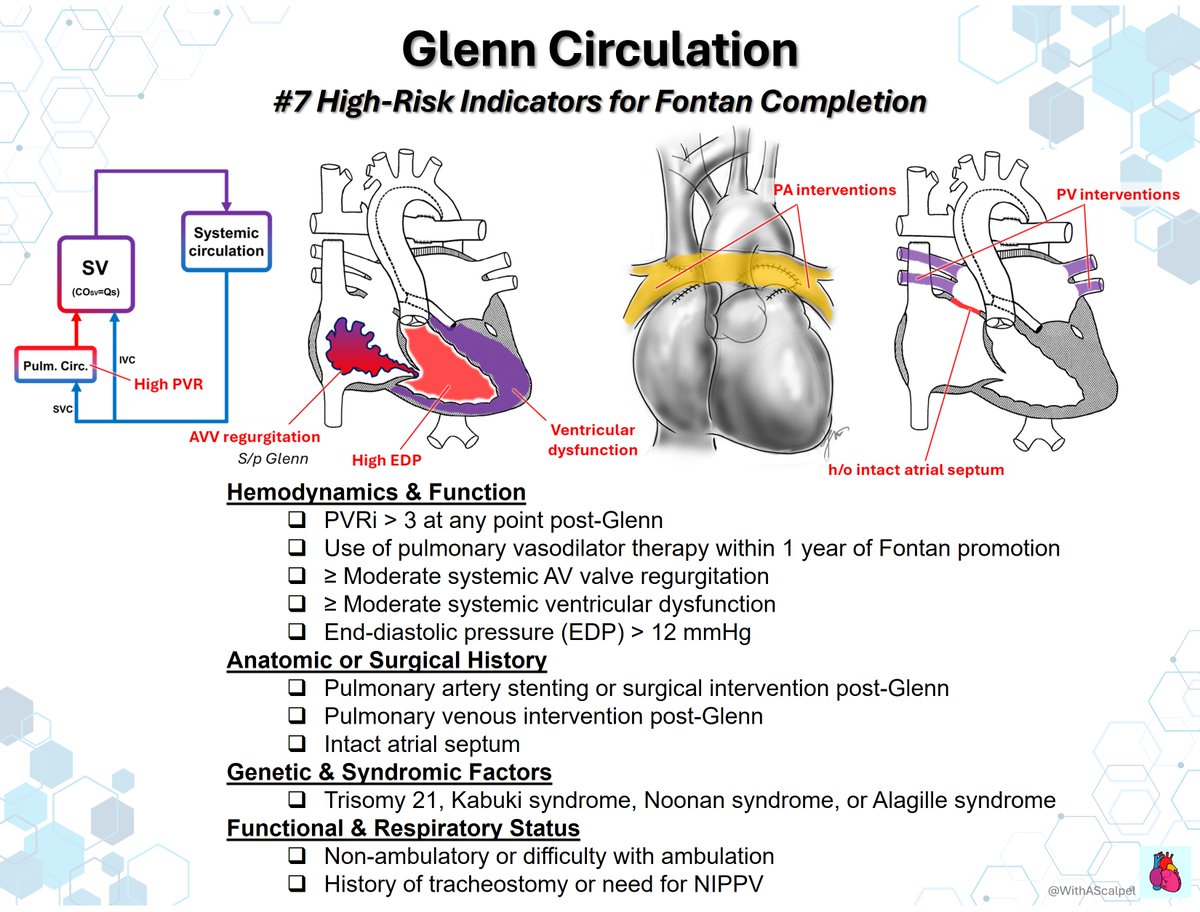
Glenn Circulation: From Flow Physiology to Fontan Risk Assessment 👉CO₂ affects Glenn flow twice: hypercapnia raises PVR but augments cerebral flow, SVC return, pulmonary flow, and SpO₂. 👉Over time, lower SVC contribution, venovenous collaterals, pulmonary AVMs, and APCs progressively reduce Glenn efficiency. 👉Pulsatile flow may support PA growth and oxygenation, but volume loading, high PVR/EDP, AVVR, or dysfunction signal Fontan risk.
WithAScalpel - Fumiya Yoneyama, MD, PhD tweet mediaWithAScalpel - Fumiya Yoneyama, MD, PhD tweet mediaWithAScalpel - Fumiya Yoneyama, MD, PhD tweet mediaWithAScalpel - Fumiya Yoneyama, MD, PhD tweet media
English
1
16
54
1.6K
WithAScalpel - Fumiya Yoneyama, MD, PhD
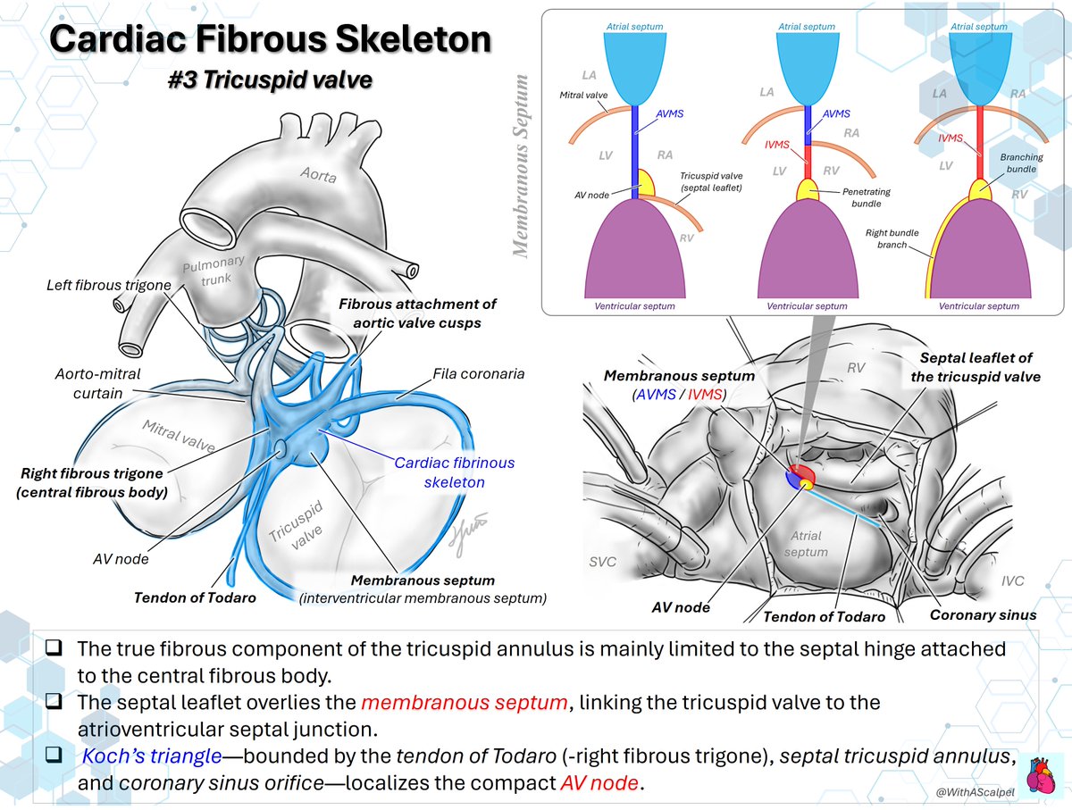
Interatrial Septum—Right Atrial Anatomy and Conduction Landmarks📌💡 👉The interatrial septum is centered on the fossa ovalis, while the surrounding rims relate closely to the venae cavae, crista terminalis, and atrioventricular junction. 👉The Triangle of Koch—bounded by the tendon of Todaro, septal tricuspid annulus, and coronary sinus ostium—localizes the AV node near its apex.
WithAScalpel - Fumiya Yoneyama, MD, PhD tweet media
Alexander Mladenow MD@alex1708ander

Beyond basic echo anatomy #echofirst 🌟Elevating the way we see the interatrial septum in TEE 🥽 The interatrial septum is more than anatomy 🫀- it’s a procedural roadmap📍 TEE guidance for: 1️⃣ M-TEER (transseptal puncture) 2️⃣ PTC of ostium secundum ASD

English
0
12
71
5K
WithAScalpel - Fumiya Yoneyama, MD, PhD
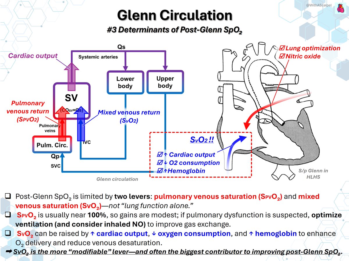
Glenn Circulation: Physiology, Anastomosis, and Postoperative Oxygenation 👉The bidirectional Glenn diverts SVC flow directly to the pulmonary arteries, unloading the single ventricle. 👉A wide, tension-free SVC–RPA anastomosis and low PVR are essential for unobstructed passive pulmonary blood flow. 👉Post-Glenn SpO₂ depends on pulmonary venous and mixed venous saturation; optimize output, oxygen use, and hemoglobin.
WithAScalpel - Fumiya Yoneyama, MD, PhD tweet mediaWithAScalpel - Fumiya Yoneyama, MD, PhD tweet mediaWithAScalpel - Fumiya Yoneyama, MD, PhD tweet media
English
0
23
71
2.6K
WithAScalpel - Fumiya Yoneyama, MD, PhD
Cardiac Conduction Axis—From the AV Node to the Bundle Branches⚡️💡 👉The AV node continues as the penetrating bundle through the central fibrous body and membranous septum before becoming the branching bundle. 👉The branching bundle gives rise to the LBB, while the RBB emerges onto the RV septal surface posterior to the muscle of Lancisi.
WithAScalpel - Fumiya Yoneyama, MD, PhD tweet mediaWithAScalpel - Fumiya Yoneyama, MD, PhD tweet media
HeartRhythm@hrs_journal

Unravelling the enigmatic morphology of the atrioventricular conduction axis using Hierarchical Phase-Contrast Tomography (HiP-CT) #OpenAccess @Chloe_Li_ @aaalmehandi @UCL_ICS heartrhythmjournal.com/article/S1547-…

English
1
18
72
6.7K
WithAScalpel - Fumiya Yoneyama, MD, PhD
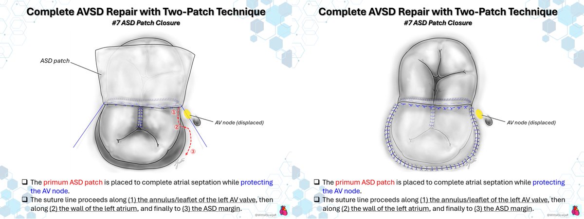
Complete AVSD Repair with Two-Patch Technique #7: ASD Patch Closure 👉The primum ASD patch completes atrial septation while protecting the displaced AV node during closure. 👉Begin along the left AV valve annulus/leaflet, then redirect the suture line onto the left atrial wall. 👉Complete closure by advancing from the left atrial wall to the ASD margin, maintaining a conduction-sparing course.
English
1
9
11
981
WithAScalpel - Fumiya Yoneyama, MD, PhD
LVAD Evaluation Checkpoints🤖☑️ 👉A systematic assessment should include the device, inflow cannula, LV unloading, RV/PVR, aortic valve opening, aortic regurgitation, and mitral valve function. 👉Integrating pump flow with RAP, LAP, and inlet pressure helps distinguish volume imbalance, cannula obstruction, RV failure, and inadequate pump support.
WithAScalpel - Fumiya Yoneyama, MD, PhD tweet mediaWithAScalpel - Fumiya Yoneyama, MD, PhD tweet media
EHJ-IMP Editor-in-Chief@EHJIMPEiC

🫀📡 Can echocardiography really “see” LVAD complications? A new study answers a key clinical question: 👉 How accurate is TTE in detecting outflow graft abnormalities? ✨ Key findings: 🎯 Specificity = 100% → if you see it, it’s real ⚠️ Sensitivity = 61% → but you may miss cases 📍 Much better for: 👉 Distal graft (71% sensitivity) ❗ Limited for: 👉 Proximal graft (only 33%) 📊 As shown in the graphical abstract (page 2): 👉 TTE can identify: 🔁 Kinking (≥90° bend) 🚫 Stenosis (≥50%) 👉 Doppler clue: ⚡ Vmax >2.0 m/s suggests obstruction 💡 Clinical message: 🩺 TTE = non-invasive, bedside, repeatable 🧠 Great for screening & follow-up 🧪 But CT remains gold standard for diagnosis 🚨 Why it matters: Outflow graft complications → ❗ HF symptoms ❗ low pump flow ❗ device alarms 🎯 Big take-home: From “echo vs CT” ➡️ to “echo for screening, CT for confirmation” 📄 Read the full paper: 👉 🔗 DOI: 10.1093/ehjimp/qyag017 #Cardiology #Echocardiography #LVAD #HeartFailure #CardioTwitter #Imaging #CriticalCare #MedTech #Ultrasound #PrecisionMedicine 🩺📊

English
0
10
31
2.8K
WithAScalpel - Fumiya Yoneyama, MD, PhD
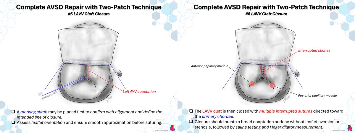
Complete AVSD Repair with Two-Patch Technique #6: LAVV Cleft Closure 👉A marking stitch confirms cleft alignment, leaflet orientation, and the intended line of LAVV closure before suturing. 👉Close the cleft with multiple interrupted sutures directed toward the primary chordae, preserving smooth leaflet approximation. 👉Create a broad coaptation surface without eversion or stenosis, then confirm competence by saline testing and Hegar sizing.
English
1
2
5
672
WithAScalpel - Fumiya Yoneyama, MD, PhD
Complete AVSD Repair with Two-Patch Technique #5: Patch–Leaflet–Patch Suture Sequence 👉The VSD patch is sized and positioned first to preserve common AV valve leaflet mobility and left AV valve geometry. 👉Interrupted sutures pass from the VSD patch through the common AV valve leaflet, establishing ventricular septation. 👉The same sutures pass through the ASD patch and are tied, creating a balanced patch–leaflet–patch sandwich.
English
1
2
13
784
WithAScalpel - Fumiya Yoneyama, MD, PhD
Lower-Extremity Arterial Anatomy🌱🐾 👉The external iliac artery becomes the common femoral artery beneath the inguinal ligament, then divides into the profunda femoris and superficial femoral arteries. 👉The superficial femoral artery continues through the adductor canal and becomes the popliteal artery beyond the adductor hiatus—an important landmark for surgical exposure and bypass planning.
WithAScalpel - Fumiya Yoneyama, MD, PhD tweet media
English
0
0
2
482
WithAScalpel - Fumiya Yoneyama, MD, PhD
Complete AVSD Repair with Two-Patch Technique #4: VSD Patch Closure 👉The displaced conduction axis runs near the inferior VSD rim; place sutures slightly toward the right-sided margin. 👉Fashion the VSD patch slightly undersized—about 80% of the A–P span—to avoid chordal or annular tension. 👉Near the AV node, keep bites shallow and incorporate right AV valve tissue rather than the vulnerable septal margin.
English
1
3
9
930
WithAScalpel - Fumiya Yoneyama, MD, PhD
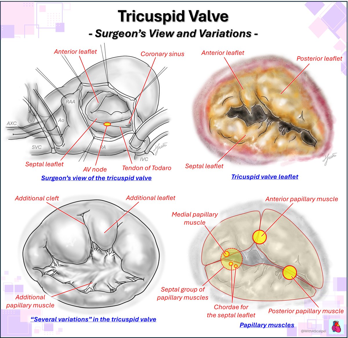
Tricuspid Valve Anatomy—A Surgeon’s Map🗺️ 👉The anterior, posterior, and septal leaflets are oriented using the coronary sinus, tendon of Todaro, and caval orifices. 👉The septal annulus overlies the membranous septum and AV node region, requiring superficial sutures and respect for variable chordal anatomy.
WithAScalpel - Fumiya Yoneyama, MD, PhD tweet mediaWithAScalpel - Fumiya Yoneyama, MD, PhD tweet media
Alexander Mladenow MD@alex1708ander

Valve imaging in TEE starts with the correct orientation. Keep in mind that during tricuspid valve surgery, the valve is typically oriented in the SURGEON’S view. This orientation differs slightly from that used for TEE-guided structural heart interventions #echofirst
English
0
18
80
8K
WithAScalpel - Fumiya Yoneyama, MD, PhD
Complete AVSD Repair with Two-Patch Technique #3: Conduction System for VSD Patch Closure 👉In complete AVSD, the AV node is displaced posteriorly and inferiorly, outside its usual position in Koch’s triangle. 👉The penetrating bundle arises from the nodal triangle and courses beneath the inferior bridging leaflet. 👉The non-branching bundle runs anteriorly just beneath the septal crest before dividing into right and left branches.
English
1
3
12
854