
Mahatma
1.6K posts

Mahatma
@DocMahatma
Serenidad y paciencia amigos. #radres Interventional Radiology / @INCMNSZMX Radiado x @IngridAlonsoRa1
Universo de Röntgen Katılım Ocak 2010
915 Takip Edilen725 Takipçiler

El fragmento de un video presentado en SAGES 2024: una complicación catastrófica en una #colecistectomía asistida con 🤖 y con verde de indocianina. La indicación quirúrgica fue #pancreatitis biliar. #Cirugía #Cirujanos
Español
Mahatma retweetledi
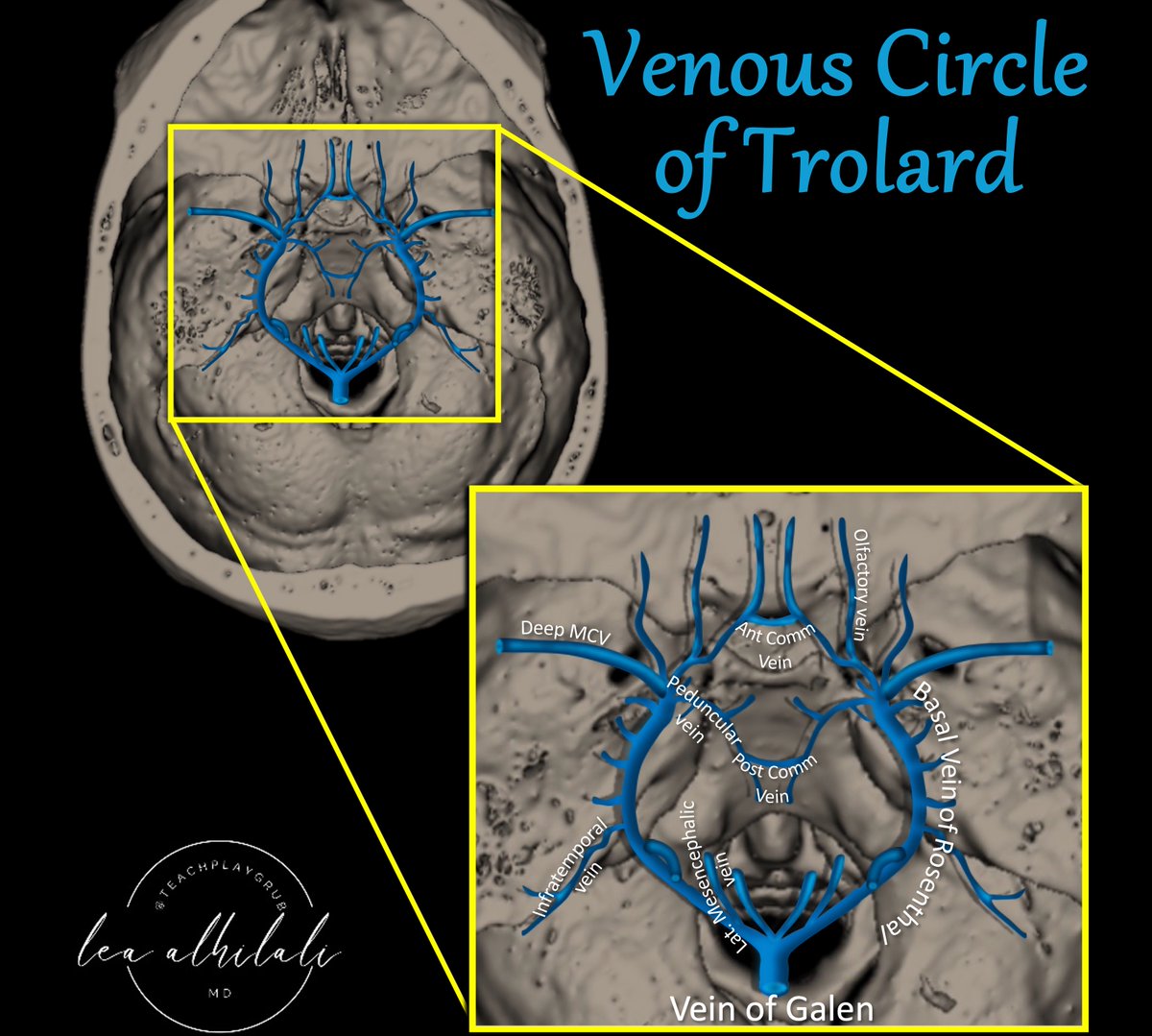
Think you know brain vascular anatomy?
How many brain vascular circles do you know?
Everyone knows the Circle of Willis, but few know its venous counterpart: Venous Circle of Trolard!
Can you see the polygon on top of a❤️??
Venous Circle of Trolard is the midline connection between the right & left venous networks.
Peduncular veins form the so-called “mesencephalic heart.”
It provides venous collaterals in both the transvesrse & AP directions
It could potentially compensate in contralateral basal occlusion from tumor or surgical injury.
On the flip side--in a dural AVF, it could result in hemorrhage on the contralateral side!
Hopefully now when it comes to venous anatomy, you will always bee in the right vein!
English
Mahatma retweetledi
1/Does PTERYGOPALATINE FOSSA anatomy feel as confusing as its spelling?
Does it seem to have as many openings as letters in its name?
Are you pterrified of the pterygopalatine fossa (PPF)?
Let this thread on PPF anatomy help you out.

English
Mahatma retweetledi

ACL reconstruction should be performed by skilled surgeons, that can guarantee anatomical tunnel positioning. Read this study to find out what can happen if the femoral tunnel is not anatomically placed.
#knee #ACLR #complications
Read here: doi.org/10.1002/ksa.12…

English
Mahatma retweetledi
Tired of stressing whether a brain tumor is progressing?
Wish you had some insurance about calling tumor recurrence?
Here’s the cheat sheet you NEED for the best signs of tumor progression!
Read on for the newest @theAJNR SCANtastic on tumor imaging:
ajnr.org/content/45/4/4…
▶️Tumor progression is like tumor escaping treatment.
🔷Just when treatment thinks it’s got tumor trapped at cliff, tumor is able to get away
🔶Think how you would get away if you were chased to a cliff’s edge---these are same signs of tumor progression:
➡️Jump off into the water:
🔸Tumor heads to the water—the ventricular surface
🔸Subependymal enhancement is very specific (93% sensitivity), but it isn’t commonly seen (38% sensitive).
➡️Cross over the rickety bridge:
🔸Tumor crosses over the brain’s main bridge: the corpus callosum
🔸Callosal involvement is only significant if combined w/multiple foci or crossing midline.
➡️Slide down the rocks:
🔸Spectroscopy looks like a slide down: Increased choline is seen w/recurrence—when combined w/low NAA, it looks like a reverse Hunter’s angle
🔸Increased Cho/NAA or Cho/Cre ratios can suggest recurrence
➡️Run fast & get your red blood hot:
🔸Blood volume is hot on perfusion: high rCBV on perfusion
🔸rCBV hotter than the normal contralateral white matter can be used as a gestalt
🔸Quantitatively, rCBV >1.75 is high suggestive of tumor & rCBV > 1.0 is concerning
➡️Try to sneak away when it’s dark:
🔸Tumor looks dark: Low ADC value (dark on T2) is concerning for tumor
🔸Quantitatively ADC value ≤ 1220 × 10-6 mm2/s is 74% sensitive & 89% specific
🔸I joke, “If the ADC is less than 1000 x 10-6, I am 1000% sure it’s tumor!”
Now you know the best signs of tumor recurrence on imaging!
Hopefully now you know when tumor progression should make your impression!

English
Mahatma retweetledi

Anatomía tomográfica del cuello: un enfoque para residentes 🔎 Este #PiperSERAM ayuda a los residentes en el conocimiento de las estructuras anatómicas del cuello, así como, en la correlación de los diferentes espacios cervicales entre ellos, y que ello les sirva para la identificación de anormalidades Accede aquí 🔗 buff.ly/4aH28VG

Español
Mahatma retweetledi
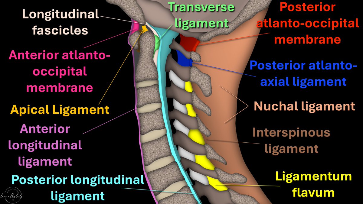
Afraid to stick your neck out when it comes to naming cervical spine ligaments?
Know the anterior longitudinal ligament (ALL) & that’s ALL?
Is the nuchal ligament a NEW CALL for you?
How many ligaments do YOU know?
Here’s are the ligaments to know & how you can remember them
➡️ALL:
🔸As it’s name implies, it runs ALL the way down the spine from the occiput to the sacrum
🔸It’s the ANTERIOR longitudinal ligament so it is ANTERIOR to the vertebral bodies
➡️PLL:
🔸PLL stands for POSTERIOR longitudinal ligament, so it’s posterior to vertebral bodies
🔸I call it “PARTIALLY longitudinal ligament” bc it only runs from C2 inferiorly
🔸Above C2 it’s the tectorial membrane.
🔸I remember this bc it has a different name but is TECT-nically part of the PLL
➡️Cranio-cervical ligaments:
🔸In the sagittal plane C2 looks like a mountain peak
🔸Ligaments here are arranged how you climb a mountain!
▶️AT Last you reach the ANTERIOR mountain (first is ANTERIOR ATLanto-occipital membrane)
▶️You hike up the APEX (next is APICAL ligament)
▶️Finally, you have to TRAVERSE the LONG way down (TRANSVERSE & LONGitudinal fibers)
➡️Posterior ligaments:
▶️Ligamentum flavum:
🔸I remember this bc Flavum & Facets both start w/F = Flavum is by the Facets
🔸F is also for FAR FIVE, so ligamentum flavum is only on the lowest/farthest five cervical vertebral bodies
🔸Above this, membranes are named for what they connect (posterior atlanto-occipital & posterior atlanto-axial membrane)
▶️Nuchal ligament:
🔸This is where we see the nuchal fold & is the equivalent of the supraspinous ligament from the occiput to C7.
Now you know the major cervical ligaments you can see on sagittal images!
Hopefully, now you will be ready when injury to these structures occurs in your neck of the woods!
English
Mahatma retweetledi

Ankle and midfoot ligaments: Ultrasound with anatomical correlation
FREE PDF
ultrasoundtraining.com.au/wp-content/upl…


English
Mahatma retweetledi
Normal fat pad anatomy vs positive fat pad signs in elbow👍 In my older tweets we've addressed anterior & posterior fat pads, and in this tweet , the supinator fat pad also explained.

English
Mahatma retweetledi
ELBOW COMMON EXTENSOR:
ECRB and the anterior edge of EDC are the most involved in tendinopathy.
Nirschl SCRATCH TEST: scraping and peeling to assess tendinosis extent during debridement.
Main components: ECRB (II), Extensor digitorum communis (IV) and Extensor digiti minimi (V)
ECU dorsal origin (VI)
ECRL (II): origins from supracondylar ridge
#elbow


English
Mahatma retweetledi
Ultrasound imaging of the posterior lateral corner of the knee
FREE PDF
insightsimaging.springeropen.com/counter/pdf/10…


English
Mahatma retweetledi

🔬 10 pasos para leer un artículo científico:
1. Comience por la introducción, no por el resumen.
2. Identifique cuál es la pregunta principal y cómo los autores intentan responderla.
3. Haga un resumen del contexto en menos de cinco frases, teniendo en cuenta trabajos previos, limitaciones y próximos pasos.
4. Identifique las preguntas específicas planteadas en el artículo, incluyendo hipótesis nulas si corresponde.
5. Identifique el enfoque utilizado por los autores para responder a las preguntas específicas.
6. Lea los resultados y resuma cada experimento, figura y tabla sin interpretarlos.
7. Analice si los resultados responden a las preguntas planteadas y qué significan.
8. Lea la discusión, conclusiones e interpretación de los autores, comparando su interpretación y considerando posibles alternativas.
9. Vuelva al resumen y verifique si coincide con lo planteado por los autores y su propia interpretación.
10. Busque las opiniones de otros investigadores sobre el artículo, identificando expertos en el campo y considerando sus opiniones y corroboraciones.
¡Recuerde que la ciencia es un proceso continuo y emocionante, nunca deje de leer y aprender! 🌟

Español
Mahatma retweetledi

Respuesta: Granulomatosis con Poliangeítis
Aprende más:
pubs.rsna.org/doi/10.1148/rg…
Traducido por: @MariaLuciaBrunV
#CasosCooky #CookyBites #104 #RGphx @cookyscan1 @RadioGraphics

Español
Mahatma retweetledi
Patella,like an inverted triangle embedded in quadriceps tendon.The posterior articulating surface of patella composed of 2facets,a medial & lateral facet,separated by a vertical ridge.
In 30% of population,there's a 3rd facet ("Odd facet") most medially.
pin.it/6CIcDlykW

English
Mahatma retweetledi

🇲🇽 Por ser un cuento de terror. Y una nueva forma de narrar. Y una novela de amor y una crítica a ese amor. Y por ser un poema sobre la nostalgia, una fábula sobre la destrucción y sobre lo que permanece, un tratado sobre la soledad y una prosa. Y por ser algo más.

Español