
Edgar Lorente retweetledi

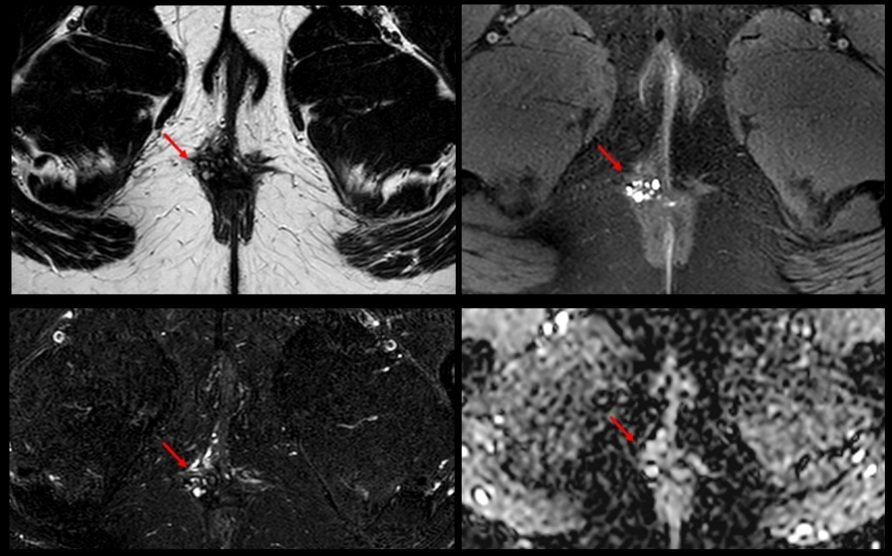
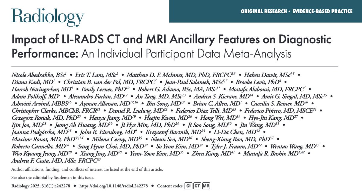
📍Impact of #LIRADS CT and MRI Ancillary Features on Diagnostic Performance: An Individual Participant Data Meta-Analysis🚨👇
@Christian_vdP @malabousi @docamitgs @dr_ash_arvind @DukeMedSchool @UofTMedIm
@RITEditor #RadInTraining #Tweetorial 🧵
@Radiology_RSNA @RadiologyEditor
English