Sameer Raniga

10.6K posts

Sameer Raniga banner
Sameer Raniga

Sameer Raniga

@samrad77

Radiologist. Trauma and Emergency Radiology. One view is NO view. #radtwitter | #FOAMrad | #radres | #radEd| #radiology | #EmergencyRad

Muscat, Oman Katılım Mart 2013
4.4K Takip Edilen19.4K Takipçiler
Sameer Raniga
Sameer Raniga@samrad77·
A large dorsal talar beak on sagittal CT or lateral ankle radiograph. Talar beak is a marker of restricted subtalar motion, not a diagnosis. In a young patient, actively search for a subtalar coalition. This case showed a fibrous coalition of Medial accessory talocalcaneal articulation at the level of the posterior sustentaculum tali, between the Talus and Calcaneus. Wisdom: A talar beak should trigger a search, not end it. — Pearls, pitfalls and wisdom from my reporting list
Sameer Raniga tweet mediaSameer Raniga tweet mediaSameer Raniga tweet mediaSameer Raniga tweet media
English
4
19
149
5.9K
Sameer Raniga
Sameer Raniga@samrad77·
One view is no view! Weber type B Lateral malleolar fractures are often only seen on lateral view.
Sameer Raniga tweet mediaSameer Raniga tweet media
English
1
7
64
3.3K
Sameer Raniga
Sameer Raniga@samrad77·
13 years on Twitter. For the first 8, I barely said a word. Then during COVID, I began posting a few thoughts. Cases. Notes. Small reflections from daily practice. I never expected it to become this. A community. A classroom without walls. A place to share ideas, learn from strangers, and meet colleagues long before meeting them in person. Today, many people recognize me first from something I posted here. That still feels surreal. Nearly 20,000 of you now follow along. Grateful for every conversation, every disagreement, every shared learning moment. The journey continues. Thank you for being part of it. 🙏
Sameer Raniga tweet media
English
4
0
38
1.5K
Sameer Raniga
Sameer Raniga@samrad77·
On portal venous phase CT, the uterus enhances more than the cervix. This differential enhancement is common and physiological. Yet it is often mistaken for cervical pathology. The relatively lower attenuation of the cervix is often overcalled a cervicitis or a cervical mass. Understanding normal enhancement patterns helps avoid these errors and keeps us from creating pathology where none exists. —Pearls, Pitfalls, and Wisdom from my reporting list
Sameer Raniga tweet mediaSameer Raniga tweet media
English
5
26
177
8.5K
Sameer Raniga
Sameer Raniga@samrad77·
Radiopaedia 2026 rPoster submissions are open. Deadline: 22 March. If you have a strong teaching idea, this is one of the highest-yield platforms to share it. Accepted rPosters: · Feature in the global virtual conference · Receive a DOI citation · Compete for editorial awards Submission details: radiopaedia.org/courses/rposte… If you are unsure what a winning poster looks like, study last year’s award-winning poster, "Physical fractures: understanding growth plate injuries” by @DrLeoLustosa A simple idea executed exceptionally well with clean design, clear teaching points, and genuine interactivity. View it here: radiopaedia.org/courses/rposte… While you are there, browse the full archive. It is all free and sets the benchmark. Good posters are not about volume. They are about clarity, structure, and one strong message. If you have been thinking about submitting, do it this year. @Radiopaedia @thexraydoctor @DrAndrewDixon @murf1990 @daniel_gewolb @teachplaygrub
Sameer Raniga tweet media
English
0
6
17
1.8K
Sameer Raniga
Sameer Raniga@samrad77·
Os terminale: A normal developmental variant of the odontoid tip. It represents non-fused secondary ossification center of the odontoid tip. Pearls: Small, well-corticated ossicle at the dens tip. Smooth margins. Normal C1–C2 alignment. Pitfall: Commonly mistaken for a type I odontoid fracture. Fractures are irregular and non-corticated and fits in like a puzzle piece with the donor site. 🧩 Wisdom: If the edges are smooth, corticated, and quiet on MRI, pause before labeling it a fracture. Reporting tip: “Findings are consistent with os terminale, a developmental variant. No imaging features to suggest acute odontoid fracture or instability.” —Pearls, pitfalls and wisdom from my reporting list
Sameer Raniga tweet mediaSameer Raniga tweet media
English
3
25
165
6.5K
Sameer Raniga
Sameer Raniga@samrad77·
A relatively prominent, T1-bright pituitary in a newborn MRI is usually normal physiology from maternal estrogen exposure. Pitfall: Applying adult size criteria and overcalling it as haemorrhage, hyperplasia or tumor. Wisdom: Homogeneous signal, thin midline stalk, no mass effect. Let context guide the call. —Pearls, pitfalls and wisdom from my reporting list
Sameer Raniga tweet mediaSameer Raniga tweet media
English
1
9
83
3.1K
Sameer Raniga
Sameer Raniga@samrad77·
This is “torus mandibularis” on CT. A benign bony exostosis arising from the lingual cortex of the mandible. Often bilateral and symmetric. Cortical attenuation (mislabelled as osteoma). Smooth margins. No adjacent soft tissue component. Why it matters: • Frequently incidental • Can be misread as a mass or aggressive lesion (Osteoid neoplasm/osteoma) • Should not trigger biopsy or further imaging Wisdom: Radiology is often about knowing what not to chase. — Pearls, pitfall and wisdom from my reporting list
Sameer Raniga tweet media
English
3
11
91
4.6K
Sameer Raniga
Sameer Raniga@samrad77·
Nothing fancy today. Just fundamentals that matter. Lipohemarthrosis on a cross-table lateral knee radiograph equals an intraarticular fracture until proven otherwise. In this case: •The cross-table lateral shows a clear fat–fluid level. •The AP view localizes the injury to the lateral tibial plateau. •This is consistent with a Schatzker type II fracture. Teaching point: In acute knee trauma: •Always obtain AP and cross-table lateral views. •A non–cross-table lateral can miss lipohemarthrosis. Missing lipohemarthrosis means missing an intra-articular fracture. Basic radiographs. Correct technique. High yield. —my reporting list
Sameer Raniga tweet mediaSameer Raniga tweet media
English
2
18
144
8.7K
Sameer Raniga
Sameer Raniga@samrad77·
Nothing fancy today. Just a chest X-ray. And those beautiful, elegant Kerley B lines at the lung bases. There is something deeply satisfying about them. An image where anatomy, physiology, and timing align. Radiology is full of noise. But every now and then, you see a sign that is clean, honest, and complete. Just the quiet beauty of a finding that says exactly what it means. —My poetic reporting list 🙃
Sameer Raniga tweet mediaSameer Raniga tweet media
English
4
17
120
9.1K
Sameer Raniga
Sameer Raniga@samrad77·
Something fancy today: Sacral nerve stimulation (SNS) device. What to recognize on imaging: •pulse generator: Located in the subcutaneous soft tissues of the upper gluteal region. •Lead: A thin electrode coursing toward the sacrum. •Tip position: The active electrode tip lies just anterior to the sacrum in the presacral pelvic soft tissues. What it does: It delivers low-amplitude electrical stimulation to sacral nerves to modulate abnormal brain–spinal cord-bladder–bowel signaling. Why it is used: A third-line therapy for refractory symptoms, including: •Overactive bladder with or without urge incontinence •Chronic fecal incontinence Radiology takeaway: Know the hardware components, confirm expected lead and tip position, and avoid mislabeling it. —My today’s reporting list
Sameer Raniga tweet mediaSameer Raniga tweet media
English
3
7
50
4.9K
Sameer Raniga
Sameer Raniga@samrad77·
When reviewing head CT or MRI in sickle cell patients, extend the search beyond the brain. Sickle cell disease often involves skull and scalp. Sagittal CT and T1-weighted MRI demonstrate: • Diffuse calvarial thickening from chronic marrow hyperplasia • Heterogeneous marrow with interspersed patchy sclerotic foci • Multifocal calvarial bone infarctions, several with acute/subacute characteristics (T1 hyperintense) • Overlying subperiosteal scalp hematoma without associated calvarial fracture • No intracranial hemorrhage or acute parenchymal lesion These findings collectively reflect an acute vaso-occlusive crisis involving the calvarium. Wisdom: In sickle cell disease, always look beyond the brain. Calvarial marrow and soft-tissue changes often tell the real story. —pearls, pitfalls and wisdom from my reporting list
Sameer Raniga tweet mediaSameer Raniga tweet mediaSameer Raniga tweet media
English
3
16
116
5.5K
Sameer Raniga
Sameer Raniga@samrad77·
Join us tomorrow at the @RSNA 2025 Date: 1st December 2025 Session: Core Concepts in Abdominopelvic Trauma Time: 9:30–10:30 AM Room: E450A I’ll be moderating the session and also speaking on "Pelvic Ring Injuries: Core Concepts Revisited." If you deal with trauma CT, this session will help sharpen your search pattern and cut blind spots. Posting a sneak peek of a few slides from my talk. Hope to see you there!
Sameer Raniga tweet mediaSameer Raniga tweet mediaSameer Raniga tweet mediaSameer Raniga tweet media
English
2
14
118
5.5K
Sameer Raniga retweetledi
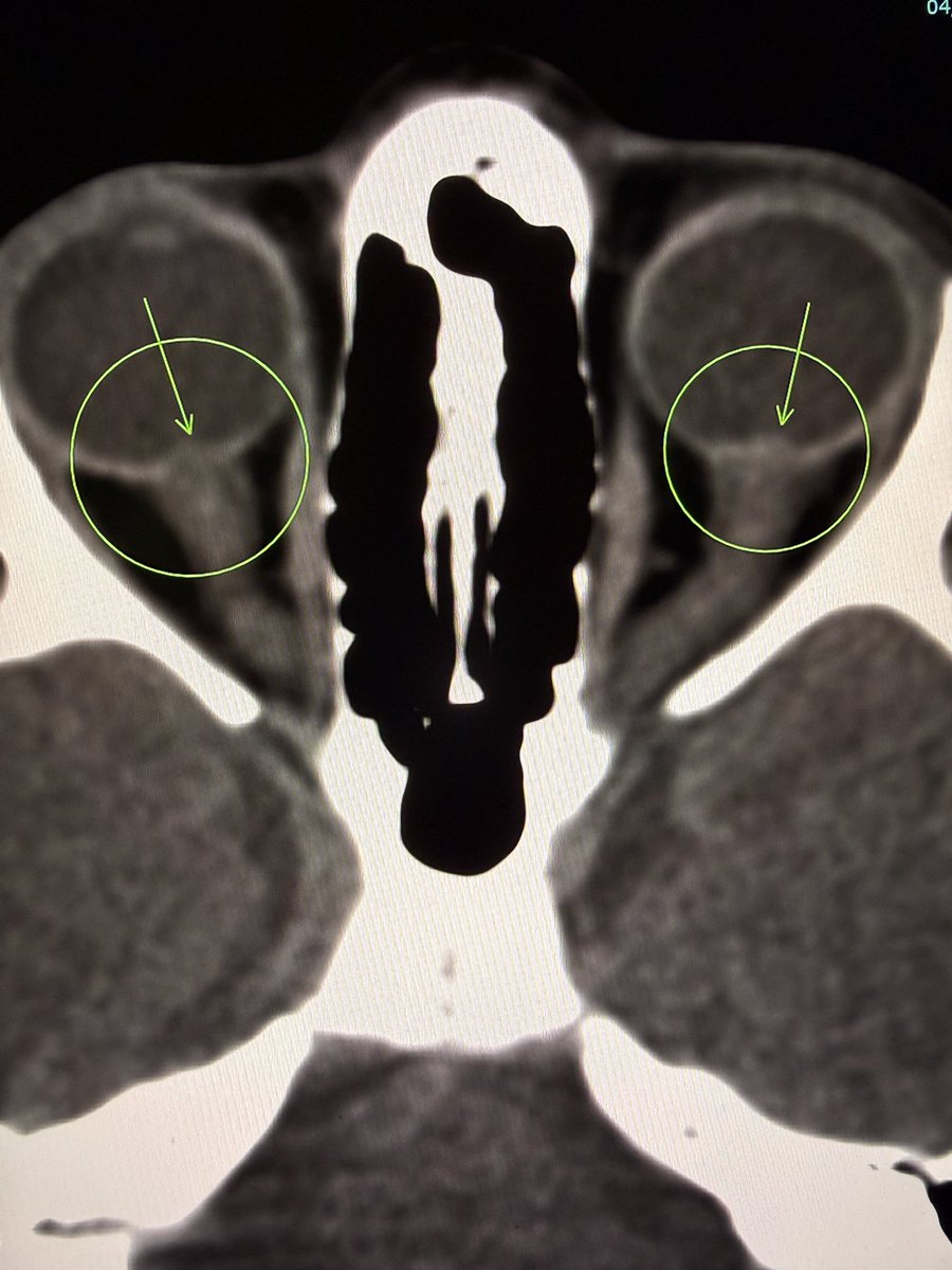
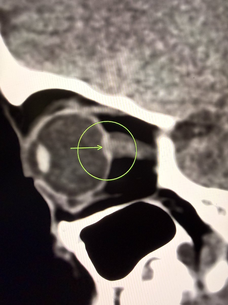
Akshay Baheti
Akshay Baheti@Dr_AkshayBaheti·
Trainees want to compare US, UK, Canada, Australia, Middle East etc based on training quality, match competitiveness, workload, academic scope, salaries, & immigration ease; and then decide where to apply. But reliable, practical guidance is hard to find! @samrad77 @ASK_MSK
English
1
1
7
818
Sameer Raniga
Sameer Raniga@samrad77·
I am honored to be part of the @RSNA Annual Meeting this year at McCormick Place, Chicago. I will be moderating and presenting in the session "Core Concepts in Abdominopelvic Trauma: Imaging Strategies and Advances." Session details Session number: M3-CER05 Date: 1 December 2025 Time: 9:30 to 10:30 AM Room: E450A Presenters • @ArjunKalyanpur - Solid Visceral Injuries: Liver and Spleen • @samrad77 - Pelvic Ring Injuries: Core Concepts Revisited • Ana Blanco-Barrio- Bowel and Mesenteric Injuries: MDCT Insights and Pitfalls • Elizabeth Dick– Urinary Tract Injuries: A Simplified Imaging Approach Looking forward to an engaging hour and meaningful discussion on high-impact trauma imaging.
Sameer Raniga tweet mediaSameer Raniga tweet mediaSameer Raniga tweet mediaSameer Raniga tweet media
English
1
1
33
2K
Orthopod
Orthopod@doc_mehta·
@samrad77 And why is that lytic lesion in banana view with fracture
English
1
0
0
185
Sameer Raniga
Sameer Raniga@samrad77·
Nothing fancy today. Scaphoid dictum: For scaphoid fractures, one view is no view. Two views are too few. Three views may still miss the injury. That is why we use all four projections. Many fractures appear on only one of them. Scaphoid waist fractures show best on the ulnar-deviation or banana view (our case) Distal pole fractures are clearer on the oblique views. Proximal pole fractures remain tricky, but the AP or banana view may reveal the cortical break. The lateral projection helps to assess deformity and alignment, not fracture detection per se. Use all four views and match the projection to the part of the scaphoid you suspect. It is the simplest way to avoid a missed fracture in a bone that hides well. —My reporting list- scaphoid is special 😅
Sameer Raniga tweet mediaSameer Raniga tweet mediaSameer Raniga tweet mediaSameer Raniga tweet media
English
4
11
79
5.4K