Hitachi Electron Microscope

3.3K posts

Hitachi Electron Microscope banner
Hitachi Electron Microscope

Hitachi Electron Microscope

@Hitachi_EM

Since 1939, Hitachi has developed and manufactured various Electron Microscopes including unique tabletop and ultrahigh voltage as well as SEM, TEM and FIB.

Katılım Mart 2018
299 Takip Edilen23.4K Takipçiler
Hitachi Electron Microscope
It’s National Renewable Energy Day ! 🌞🌊🌀💡🌱 (March 21) 
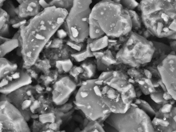
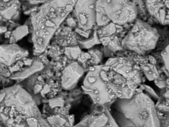
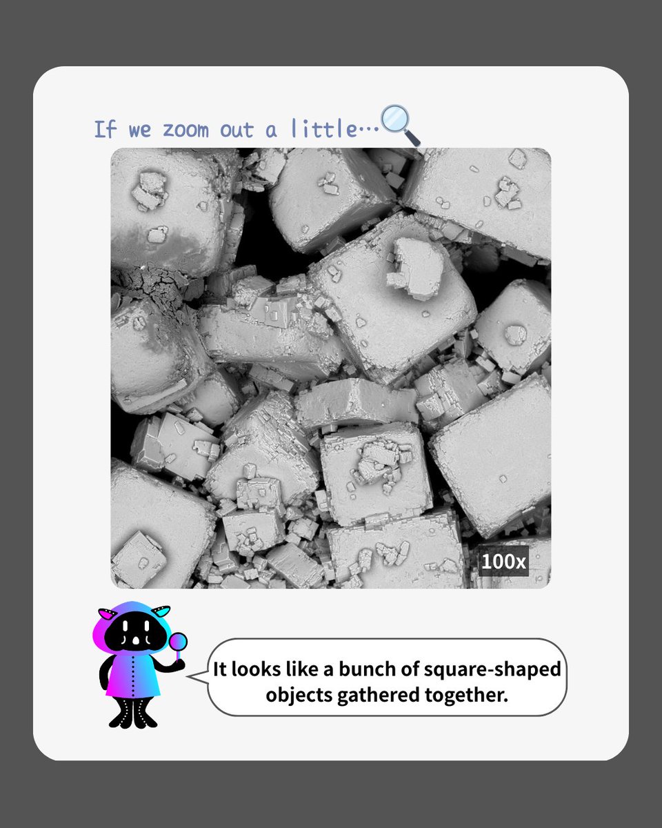
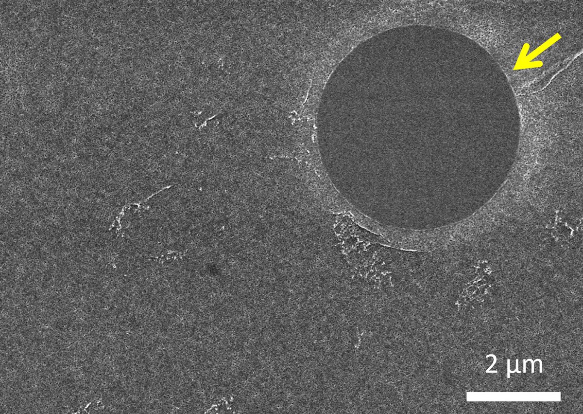
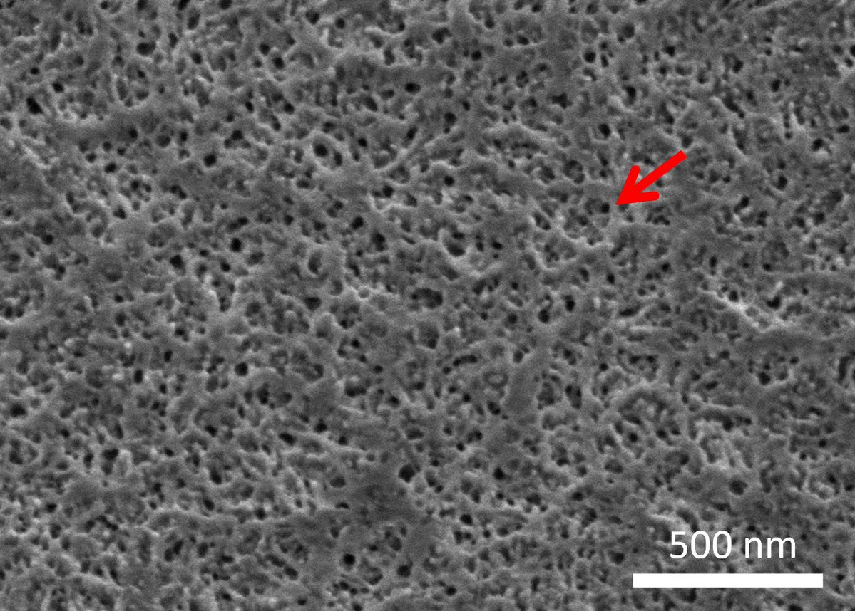
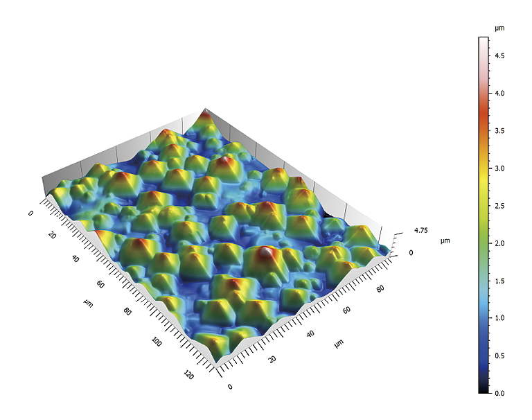
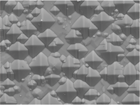
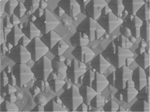
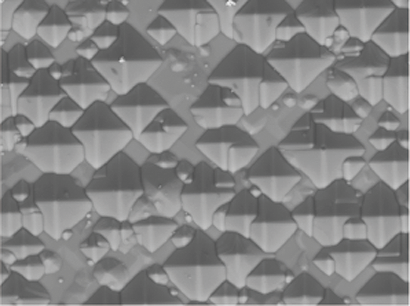
3D image of Solar Cell generated from multi-directional BSE images using Hitachi map 3D 
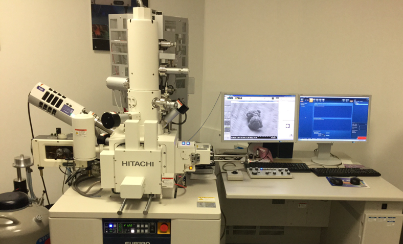
Hitachi map 3D automatically combines 4 images acquired from different directions using the backscattered electron detector to construct a 3D model. Measurements such as height between two points, volume, and simple surface roughness (area roughness, line roughness, etc.) are possible. Since all backscattered electron data is collected in a single acquisition, it is not necessary to tilt the sample or adjust the field of view. To know about the 3D imaging, check out the following post !
Hitachi Electron Microscope tweet mediaHitachi Electron Microscope tweet mediaHitachi Electron Microscope tweet mediaHitachi Electron Microscope tweet media
English
0
0
2
136
Hitachi Electron Microscope
Happy World Oral Health Day ! 😬🦷🪥👄 (March 20) Toothpaste at 15 kV (giving the best resolution) and 5 kV (emphasizing surface details) About the microscope, check out the following post !
Hitachi Electron Microscope tweet mediaHitachi Electron Microscope tweet mediaHitachi Electron Microscope tweet media
English
0
1
3
232
Hitachi Electron Microscope
A partially-manufactured GAA structure device with three layers of Si/SiGe stacked structure was evaluated using FIB-SEM and STEM. 41 DF-STEM images, each showing 5 pillars of Si/SiGe stacked structure, were acquired to measure the horizontally etched amount (Recess) of Si/Ge layer. Recess values of every layer, “Top”, “Middle”, and “Bottom”, of 205 pillars were automatically measured by an off-line measurement software, the Terminal PC. Recess values at “Top” and “Middle” were almost the same while those of “Bottom” were smaller. The variation of the Recess at “Bottom” was the largest. About the instruments, check out following post !
English
0
1
7
423
Hitachi Electron Microscope retweetledi
Hitachi High-Tech Corporation
This image looks just like a sugar cube. But somehow, something feels slightly off. The shape is square, yet parts of it seem rounded and a little crumbled. As you change the magnification under an electron microscope, it may start to look like something entirely different. The things we’re so familiar with that we barely notice them can reveal surprisingly unexpected forms in the microscopic world. Take a closer look and discover what it really is.
Hitachi High-Tech Corporation tweet mediaHitachi High-Tech Corporation tweet mediaHitachi High-Tech Corporation tweet mediaHitachi High-Tech Corporation tweet media
English
0
1
1
160
Hitachi Electron Microscope
Happy National 3D Day ! 🧬🎲🖥️🥽 (The 3rd day of the 3rd week of the 3rd month of the year) Array Tomography of a Rat Hepatocyte 3D model of a rat hepatocyte was reconstructed using Auto Capture for Array Tomography software (ACAT). High-resolution images of over one hundred serial ultrathin sections were acquired thanks to stable auto focus performance. Obtained images were then imported into Image-Pro, image analysis software, for 3D reconstruction and segmentation. About the array tomography, check out the following post !
English
0
0
3
409
Hitachi Electron Microscope
Happy Lips Appreciation Day ! 💄💋 (March 16) In recent cosmetics, polymer gels are used as materials with functions such as water retention, permeability, and breathability. Cryo-SEM, a method that observes the material while it contains water and oil, is used to observe its microstructure. A cryo-system was attached to an FE-SEM to perform cryo-SEM observation of a rapidly frozen gel cosmetic material, observing the submicron network structure. Cryo-fractured cross sections show a liquid sphere with a diameter of 4 μm (yellow arrows) in uniform network structure as well as pores of 5–50 nm (red arrows) and fibers of several tens of nanometers forming the network structure. To know about the microscope, check out the following post !
Hitachi Electron Microscope tweet mediaHitachi Electron Microscope tweet mediaHitachi Electron Microscope tweet media
English
0
0
2
271
Hitachi Electron Microscope
Hitachi Electron Microscope@Hitachi_EM·
Happy Pi Day ! π○●🥧 (March 14) Automated multi-sample particle analysis using a Circular multi sample holder with 33 pcs of 15 mm Circular stub Metal particles dispersed on 33 stubs were analyzed as pseudo foreign particles using “Macro Function” software. SEM images of 3X4 fields in the central area of 5 mm square on each stub were automatically captured. Number of particles, maximum dimension, and aspect ratio of each particle on acquired 396 BSE images were automatically measured with an image analysis software, Image-Pro. If you want to know more about the particle analysis, check out the following post !
English
0
1
4
366
Hitachi Electron Microscope
Hitachi Electron Microscope@Hitachi_EM·
It’s World Kidney Day 🩺🥼🔬 ! (March 12, 2026) “Examination of the light microscopic slide of renal biopsy specimens by utilizing Low-vacuum scanning electron microscope” Histopathological diagnosis is now widely practiced in addition to general clinical tests for the diagnosis of renal disease. In the established methodology, the kidney tissue obtained from the needle biopsy are cut into three pieces, two of them are small and processed for examination by immunofluorescent microscopy and by transmission electron microscopy (TEM). The large remainder portion is embedded in paraffin and finely sliced, a stained specimen for light microscopy (LM) is prepared, and the histological changes are examined. The information obtained from these three methodologies is consolidated for the pathological diagnosis of the renal biopsy specimen. TEM is widely used for kidney tissue examination, but the use of scanning electron microscopy (SEM) for examination of renal biopsy has remained largely unexplored. Reports on its use for biopsy specimen are small in number, and generally limited to research studies, due to the complexity of the sample preparation and the restricted information only on the images of tissue surfaces. With the recent development of tabletop low-vacuum scanning electron microscope (LV-SEM), a new observation method has been investigated by utilizing their magnification and resolution levels, which are substantially higher than those of LM. This new technology in LV-SEM is direct observable means of ordinary glass-slide LM specimens with no further treatment, and it also provides the possibility of histopathological examination in 3D level according to the LM specimen thickness. Establishment of the examination method using LV-SEM holds promise of enhancing the quality of pathological diagnosis. Here we present some of the results of our investigation on examination of renal biopsy specimens by using LV-SEM, which may serve as reference for further progress. If you want to know more about the examination, check out the following post !
Hitachi Electron Microscope tweet mediaHitachi Electron Microscope tweet mediaHitachi Electron Microscope tweet media
English
0
0
5
248