servando lopez retweetledi

Felices Festas!
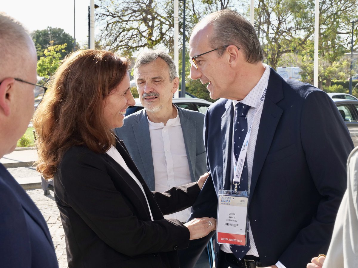
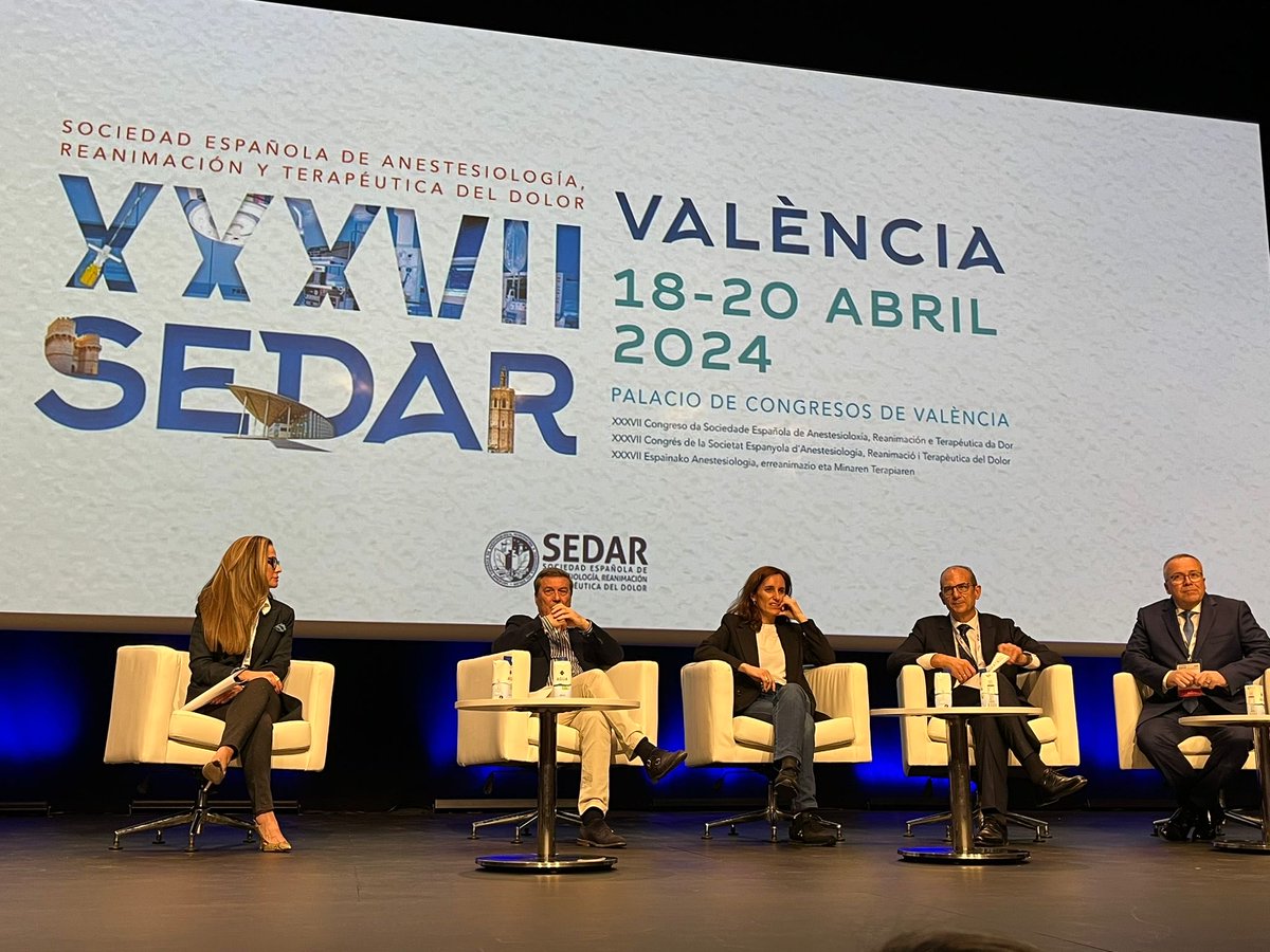
@Xunta @SanidadeXunta @sedar_es @JG_Anestesia @MarinaVarelaDu1 @sabe_dr @AnestChuac @LopezAlais @anestesio610 @AnestesiaGuada1 @anestesiamar @TVGalicia

Español
servando lopez
323 posts


@LopezAlais
Medicina perioperatoria. #Anestesia ambulatoria. Amor pola #Ribeira Sacra. Bon paladar


Reclamamos al Ministerio @sanidadgob y a las Consejerías de Sanidad que se actualice nuestro programa de formación MIR al mismo nivel que la formación europea. Llevamos 28 años de retraso en la actualización del programa MIR. ¡NO, sin el quinto año! bit.ly/4e5iqc8