Sabitlenmiş Tweet

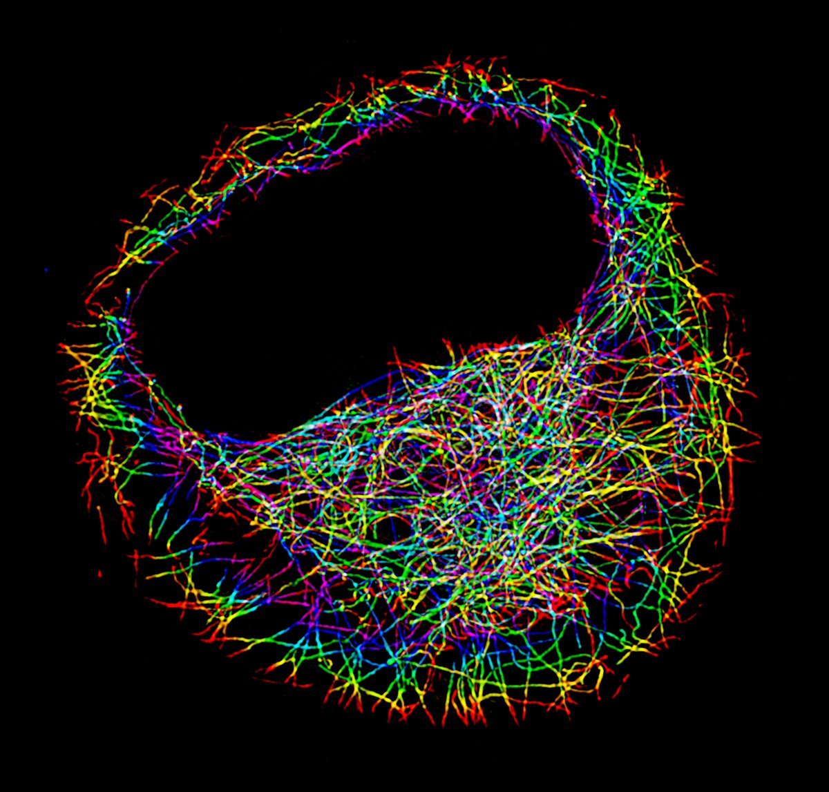
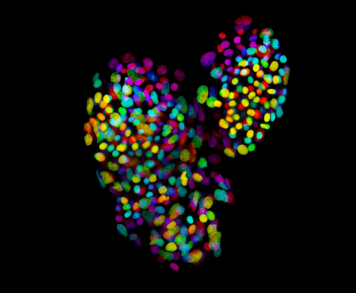
I am excited to finally be able to share with you our work reporting the first Extracellular Vesicle with a personality! This video does not show a cell, it is a Blebbisome! #CellBiology
nature.com/articles/s4155…
English
Dylan Burnette
3.3K posts
@MAG2ART
Cell biologist studying how a heart grows and dies; also Blebbisomes. Associate Professor at Vanderbilt. Married to @gillianhoo.


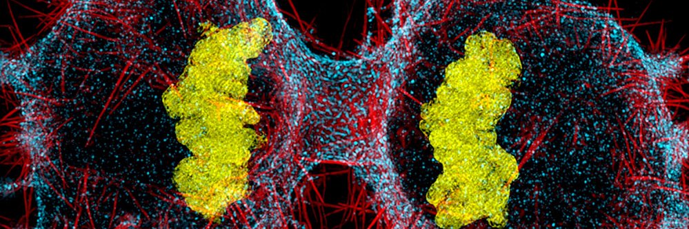
When key “housekeeper” brain cells grow in lab dishes, they spawn unusual microscopic vesicles that can move on their own and carry energy-generating organelles, a research team revealed. The biologists have dubbed their discovery zombosomes because the blobs can move like cells for a period despite lacking a nucleus, which acts as a cell's control center. The group also showed the membrane-bound messengers ferry proteins related to Parkinson’s disease, suggesting they may contribute to it and other brain disorders. Learn more: scim.ag/49WB4Um @NewsfromScience