Sabitlenmiş Tweet
Dr M.AL-gradey
1.3K posts

Dr M.AL-gradey
@MAbdullah58266
Medical Doc,Health content, follow for medical CASES &MCQS daily Medical pearls Questions alternative account
Katılım Nisan 2026
135 Takip Edilen83 Takipçiler

@DrsansariOrd ✅ Bacteria
➡️Clostridium tetani
✅Important characteristics
➡️Gram-positive rod
➡️Anaerobic
➡️Spore-forming bacterium
➡️Produces tetanospasmin toxin
🟢The toxin blocks inhibitory neurotransmitters (GABA and glycine) causing muscle rigidity and spasms.
English

Dx: Erb’s palsy
A newborn with a weak or absent Moro reflex on one side after a difficult delivery suggests injury to the brachial plexus (C5–C6 roots), commonly caused by shoulder dystocia or traction during birth.
Haji Siyamuddin ANSARI@DrsansariOrd
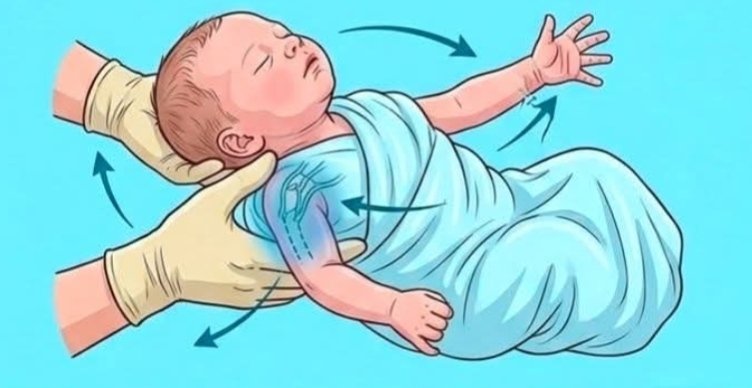
A newborn with weak Moro reflex on one side after difficult delivery may have❓ A. Erb’s palsy B. Hydrocephalus C. Neonatal sepsis D. Hypoglycemia
English

@kh505043 Dx: Drug Body Packing
Clinical presentation
May be:
➡️Asymptomatic
➡️Intestinal obstruction
➡️Drug toxicity if a packet ruptures
English

@ajlkfad2333 ✅ B) Kinky, brittle hair
🟢features of Menkes disease:
•Defect in copper transport
Leads to copper deficiency
➡️Characteristic:
Kinky/coarse brittle hair
Hypotonia
Developmental delay
Seizures
Failure to thrive
➡️The hair is often described as:
“Steel wool hair”
Pili torti
English

@abdulra_hman11 ✅ داء كرون (Crohn’s disease)
لماذا يسمى Cobblestone؟
لأن المخاطية تصبح:
فيها تقرحات طولية وعرضية
مع مناطق مخاطية متورمة بينها
فتعطي شكلاً يشبه الحجارة المرصوفة (cobblestones).
العربية

@modernHealthMe Noma (also called Cancrum oris) is a rapidly progressive gangrenous infection of the mouth and face, mainly affecting malnourished children in poor-resource settings.
Begins as gingivitis or oral ulcer
English

@DrsansariOrd ✅ A. Erb’s palsy
A newborn with a weak or absent Moro reflex on one side after a difficult delivery suggests injury to the brachial plexus (C5–C6 roots), commonly caused by shoulder dystocia or traction during birth.
English

Two identical twin doctors joined the same hospital on the same day.
Both wear the same scrubs, same watch, same hairstyle, even the same perfume. 😭
One is a radiologist.
The other is a gynaecologist.
The nurses say there’s only ONE tiny detail that exposes the gynaecologist twin… 👀
Can you spot which doctor is the gynaecologist?

Sukkur, Pakistan 🇵🇰 English

🔴Ameloblastoma is a benign odontogenic tumor that originates from the enamel-forming cells of the jaw.
➡️Robinson's Definition:
🔴Ameloblastoma is usually Unicentric, Nonfunctional," Intermittent in growth, Anatomically benign, and Clinically persistent.
(Mnemonic: UNIAC)
Dr Ihab Suliman@IhabFathiSulima
What is the diagnosis ⁉️
English

@IhabFathiSulima Ameloblastoma is a benign odontogenic tumor that originates from the enamel-forming cells of the jaw.
Robinson's Definition:
➡️Ameloblastoma is usually Unicentric, Nonfunctional," Intermittent in growth, Anatomically benign, and ".Clinically persistent
(Mnemonic: UNIAC)


English

@IhabFathiSulima Dx:Superior Vena Cava Obstruction
➡️Common causes include lung cancer, lymphoma, and thrombosis from central venous catheters.
➡️Patients present with facial swelling, distended neck veins, dyspnea, cough, and upper limb edema.


English

@docakx This is sometimes called aleukaemic leukaemia. Bone marrow infiltration and systemic symptoms occur later.
Sometimes myeloid leukaemia relapses in the skin after apparently successful treatment of bone marrow. It is then called ‘extramedullary’ leukaemia.
English

@docakx Leukemia cutis occurs when malignant white blood cells infiltrate the skin. It typically presents as firm,painless,and rubbery bumps,flat patches,or thickened plaques.
The skin lesion is the very first sign of the haematological malignancy in 7% of patients with leukaemia cutis.

English

Answer: Hemorrhagic brain tumor (most likely Glioblastoma)
➜ Key imaging feature:
Heterogeneous lesion with mixed density → hemorrhage within tumor.
➜ Edema pattern:
Marked perilesional edema → suggests chronic neoplastic process, not acute bleed.
➜ Mass effect:
Significant midline shift and compression → expansive space-occupying lesion.
➜ Morphology:
Irregular infiltrative margins → consistent with high-grade glioma (GBM).
English


@DrsansariOrd @IhabFathiSulima @docakx Exactly, hemorrhagic stroke
Thank you so much for your detailed information
English
Dr M.AL-gradey retweetledi

This is far more than just “stroke-like symptoms” — this is a large acute lobar intracerebral hemorrhage with dangerous mass effect.
A few additional high-yield points:
🚨 Why this is immediately life-threatening
The major killer here is not only the bleed itself, but:
Rapid rise in intracranial pressure (ICP)
Progressive midline shift
Risk of subfalcine or transtentorial herniation
The compressed ventricle and distorted anatomy already suggest severe intracranial hypertension.
🧠 Important localization clue
Because the hemorrhage is:
Lobar
Cortical/subcortical
Not centered in basal ganglia
…it becomes less typical for chronic hypertension alone.
That is why:
CAA becomes highly likely in elderly patients
AVM/tumor/cavernoma must be excluded in younger patients
📌 CT appearance pearl
Acute blood appears hyperdense (bright white) on non-contrast CT because freshly clotted blood has high protein and iron concentration.
⚡ “Stroke mimic” reminder
Not all sudden focal deficits are ischemic strokes. A non-contrast CT is critical before thrombolysis because giving tPA to this patient would be catastrophic.
🚑 Neurosurgical red flags present here
Large hematoma volume
Midline shift
Mass effect
Cortical superficial location
These are exactly the features that trigger urgent neurosurgical consultation.
🧠 Classic teaching:
> “Deep hemorrhage = hypertension until proven otherwise.
Lobar hemorrhage = think beyond hypertension.”
Dr M.AL-gradey@MAbdullah58266
Came in for stroke like symptoms. What's the diagnosis?🤔 @IhabFathiSulima
English

@IhabFathiSulima Often associated with pulmonary contusion (bruising of the lung), which further compromises respiratory function.
English

@IhabFathiSulima ✅Pathophysiology:
The flail segment moves paradoxically during respiration:
🔸Inspiration: flail segment moves inward (opposite to normal chest wall movement).
🔸Expiration: flail segment moves outward.
This paradoxical movement impairs ventilation and oxygenation.
English


