MTD 🩺

498 posts

MTD 🩺 banner
MTD 🩺

MTD 🩺

@MTDMED

Medicine explained through cases & visuals. Posts ≠ medical advice.

Katılım Mayıs 2026
226 Takip Edilen2.5K Takipçiler
MTD 🩺
MTD 🩺@MTDMED·
You are managing a critically ill patient in the ICU with severe sepsis who has now developed active bleeding from their IV sites. What is your differential diagnosis?
MTD 🩺 tweet media
English
0
1
1
87
MTD 🩺
MTD 🩺@MTDMED·
Atrial septal defect closure device💡
English
0
2
8
889
MTD 🩺
MTD 🩺@MTDMED·
🩺 Chest Tube (Tube Thoracostomy) A chest tube (tube thoracostomy) is inserted into the pleural space to evacuate air, blood, pus, or fluid, allowing the lung to re-expand. Indications Pneumothorax Tension pneumothorax (after immediate needle/finger decompression) Large or symptomatic spontaneous pneumothorax Traumatic pneumothorax Hemothorax Hemopneumothorax Empyema Complicated parapneumonic effusion Malignant pleural effusion (selected cases) Chylothorax Safe triangle The chest tube is inserted through the safe triangle, bounded by: Lateral border of pectoralis major Anterior border of latissimus dorsi Horizontal line at the 5th intercostal space (nipple level in males) Apex below the axilla Usual insertion site: 4th or 5th intercostal space, just anterior to the mid-axillary line. Procedure Position the patient (arm abducted above the head). Sterilize and drape. Infiltrate local anesthetic. Make a 2–3 cm skin incision over the rib. Perform blunt dissection with a curved clamp. Enter the pleural space just above the upper border of the rib (avoids the intercostal neurovascular bundle). Perform finger thoracostomy to confirm entry and clear adhesions. Advance the chest tube into the pleural cavity. Connect to an underwater seal drainage system (± suction if indicated). Secure with sutures and apply an occlusive dressing. Confirm placement with a chest X-ray. Tube direction Pneumothorax: Direct the tube anteriorly and superiorly (toward the apex). Fluid (hemothorax, pleural effusion, empyema): Direct the tube posteriorly and inferiorly (toward the base). Complications Bleeding (intercostal vessels) Lung laceration Injury to diaphragm, liver, spleen, or stomach Infection Tube malposition or blockage Persistent air leak Re-expansion pulmonary edema (after rapid drainage of a large pneumothorax or effusion)
English
0
0
2
307
MTD 🩺
MTD 🩺@MTDMED·
What is the name of this procedure?
English
10
9
81
30K
MTD 🩺
MTD 🩺@MTDMED·
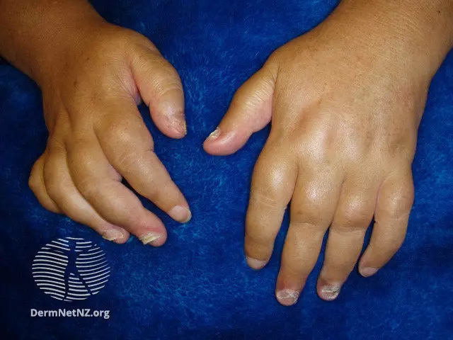
🩺 Dupuytren's contracture (Palmar fibromatosis) A progressive fibroproliferative disorder of the palmar fascia that leads to fixed flexion contractures of the fingers due to shortening of fibrous cords beneath the skin. It is not a tendon disorder. High-yield facts Most commonly affected fingers: Ring (4th) → Little (5th) Usually bilateral, though severity may differ. Classically painless. Begins as a firm palmar nodule → develops into a fibrous cord → progressive finger flexion. Risk factors ("Viking disease") Male > Female Age >50 years Northern European ancestry Family history (strong genetic component) Diabetes mellitus Smoking Alcohol use Epilepsy (possibly related to long-term antiepileptic therapy) Clinical features Palmar nodules Skin puckering/dimpling Thick fibrous cords Progressive inability to extend the fingers Difficulty shaking hands, wearing gloves, or placing the hand flat Diagnosis Clinical Hueston's tabletop test: Positive if the patient cannot place the palm flat on a table. Histology Proliferation of myofibroblasts Excess type III collagen deposition Associated fibromatoses Peyronie disease (penile fibromatosis) Ledderhose disease (plantar fibromatosis) Garrod knuckle pads Treatment Observation if mild and functional. Collagenase clostridium histolyticum injection (selected patients). Percutaneous needle fasciotomy/aponeurotomy. Limited fasciectomy is the standard surgical treatment for significant contracture. Surgery is generally considered when:MCP contracture ≥30°, or Any functionally significant PIP contracture
English
0
0
0
83
MTD 🩺
MTD 🩺@MTDMED·
What is the name of this condition?
MTD 🩺 tweet media
English
7
3
20
2.6K
MTD 🩺
MTD 🩺@MTDMED·
Pot belly Protruding umbilicus (umbilical hernia) Poked-out tongue (macroglossia) Puffy face Pallor (pale, dry, or yellowish skin) What is the diagnosis? 🚨
MTD 🩺 tweet media
English
0
1
2
258
MTD 🩺
MTD 🩺@MTDMED·
"Watermelon stomach" is seen in which condition?
MTD 🩺 tweet mediaMTD 🩺 tweet media
English
11
7
63
9.4K
MTD 🩺
MTD 🩺@MTDMED·
Which organ damage causes this?
MTD 🩺 tweet media
English
10
1
14
1.5K
MTD 🩺
MTD 🩺@MTDMED·
🩺 Answer: Electrolysis. Details: Electrolysis is the only FDA-approved method for permanent hair removal. A fine probe is inserted into each hair follicle, and an electric current destroys the hair follicle and germinative cells, preventing regrowth. It is effective for all hair colors and skin types, unlike laser hair removal, which is most effective on dark hair. Common indications include hirsutism, removal of isolated coarse hairs, and patients who are not good candidates for laser therapy. Temporary side effects include erythema, edema, and discomfort; rare complications include scarring and post-inflammatory pigment changes.
English
0
0
0
101
MTD 🩺
MTD 🩺@MTDMED·
Only FDA-approved method for permanent hair removal. What's the name of this procedure?
English
5
1
39
27K
MTD 🩺
MTD 🩺@MTDMED·
🩺 Answer: Senna (an anthraquinone-containing stimulant laxative). Details: Melanosis coli is classically caused by chronic use of anthraquinone stimulant laxatives, most commonly:Senna Cascara sagrada Aloe Rhubarb-derived laxatives Anthraquinones induce apoptosis of colonic epithelial cells. Macrophages in the lamina propria phagocytose the apoptotic cells, leading to accumulation of lipofuscin pigment (not melanin), which causes the characteristic brown-black discoloration of the colonic mucosa. It is a benign, reversible condition that typically fades within months after stopping the laxative. Not associated with an increased risk of colorectal cancer.
English
0
0
0
67
MTD 🩺
MTD 🩺@MTDMED·
Chronic use of which laxative causes this?
MTD 🩺 tweet mediaMTD 🩺 tweet media
English
4
1
10
2.5K
MTD 🩺
MTD 🩺@MTDMED·
🩺 Intraosseous (IO) access is an emergency vascular access technique in which a needle is inserted into the medullary cavity of a bone, allowing rapid administration of fluids, blood products, and medications when intravenous access cannot be obtained quickly. Indications Cardiac arrest Hemorrhagic or septic shock Major trauma Status epilepticus Any critically ill patient when IV access cannot be established within 60–90 seconds or after 2 failed IV attempts Common insertion sites Proximal tibia (most common in children) Proximal humerus (preferred in adults during resuscitation due to higher flow rates) Distal tibia Sternum (with dedicated devices) What can be given through IO? Crystalloids Blood products Vasopressors Epinephrine Antibiotics Calcium Any medication that can be given IV Contraindications Fracture of the target bone Previous IO attempt in the same bone within 24–48 hours Infection or burn over the insertion site Bone disorders affecting integrity (e.g., Osteogenesis imperfecta) Prosthetic joint near the insertion site Complications Extravasation Compartment syndrome Osteomyelitis (rare) Growth plate injury (children) Fat embolism (rare) Fracture High-yield facts IO access is as effective as IV access for most emergency medications. It can usually be established in under 1 minute. Conscious patients require a lidocaine flush before infusion because marrow infusion is painful. IO access is a temporary bridge and should generally be replaced with definitive IV or central venous access within 24 hours. Classic exam pearl A patient in cardiac arrest has had two unsuccessful peripheral IV attempts. The next best step is immediate intraosseous access rather than delaying resuscitation for central venous catheter placement.
English
0
0
1
134
MTD 🩺
MTD 🩺@MTDMED·
What is going on here?
English
9
8
48
9.6K
MTD 🩺
MTD 🩺@MTDMED·
🩺 "Pizza pie" retinopathy is classically seen in Cytomegalovirus (CMV) retinitis. Typical vignette: Advanced HIV/AIDS (usually CD4 <50 cells/µL) Painless, progressive visual loss or floaters Fundoscopy: fluffy yellow-white retinal necrosis with extensive retinal hemorrhages, producing the classic "pizza pie" or "cottage cheese and ketchup" appearance. Answer: ✅ CMV retinitis
English
0
0
0
54
MTD 🩺
MTD 🩺@MTDMED·
"Pizza pie" retinopathy is seen in which condition?
MTD 🩺 tweet mediaMTD 🩺 tweet media
English
4
3
33
3.9K
MTD 🩺
MTD 🩺@MTDMED·
🩺 Dural Tail Sign (Meningioma) Definition The dural tail sign is a thickened, tapering, contrast-enhancing segment of dura mater that extends away from a dural-based mass on contrast-enhanced MRI. Classic Association Meningioma (most common and classic association) The dural tail is seen in ~60–70% of meningiomas. Why Does It Occur? The enhancing "tail" represents a combination of: Reactive vascular congestion Fibrosis Inflammation Occasionally, tumor infiltration of the adjacent dura It is not always due to direct tumor spread. MRI Features of Meningioma Extra-axial, dural-based mass Homogeneous, avid contrast enhancement Dural tail sign CSF cleft between the tumor and brain Broad dural attachment May cause hyperostosis of the adjacent skull Usually causes vasogenic edema in the adjacent brain Is the Dural Tail Sign Specific? No. Although strongly associated with meningioma, it is not pathognomonic. Other causes include: Dural metastases Solitary fibrous tumor Primary CNS lymphoma Schwannoma Neurosarcoidosis Granulomatous infections (e.g., tuberculosis) Idiopathic hypertrophic pachymeningitis High-Yield Exam Pearls ✅ Most likely diagnosis: Meningioma. ✅ Best visualized on post-contrast T1-weighted MRI. ✅ Indicates enhancing adjacent dura, not necessarily tumor invasion. ✅ Remember other classic imaging clues for meningioma: extra-axial location, broad dural base, homogeneous enhancement, and hyperostosis.
English
0
0
0
78
MTD 🩺
MTD 🩺@MTDMED·
What’s the diagnosis?
MTD 🩺 tweet media
English
8
4
42
5.8K
MTD 🩺
MTD 🩺@MTDMED·
🩺 Twin-to-Twin Transfusion Syndrome (TTTS) Definition Twin-to-Twin Transfusion Syndrome (TTTS) is a complication of monochorionic (shared placenta) twin pregnancies, caused by unbalanced blood flow through placental vascular anastomoses. Occurs in monochorionic diamniotic (MCDA) twins. Affects approximately 10–15% of MCDA pregnancies. Usually develops between 16–26 weeks of gestation. Pathophysiology The placenta contains artery-to-vein vascular connections. Donor twinPumps blood to the recipient Hypovolemia Anemia Decreased renal perfusion → ↓ urine production Oligohydramnios Growth restriction Recipient twinReceives excess blood Hypervolemia Polycythemia Increased urine production Polyhydramnios Cardiac overload → heart failure → hydrops fetalis Classic Ultrasound Findings Donor twin Oligohydramnios (deepest vertical pocket <2 cm) Small or absent bladder "Stuck twin" appearance (immobile against the uterine wall) Recipient twin Polyhydramnios (deepest vertical pocket >8 cm before 20 weeks or >10 cm after 20 weeks) Large bladder Cardiomegaly Ventricular dysfunction Hydrops (late) Treatment Depends on gestational age and stage. Fetoscopic laser photocoagulation of placental vascular anastomoses (treatment of choice for Stage II–IV before viability) Serial amnioreduction (less preferred) Close surveillance in mild disease Delivery when appropriate if fetal maturity is achieved or deterioration occurs Complications Donor twin Severe growth restriction Anemia Renal insufficiency Intrauterine demise Recipient twin Heart failure Hydrops fetalis Polycythemia Neurologic injury Intrauterine demise
English
0
0
0
53
MTD 🩺
MTD 🩺@MTDMED·
What is the most likely diagnosis? A) Twin-to-twin transfusion syndrome (TTTS) C) Placental abruption D) Selective intrauterine growth restriction E) Preeclampsia with fetal growth restriction
MTD 🩺 tweet media
English
7
1
13
1.7K