Brookes Bioimaging retweetlediDr Louise Hughes@DrLouiseCHughes·2 Ara
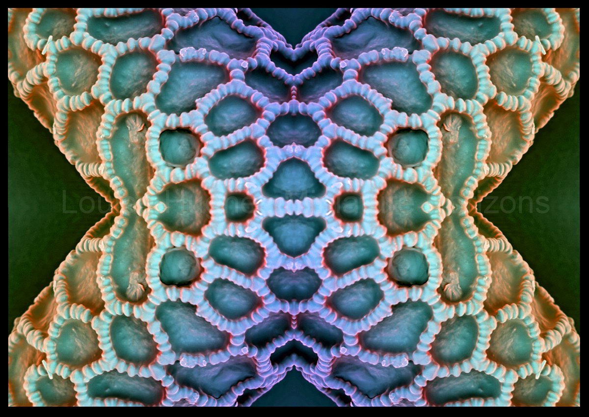
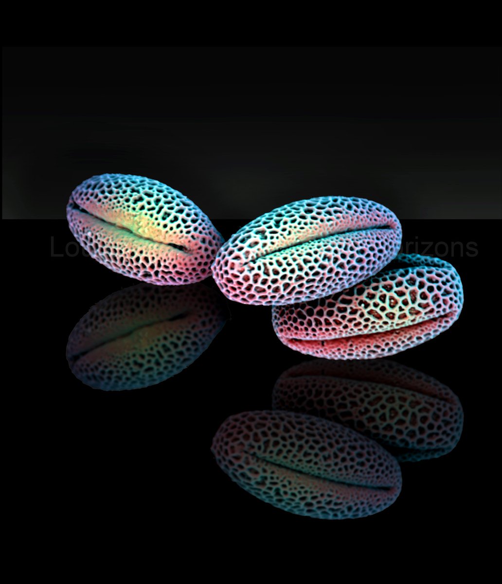
Because #fridayfeeling i am also going to post the abstract art stemming from today's #microscopyadvent of lily pollen. #sciartÇevir English011130
Brookes Bioimaging retweetlediDr Louise Hughes@DrLouiseCHughes·2 Ara2/12 Close up of a lily pollen grain captured with SEM. #microscopyadvent #microscopyart #sciart @MicroscopySoc @obu_bms @OBBU_microscopyÇevir English015200
Brookes Bioimaging retweetlediDr Louise Hughes@DrLouiseCHughes·1 AraIt is that time of year again! #microscopyadvent #1stdecember ##AdventCalendar SEM of Iris pollen grains #sciart @obu_bms @oxford_brookesÇevir English09110
Brookes Bioimaging retweetlediDr Louise Hughes@DrLouiseCHughes·23 KasSEM image (after some editing) of willow pollen, combining BSE and SE signals. #sciart #biology @obu_bms @oxford_brookes @OBBU_microscopyÇevir English0880
Brookes Bioimaging@OBBU_microscopy·11 Eki#biologyweek @obu_bms twitter.com/DrLouiseCHughe…ÇevirDr Louise Hughes@DrLouiseCHughes3D reconstruction of a Golgi apparatus from Arabidopsis root cells. #biologyweek #sciart @brookeshls @zeiss_micro @OBBU_microscopy #3DEM QME0010
Brookes Bioimaging@OBBU_microscopy·11 EkiNew article @obu_bms twitter.com/DrLouiseCHughe…ÇevirDr Louise Hughes@DrLouiseCHughesWant to know more about 3D SEM? bitesizebio.com/30796/three-di… @zeiss_micro @brookeshls #ghostwritting English0000
Brookes Bioimaging@OBBU_microscopy·11 Eki@obu_bms twitter.com/DrLouiseCHughe…ÇevirDr Louise Hughes@DrLouiseCHughesInside a root looking at the nuclei. 3D SEM data for VR #virtualreality #realdata #biologyweek #sciart @zeiss_micro @brookeshls QME0000
Brookes Bioimaging@OBBU_microscopy·6 EkiScicomm award! @brookeshls @hls_research @Brookes_Impact @oxford_brookes @obu_bms @BrookesStaff twitter.com/DrLouiseCHughe…ÇevirDr Louise Hughes@DrLouiseCHughesI have won the @RoyalSocBio Science Communication Award for an established researcher! #thrilled #scicomm rsb.org.uk/news/14-news/1… English0200
Brookes Bioimaging retweetlediThe Royal Society@royalsociety·22 TemOur very special collection of the life & works of Robert Hooke - Under the microscope ow.ly/NUZQ302lQDIÇevir English142440
Brookes Bioimaging@OBBU_microscopy·3 MarIncredible book and images. twitter.com/RoyalMicroSoc/…ÇevirRMS@RoyalMicroSoc#RobertHooke died on this day in 1703. We're celebrating his greatest work #Micrographia for #WorldBookDay English0110
Brookes Bioimaging@OBBU_microscopy·3 MarSome of the work done here at OBBU. #brookeshls twitter.com/DrLouiseCHughe…ÇevirDr Louise Hughes@DrLouiseCHughesHow to get 3D data using electron microscopy. Diagram showing some of the different techniques. #sciart English0210
Brookes Bioimaging@OBBU_microscopy·25 ŞubMicroscopy club for BMS undergraduates every other Wednesday afternoon! twitter.com/DrLouiseCHughe…ÇevirDr Louise Hughes@DrLouiseCHughesFlea mite imaged with SEM. Taken during @OBBU_microscopy microscopy club for undergrads. #brookeshls #sciart English0000
Brookes Bioimaging@OBBU_microscopy·19 ŞubUndergrad teaching this pm! twitter.com/DrLouiseCHughe…ÇevirDr Louise Hughes@DrLouiseCHughesTEM prac. with undergrads this pm. Image of plant Golgi bodies. #brookeshls #sciart @HawesChris @jakerich101 English0000
Brookes Bioimaging@OBBU_microscopy·18 AraThis was captured using serial block face scanning electron microscopy! Beautiful data. @zeiss_micro twitter.com/DrLouiseCHughe…ÇevirDr Louise Hughes@DrLouiseCHughes#microscopyadvent day 18. 3D data - stoma and the surrounding guard cells in a leaf @zeiss_micro #sciart #brookeshls English0010
Brookes Bioimaging retweetlediZEISS Microscopy@zeiss_micro·17 Ara#ZEISS #Airyscan: confocal #microscopy with improved signal-to-noise ratio & super-resolution @naturemethods nature.com/nmeth/journal/…Çevir English0120
Brookes Bioimaging@OBBU_microscopy·17 AraTransmission electron microscopy image. Good to see some traditional EM going on. :) twitter.com/DrLouiseCHughe…ÇevirDr Louise Hughes@DrLouiseCHughes#
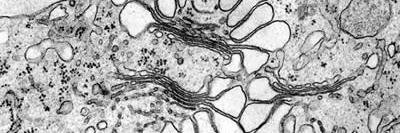
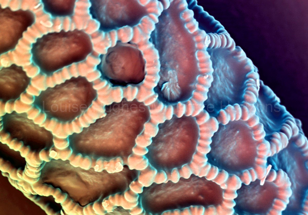
microscopyadvent day 17. Another Golgi body. This is a coloured TEM image of an animal cell. #sciart #brookeshls English0100
Brookes Bioimaging retweetlediOxford Brookes Health, Science and Techology@OBUresearchHST·11 AraEver wondered how a Scanning Electron Microscope works? Prof @HawesChris explains youtu.be/xY4YSjPipKE @OBBU_microscopy @zeiss_micro #SEMÇevirYouTube English0670
Brookes Bioimaging@OBBU_microscopy·15 AraSEM #microscopyadvent twitter.com/DrLouiseCHughe…ÇevirDr Louise Hughes@DrLouiseCHughes#microscopyadvent day 15. Compound eye on a true bug. SEM image. #sciart #brookeshls0000
Brookes Bioimaging retweetlediDr Louise Hughes@DrLouiseCHughes·14 Ara#microscopyadvent day 14. SEM image of the tip of a Drosophila proboscis, fruit fly mouthparts. #sciart #brookeshlsÇevir English2690
Brookes Bioimaging retweetlediDr Louise Hughes@DrLouiseCHughes·11 AraHow a SEM works as described by @HawesChris showing microscopes in our bioimaging unit as well as some of my data. youtube.com/watch?v=xY4YSj…ÇevirYouTube English0630