
Pathologie_MedUniWien
60 posts

Pathologie_MedUniWien
@Pathologie_MUW
The Department of Pathology performs essential functions in diagnostics, medical education and research at the Medical University of Vienna.
Katılım Şubat 2021
33 Takip Edilen188 Takipçiler
our own Dr. Kozakowski participated in this Expert-Publication on <Renal Pathology Society/International Kidney and Monoclonal Gammopathy Research Group Consensus on Pathologic Definitions and Terminology of Monoclonal Gammopathy Associated Kidney Lesions> excited to share~
English
A publishes outreach article in the leading science communication publication, Scientia
Doi: doi.org/10.33548/SCIEN…
Scientia@scientia_social
Fighting systemic viral infections! Dr Hermann Salmhofer explores new ways to minimize damage & improve treatments. doi.org/10.1093/ckj/sf… more: scientia.global/dr-hermann-sal… #Virology #MedicalResearch
English
What is the renal pathology here?
If you want to know it, well then, look up the following publication from our renal pathology team:
Kozakowski N, et al. Kidney Int Rep. 2024;10(1):256-259.
doi:10.1016/j.ekir.2024.10.014

English
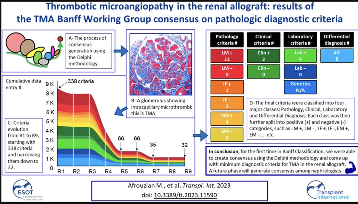
@kidneypathology Do you want to know how we did that? Check this one out: doi.org/10.3389/ti.202…

English
Pathologie_MedUniWien retweetledi

Do you want to know the Minimal Diagnostic Criteria for TMA?. Finally a consensus for the morphologic diagnosis of TMA frontierspartnerships.org/articles/10.33…
English
Interested in spatial transcriptomics? Consider reading our pilot study on spatial profiling of glomeruli in pauci-immune focal necrotizing glomerulonephritis!
kidney360.asnjournals.org/content/early/…
English
ASN Kidney360@ASNKidney360
Pauci-immune focal necrotizing glomerulonephritis entails heterogeneous glomerular lesions. This study shows benefits of spatial profiling heterogeneous glomerular injury characterization, indicating molecular correlates of glomerular injury in piFNGN bit.ly/KID0004612022
ZXX
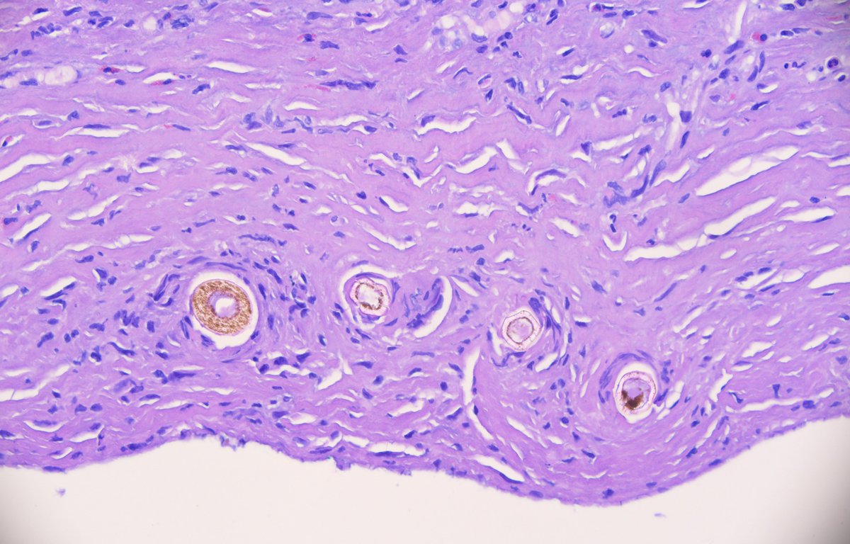
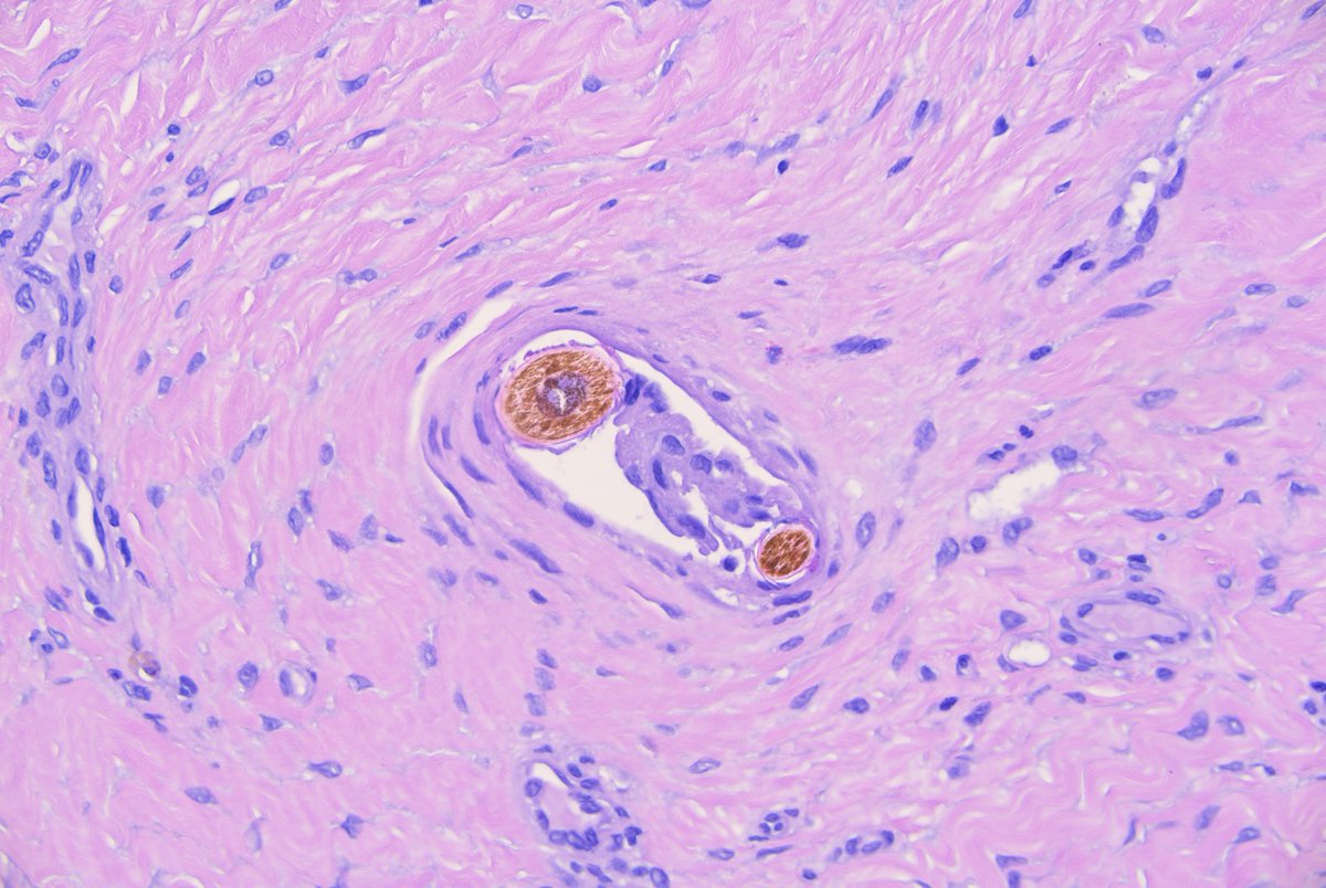
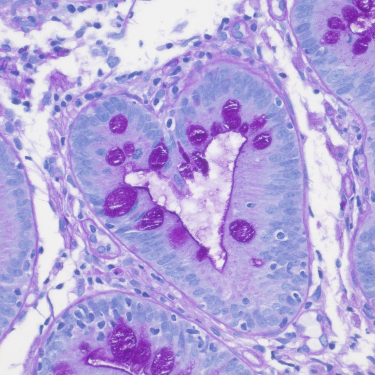
What are these structures doing in a piece of peritoneal tissue from a continuous ambulatory peritoneal dialysis catheter site? #PathTwitter #pathology
English

English
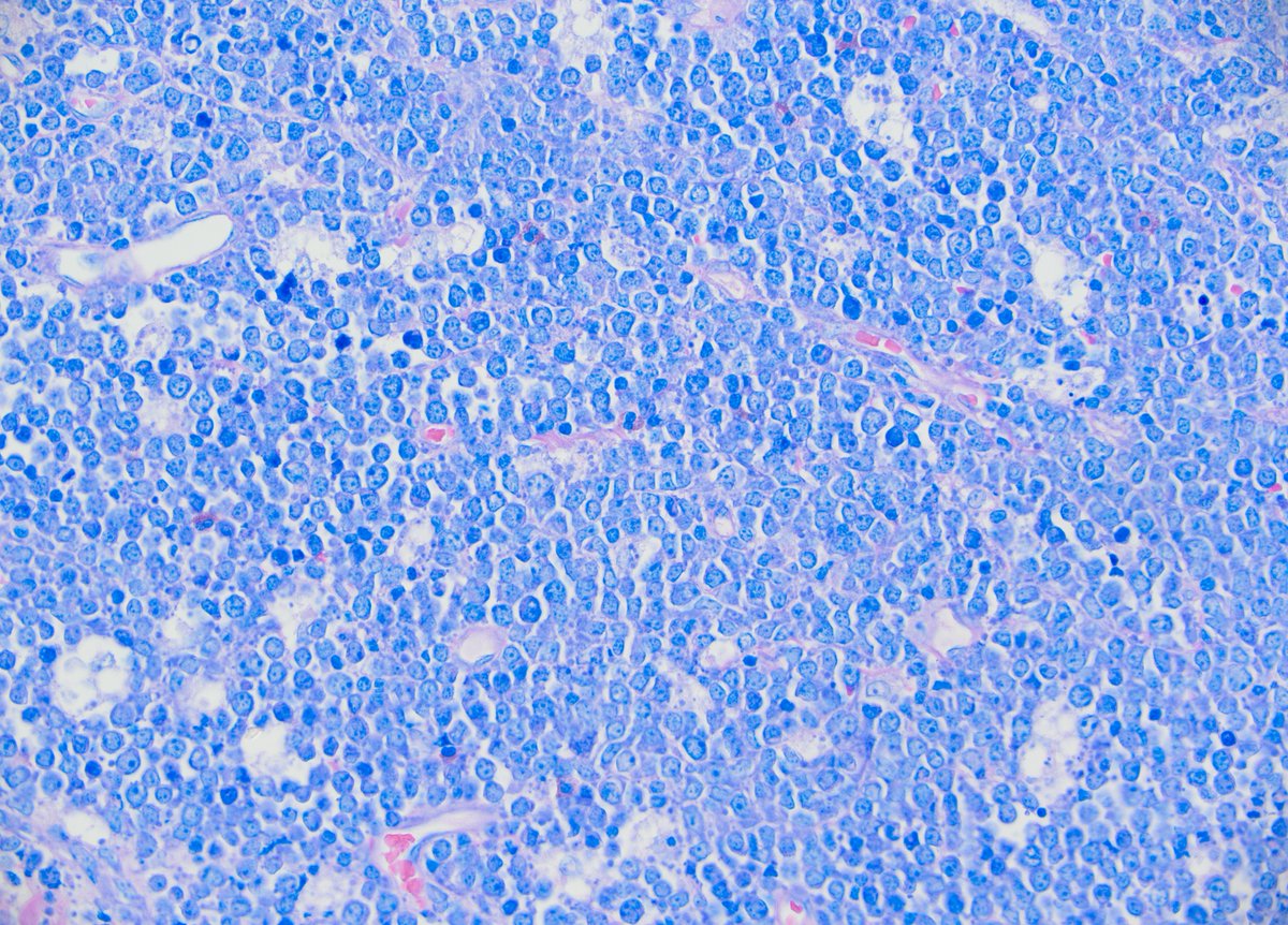
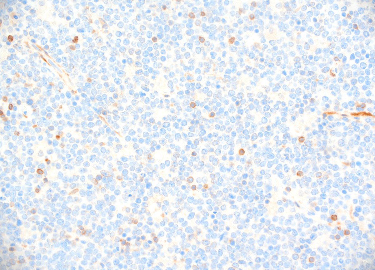
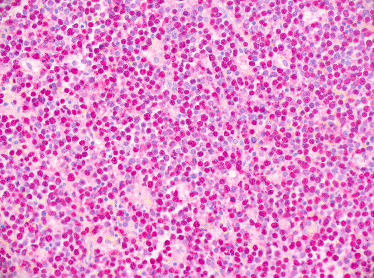
While gazing up at the night's sky hematopathologists don't search for the brightest star. They're looking for Burkitt's lymphoma. Classical starry sky pattern, medium sized, monomorphic tumor cells, EBV associated case (ileocecal resection, 3a, m), Giemsa, bcl6+, blc2-, EBER+

English
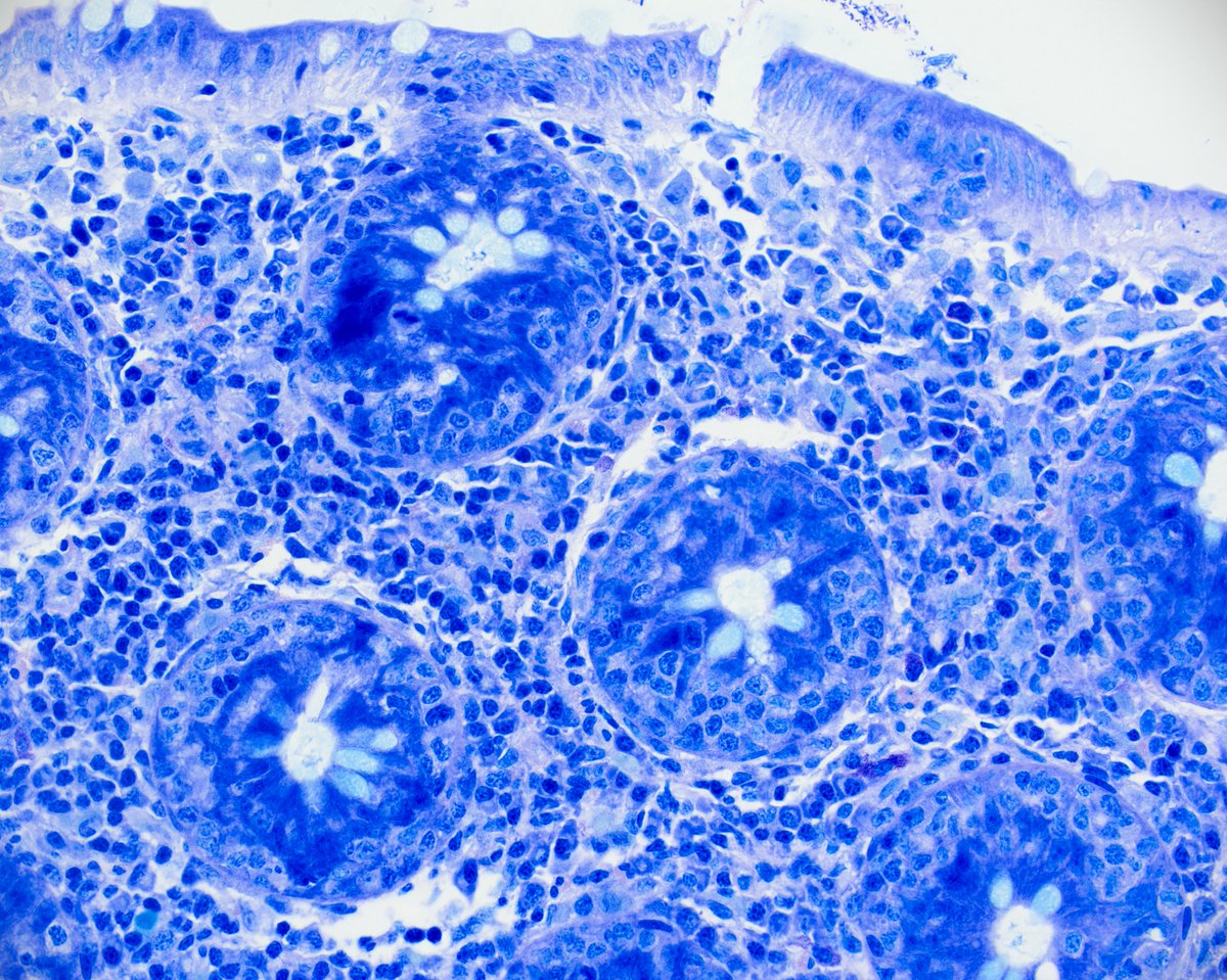
jejunal biopsy; marked villous atrophy, IEL increase, crypt hyperplasia, absent plasma cells in LP- your Dg?
❤️❤️#gipath

English
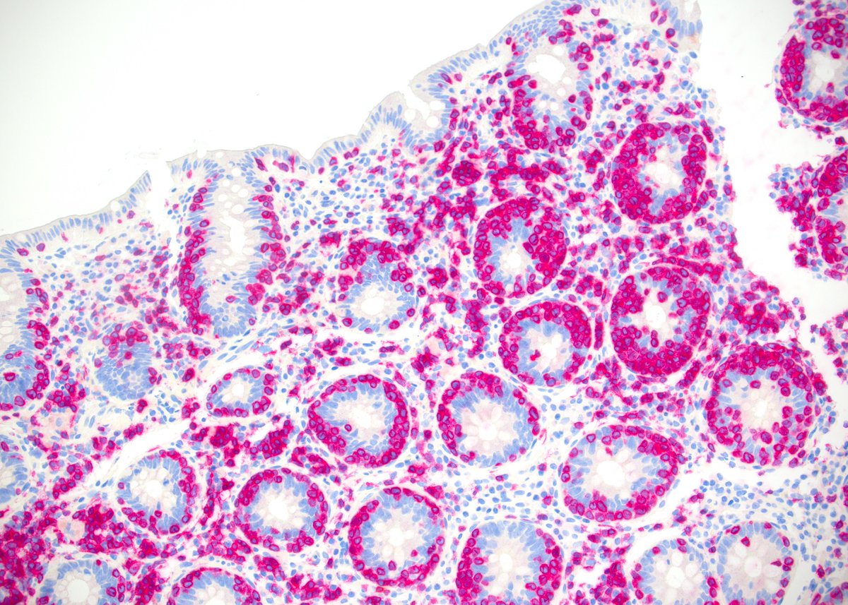
57a, w, colon biopsy, Giemsa, CD3+, CD5-, CD56+, patient with monomorphic epitheliotropic intestinal T-cell lymphoma (MEITL). The neoplastic cells are medium in size, uniform and show a prominent epitheliotropism.
English
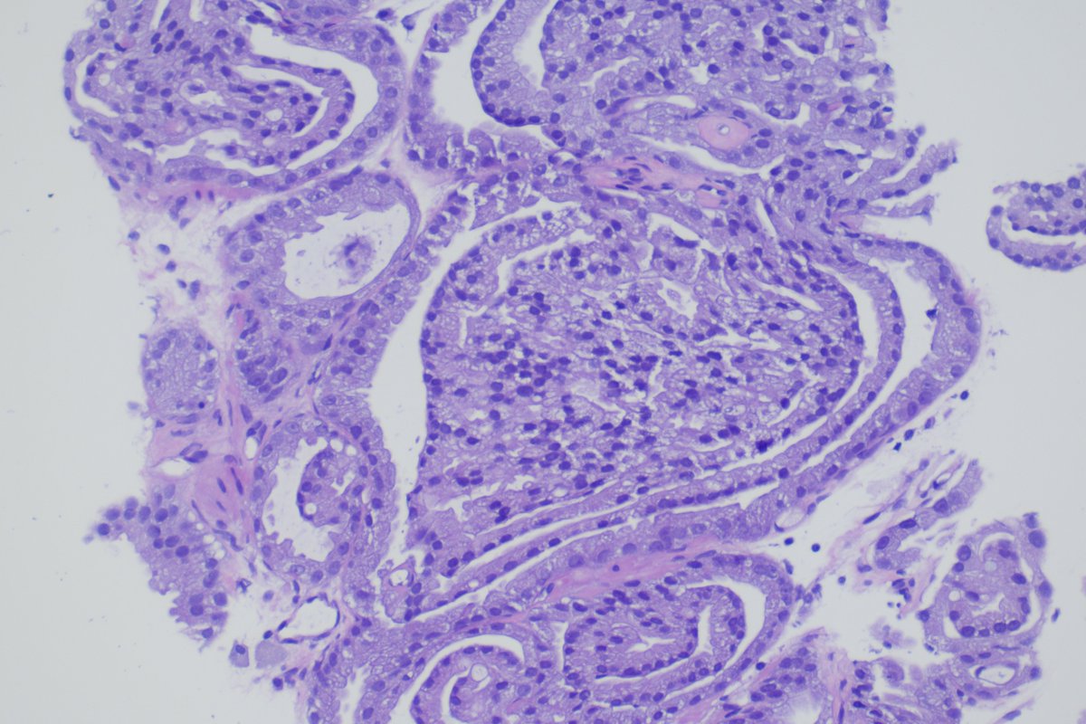
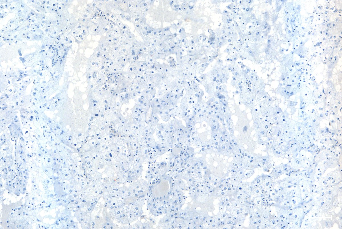
Eosinophilic renal tumor, CD117 negative: What is your diagnosis?


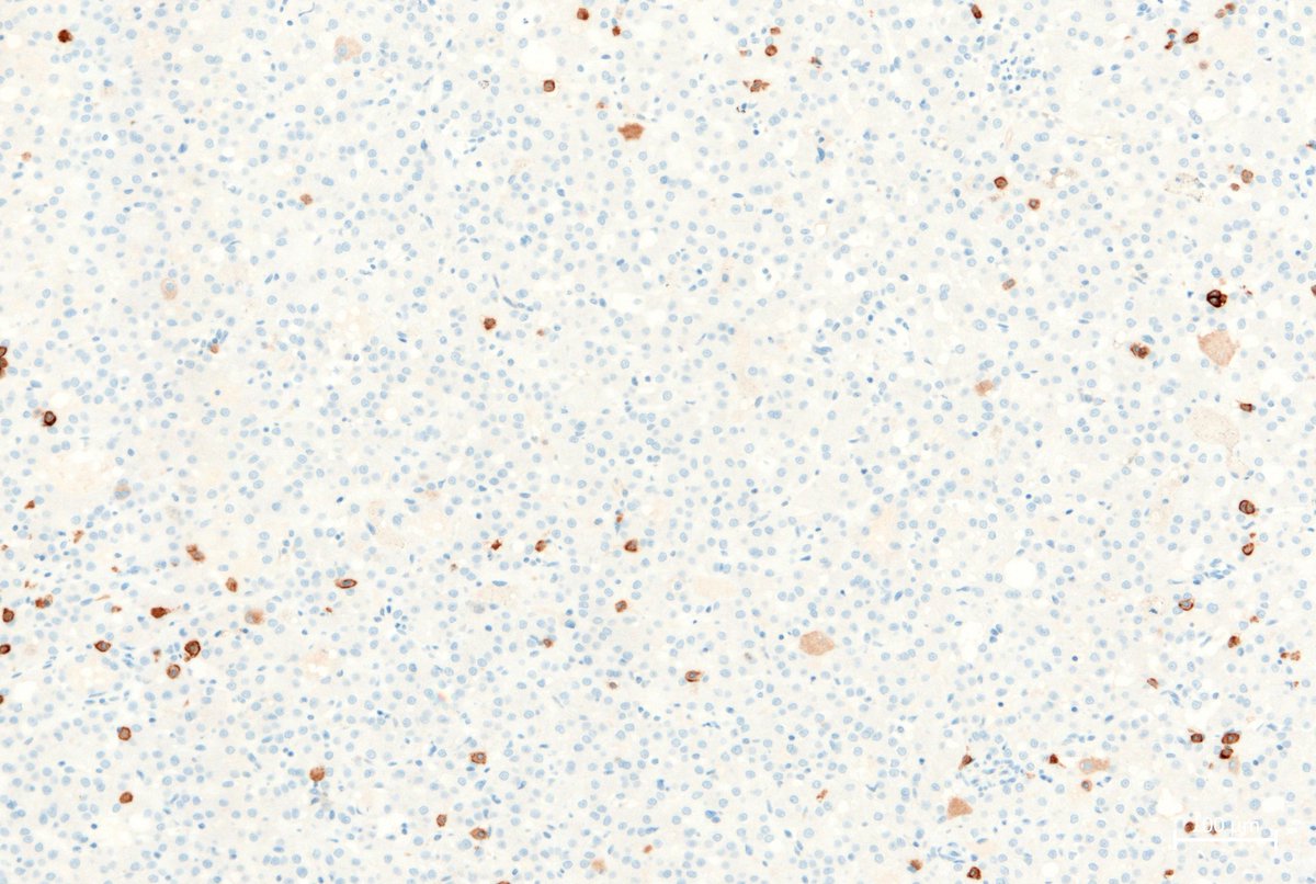
English
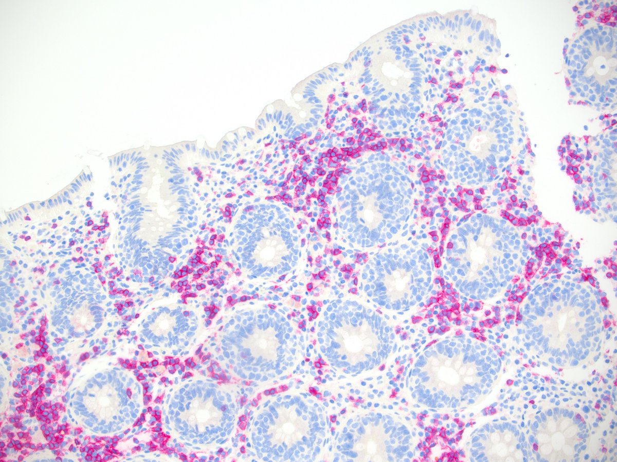
Friday afternoon quiz: Lesion in small intestine, CK7 (top right), GATA3 (bottom left), SOX10 (bottom right) ... ?


English

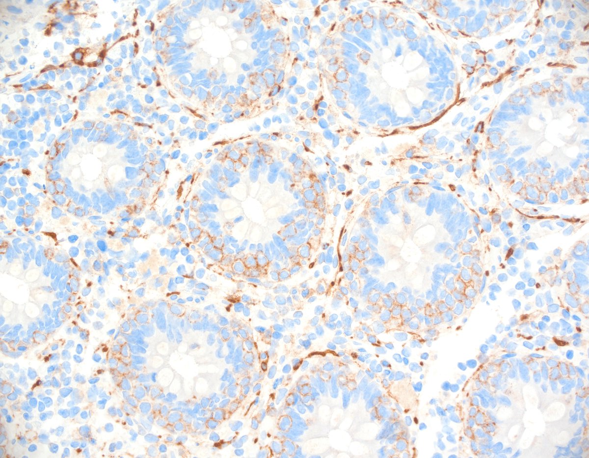
Clear cell renal carcinoma with rhabdoid features - could be challenging at first sight but shows quite typical ccRCC nuclei and positive CAIX (bottom left), negative CK7 (bottom right). #pathology


English
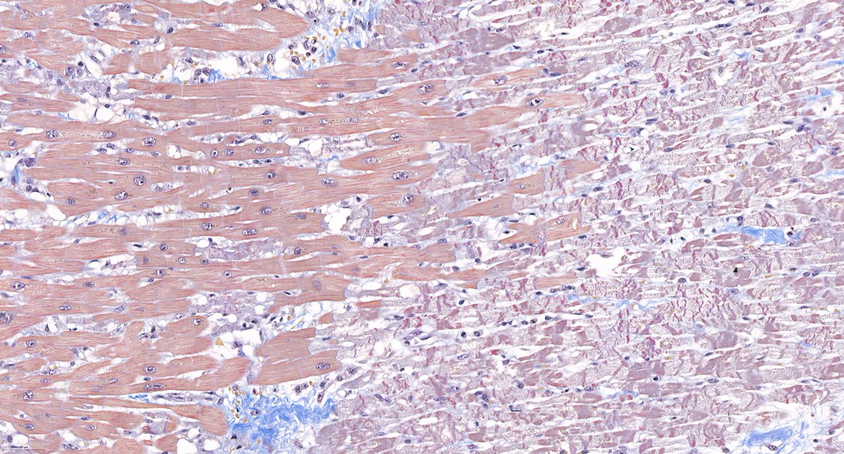
@goziemnweke @AmyHDeekenMD Acid Fuchsine Orange G - one of our favorites for renal pathology because it highlights connective tissue in blue and fibrin in bright red.
English

Contraction band necrosis and juxtaposed vital myocardium in transmural myocardial infarction. AFOG, 20x.
English