Sabitlenmiş Tweet
Yan Chu, MD
653 posts

Yan Chu, MD
@YanChuMD
⚕️GI Fellow @UKGIHep 💩; Former Chief Resident @IM_SLU via @UKYMedicine @TuftsMedicalCtr & @BrandeisU; 🇺🇸🇨🇳 #WomenInGI
Kentucky, USA Katılım Ağustos 2013
351 Takip Edilen434 Takipçiler
Yan Chu, MD retweetledi
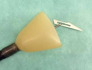
Endoscopic rubber hood:
•Bell-shaped .Protects the mucosa of the pharynx, esophagus, and cardia during extraction.
•Hood is flipped back during insertion and reverts when pulled back against the cardia- covering the foreign object
•Excellent for short sharp objects
English
Yan Chu, MD retweetledi

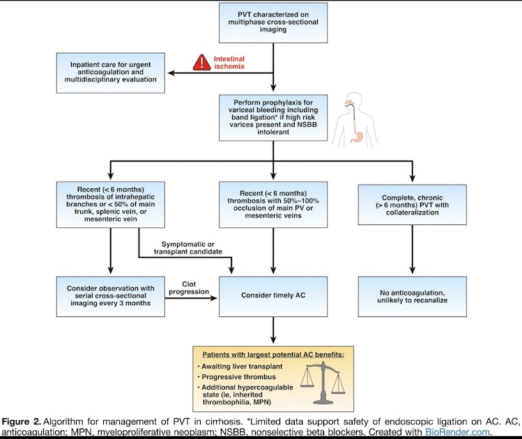
Portal vein thrombosis in cirrhosis: anticoagulation may reduce mortality, but the bleeding tradeoff is real.
In borderline cases, what tips your decision — acuity, thrombus extent, transplant candidacy, or something else?
English
Yan Chu, MD retweetledi

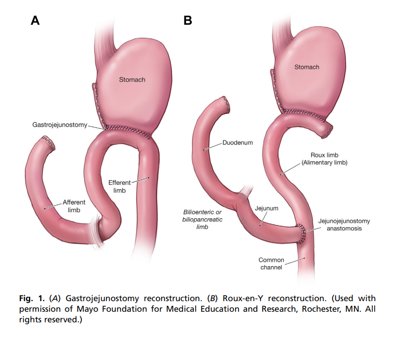
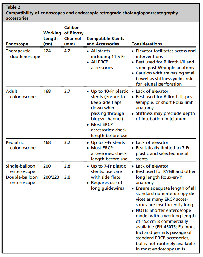
Basics of altered GI anatomy:
pubmed.ncbi.nlm.nih.gov/26431595/
🪙Efferent limb: Away from anastomosis
🪙Afferent limb: bile, pancreas juice, draining towards the anastomosis.
🪙Roux limb: savior limb coming to save obstructed/ distended stomach (in bariatrics).
🪙Scopes: Figure 2!
English
Yan Chu, MD retweetledi

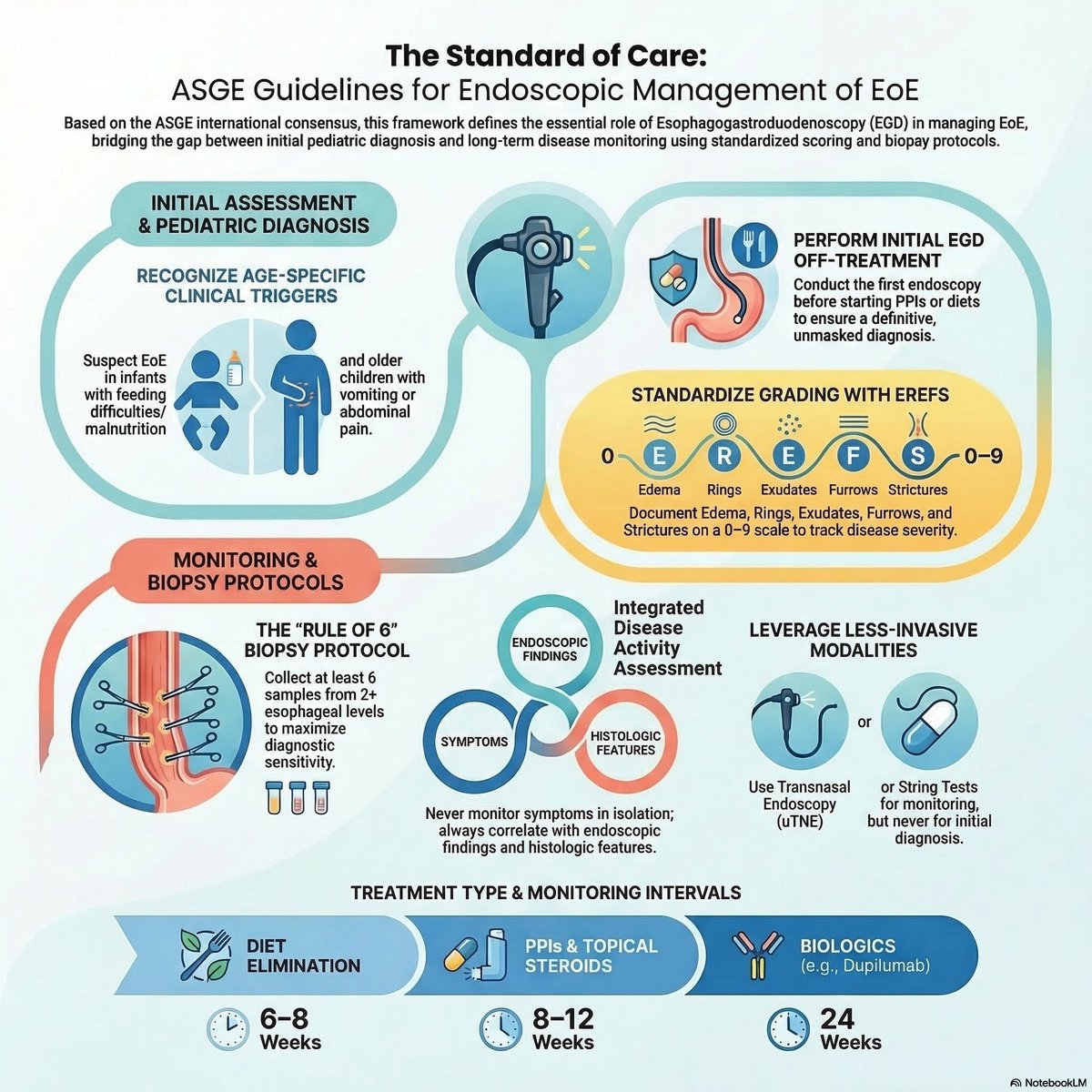
Management of Eosinophilic Oesophagitis (EoE) - 2026 ASGE guidelines
📸: sciencedirect.com/science/articl…
CY
Yan Chu, MD retweetledi
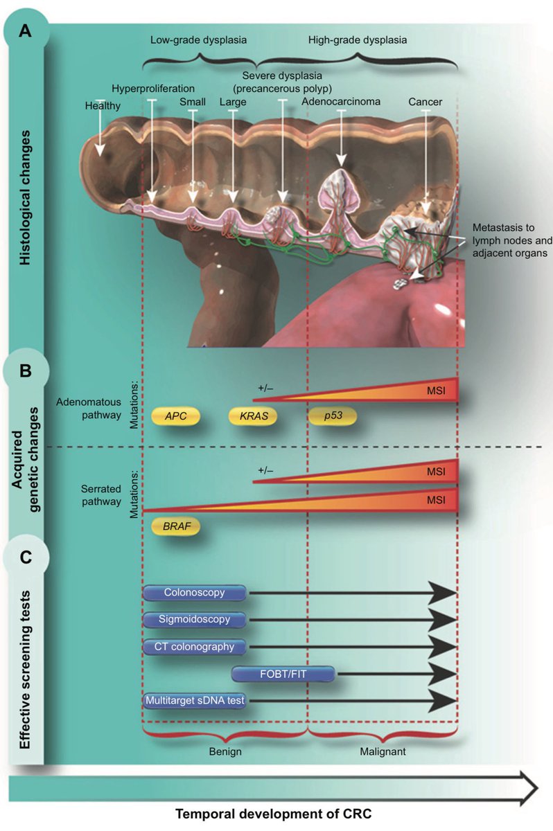
Colorectal cancer development and opportunities for screening 💡🔬
#ColorectalCancerAwarenessMonth
📸: dovepress.com/colorectal-can…
English
Yan Chu, MD retweetledi
Is it Barrett’s? Or just "Burnt Salmon"? 🍣
We are taught to look for "salmon-colored" mucosa. But as this slide from EndoCollab shows, there are 50+ shades of salmon.
The Clinical Reality: Identifying the color change is only Step 1.
To confirm the diagnosis, you must map the landmarks:
1️⃣ Locate the Gastric Folds (the true GEJ).
2️⃣ Look for the palisade veins (which exist only in the esophagus).
3️⃣ If the "salmon" color extends above these landmarks, you are in the danger zone.
Don't rely on color alone. Trust your anatomy.
Watch the full video in the comments.

English
Yan Chu, MD retweetledi

Yan Chu, MD retweetledi
Yan Chu, MD retweetledi

Most patients don't realize their doctors and nurses think about them in the spaces between visits. While driving home, getting ready for bed, while falling asleep.
Our work is a part of us, and we care about you. That's all.
English
Yan Chu, MD retweetledi

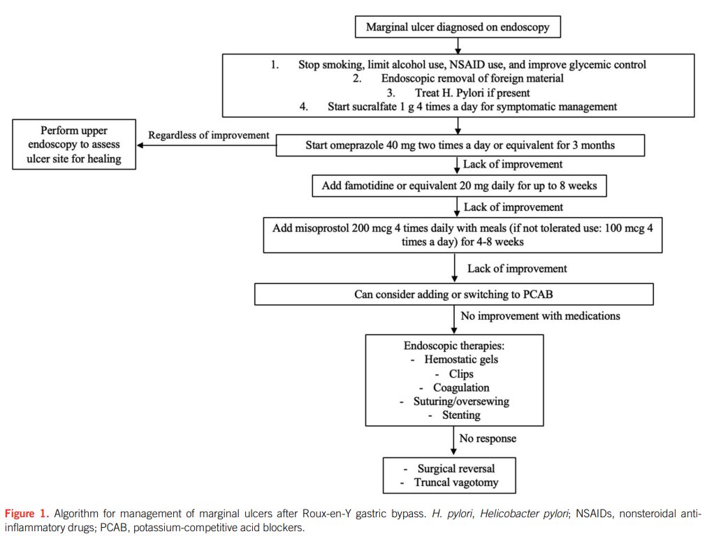
Marginal ulcer after RYGB Mgmt
▪️Stop smoking/NSAIDs
▪️Treat H. pylori
▪️Remove foreign body
▪️Open-cap PPI BID x3m
▪️Sucralfate 1g QID
▪️No response ➡️ H2RA ➡️ misoprostol ➡️ PCAB
▪️Refractory ➡️ endo therapy
▪️Failure ➡️ surgical revision
🔗 journals.lww.com/ajg/citation/2…
English
Yan Chu, MD retweetledi

📢 Roll Call! ACG Guidelines Edition
🧵👇: ACG Guidelines published in the #RedJournal in 2025, with links to the 📕guidelines, 🎙️podcasts, and ℹ️highlights!
➡️ gi.org/guidelines ⬅️
@MLongMD @JasmohanBajaj

English
Yan Chu, MD retweetledi
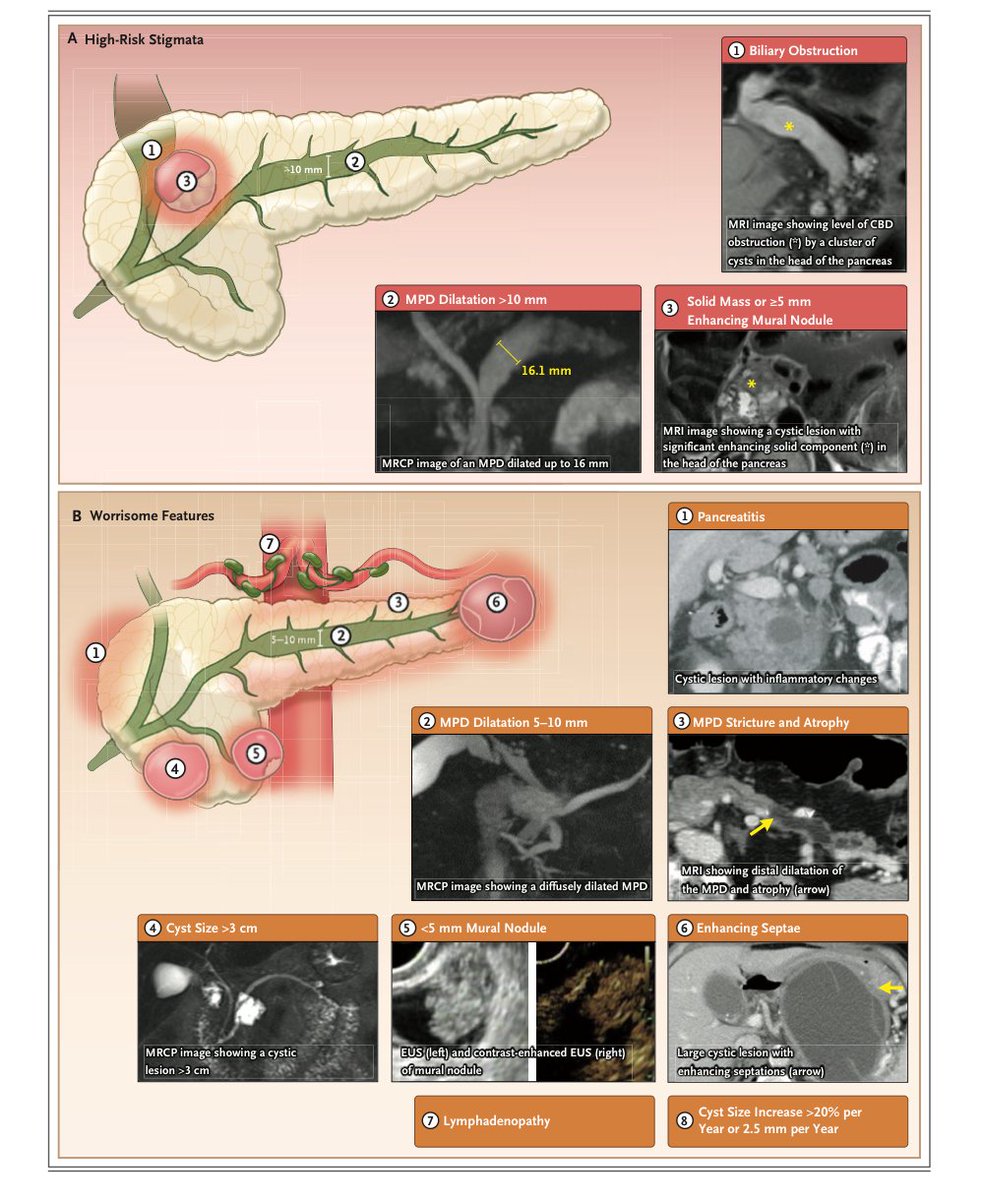
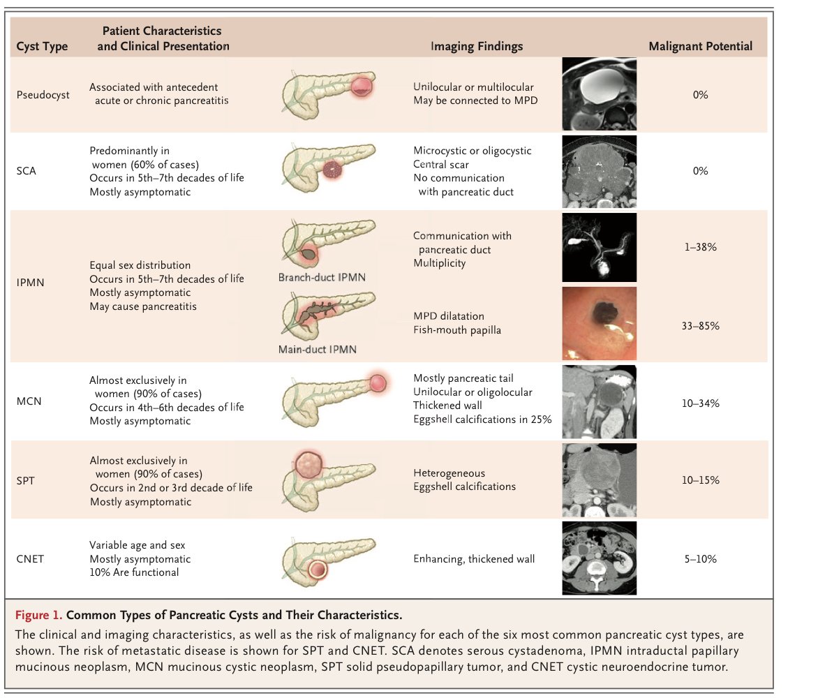
Excellent illustrations, review of pancreas cysts: nejm.org/doi/full/10.10…
➡️ 15% panc cancer arise from cysts
➡️ EUS>MRI: connection with ducts, ampulla evaluation, mural nodules
➡️VHL: Serous, MEN: NET, KRAS or GNAS: IPMN, GTNNB1: SPNT
➡️Minimal surgical excision is an option!
English
Yan Chu, MD retweetledi
An inventive way to understand liver anatomy:
This clever illustration uses the human hand to represent liver segments and vascular structures, providing a quick and memorable visual reference for healthcare professionals and students alike.

English
Yan Chu, MD retweetledi

The new blood-based test is NOT RECOMMENDED as a first line screening tool for colorectal cancer.
English
Yan Chu, MD retweetledi

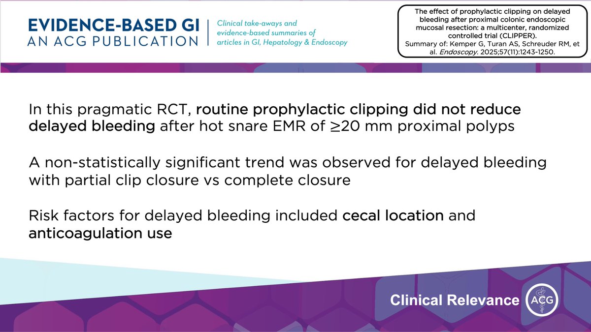
@ACG_EBGI @endoscopyjrnl @BilalMohammadMD @UNCGastro @JosephHabibi_MD @clivejmiranda @seanpattyp @AndrewMMoon @AmCollegeGastro @Neer_Dutta1 @EBGIdoc @UMN_GIHep @AmerGastroAssn @DarylRamai @KimberlyHoMD @isabelle_byers @DannyIssaMD @NickMcDonaldMD @RCaseyTurnerMD @BenClement_MD @MaryamBHaiderMD @MItaniMD @achokshi1 @menon_mythili @jalpa_devi @SLUAdvancedEndo @SuchapaAraya @NichaWongja @jordanmaloneDO @EricaLoonDO @JameelAlp @VijaySAre9 @KawtharAMohamed @WalkerReddMD @SusanLouMD @BJHansonMD @mspibddoc @DVinsard @ReutDanieliMD @ReezwanaCMD @LizzieAbyMD @TGHermanMD @RahulKarnaMD @gioroldanc @JosephKumka @annaunchaleeMD @ElfertKhaled @khalid_ahmedMD @TolgaGidenerMD @bengnonny 9/ Clinical Relevance
For routine EMR of ≥20 mm proximal polyps using hot snare: ➡️ Prophylactic clipping did NOT reduce delayed bleeding
⚠️ Key Risk Factors for Delayed Bleeding:
• Cecal polyps (RR 2.23)
• Anticoagulant use (RR 3.23)
English
Yan Chu, MD retweetledi

1/ ⏰ for #EBGI Tweetorial🧵w/ @nataliejowilson!
“Does Prophylactic Clipping Prevent Delayed Bleeding After EMR of Large Proximal Colonic Polyps?”
📜Summary bit.ly/3KzeNSz
📰Article bit.ly/3XuwjKJ
English
Yan Chu, MD retweetledi
BARRETT'S NEOPLASIA CLOCKFACE DISTRIBUTION
Circumferential spatial prediction CONFIRMS lesions cluster by quadrant
• RIGHT WALL (1–5 o'clock) DOMINATES → 68% of all dysplastic foci
• 3 o'clock position = HOT SPOT → highest density single + multifocal lesions
• Left wall (7–11 o'clock) → coldest zone, almost zero hits
PRO TRICK → Always start targeted biopsies at 2–4 o'clock position first → catches 92% of visible abnormalities in <90 sec
Clockface mapping → predicts 87% of lesion location before you even see them
Source: Ishimura N, Digestion 2014;89:291-8.
Your unit still doing random 4-quadrant biopsies first?

English
Yan Chu, MD retweetledi

Yan Chu, MD retweetledi

“The observational study found that women who reported consuming 9 to 10 daily servings of ultra-processed foods seemed to have a 45% greater likelihood of getting colon polyps before the age of 50 — as compared with women who had the least amount.” washingtonpost.com/wellness/2025/…
English
Yan Chu, MD retweetledi
1/8 ⏰ for #EBGI Tweetorial🧵w/ @ceomaliko1
“[Semaglutide for the Treatment of Metabolic Dysfuction-Associated Steatohepatitis - MASH🚀]”
📜Summary: tinyurl.com/semaebgi
📰 tinyurl.com/semamash
#GITwitter

English