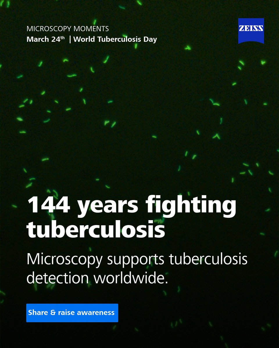
ZEISS Microscopy
10.5K posts

ZEISS Microscopy
@zeiss_micro
We are #Microscopy. All about microscopes from #ZEISS. LinkedIn: https://t.co/4jXTmJoUMF Facebook: https://t.co/ivuXIt7d1y | Imprint & Data Privacy: https://t.co/bvddlc6GZ2
Jena, Germany Katılım Haziran 2009
3.4K Takip Edilen22.7K Takipçiler

144 years later: Microscopy still fights tuberculosis 🔬
In 1882, Dr. Robert Koch discovered the bacteria causing TB using #microscopes supplied by Carl Zeiss Jena.
Microscopy bridges history & innovation. Raise awareness this #WorldTuberculosisDay!

English

@microxchile @microxchile What a great example, thanks for sharing! 🔬 Soil really is a powerful piece of forensic evidence, and it’s fascinating to see how advanced microscopy can reveal those hidden details.
We love seeing applications like this, feel free to keep them coming! 😊
English

El suelo es uno de los indicios forenses más poderosos: se transfiere fácilmente y se retiene en diferentes tipos de superficies.
Con la microscopía óptica avanzada de @zeiss_micro los #investigadoresforenses pueden identificar evidencia fundamental!👉shorturl.at/Ge0wg

Español

@Microscopy_SA @Microscopy_SA What a beautiful way to put it, “a rock as a 3D archive” is just perfect 💙 Thanks for sharing this, it’s great to see VersaXRM contributing to such insightful geoscience work!
English

Una roca no es solo una roca, es un archivo en 3D esperando ser leído.
El🔬VersaXRM de @zeiss_micro permite tomografía nanoescala de #muestrasgeológicas: flujo de fluidos, reactividad mineral, segmentación de fases y mineralogía cuantitativa con IA
👉shorturl.at/bM1oA

Español

@CommentLuisG @PlanktonPundit @NatGeo @CommentLuisG @PlanktonPundit @NatGeo Thank you for highlighting this important issue 🌊🔬 We’re proud to support researchers working to better understand microplastics and their impact on marine ecosystems.
English

@PlanktonPundit @NatGeo @zeiss_micro I refuse to accept invisible assassins choking our oceans. We conquered poles and Everest in wool. We never needed these petrochemical fibers. Return to natural fibers now.
English

Modern plastic microfibres choke to death the smallest of wildlife at the base of the marine food chain. We conquered the North and South Poles and Everest wearing just non polluting natural materials like wool. @natgeo @zeiss_micro
English

Les microfibres plastiques modernes étouffent les plus petits animaux sauvages à la base de la chaîne alimentaire marine.
Nous avons conquis les pôles Nord et Sud ainsi que l'Everest vêtus uniquement de matières naturelles non polluantes comme la laine.
@natgeo
@zeiss_micro
Dr Richard Kirby@PlanktonPundit
Modern plastic microfibres choke to death the smallest of wildlife at the base of the marine food chain. We conquered the North and South Poles and Everest wearing just non polluting natural materials like wool. @natgeo @zeiss_micro
Français

@microxchile @microxchile Thanks for sharing! 🔬 PathoPlex is a really exciting step forward for multiplexed tissue analysis. Looking forward to the webinar on March 17!
English

El futuro del análisis de tejidos tiene nombre: 𝗣𝗮𝘁𝗵𝗼𝗣𝗹𝗲𝘅🔬
📢Te invitamos a este #Webinar de @zeiss_micro con la co-autora del paper publicado en Nature: imagen multiplexada a resolución subcelular, escalable y aplicada a casos reales!
📅17/3👉shorturl.at/MlRd9

Español

@_Viroscope @_Viroscope Beautiful capture! 🔬 The symmetry breaking and actin dynamics here are fascinating to watch. Thanks for sharing this great example for Fluorescence Friday! 💙
English

A solar flare erupting on a glass coverslip☀️
This mouse cell has broken symmetry, erupting to drive itself across the surface.
🧡 Actin: Sweeping golden kinetic energy
🩵 Septin 7: Swirling regulation
🩷 Lipids: Metabolic fuel
💙 DNA: Anchored core
#SciArt #FluorescenceFriday

English

@PPBioImaging @turismoportugal @PPBioImaging @turismoportugal Thank you for the warm welcome! We’re proud to support ELMI 2026 and look forward to a great event with the bioimaging community. 🔬
English

Proud to welcome @zeiss_micro as a Gold Sponsor of #ELMI2026!
With 175+ years advancing imaging in life & materials sciences, ZEISS continues to empower researchers worldwide!
Thank you, @zeiss_micro, for your support! 🔬✨
#GoldSponsors
#meetoursponsors
@turismoportugal
English

@_Viroscope @_Viroscope what a beautiful cellular landscape! Thanks for sharing this fascinating glimpse into the complexity of a fibroblast cell.
English

🔬Imaged with a @zeiss_micro LSM980 + Airyscan 2 confocal microscope.
English

Glimpse of an alien shoreline
Not a topographical map of a distant planet, but the extreme boundary of a fibroblast cell!
🟣Actin: Structural cables
🟢Septin7: Dynamic scaffolds
🔵Lipids: Metabolic pools
An entire universe of complexity right under the objective lens.
#SciArt

English

@microxchile @microxchile Thank you for sharing this! 🔬 Polarized light microscopy is a powerful tool for forensic analysis—great to see how ZEISS Axio Imager, Axioscope 5 and Axiolab 5 are supporting such precise investigations.
English

La evidencia más valiosa muchas veces no es visible Con microscopía de luz polarizada, los peritos forenses analizan índices de refracción, fracturas y recubrimientos con gran precisión
Con🔬Axio Imager, Axioscope 5 y Axiolab 5 de @zeiss_micro es posible
👉shorturl.at/e9c0C

Español

@Microscopy_SA @Microscopy_SA Great summary! 🔬 It’s impressive how ZEISS microscopy enables reservoir characterization down to the nanometer scale.
English

🔬@zeiss_micro lleva la caracterización de reservorios al nanómetro:
🧲GeminiSEM: nanoporos en shales, sin dañar la muestra
🔩Crossbeam FIB-SEM: modelos 3D del sistema poroso
🔭Xradia Versa: 3D no destructivo de plug a microporo
⚗️Xradia Ultra: fluidos in-situ en tiempo real!

Español

Un destin tragique et un héritage consternant.
Les microfibres de nos vêtements en plastique finissent par se retrouver parmi le plancton.
@NatGeo
@zeiss_micro
Dr Richard Kirby@PlanktonPundit
A tragic destiny and our shocking legacy. The microfibres from our plastic clothes end up among the plankton. @NatGeo @zeiss_micro
Français

@ZEISS_Group @ZEISS_Group Great to see ZEISS Microscopy helping make CAR-T therapy safer and faster with our partners! 🔬💙
English

19 million cancer diagnoses each year. With @zeiss_micro inside the OPTIMA system, and strong partners in Heidelberg and at Bosch Health Campus, personalized CAR-T therapy is becoming safer, faster and more scalable.
English

@confocal_nl @confocal_nl Exciting news! We’re proud to collaborate and bring these new confocal imaging possibilities to researchers across Benelux. Looking forward to what scientists will discover with this partnership. 🔬
English

We’re excited to announce our new partnership with @zeiss_micro Benelux!
We are combining our fast Line REscan confocal imaging modules with ZEISS’s renowned microscopy solutions, expanding the imaging possibilities for researchers across Benelux.
Through this partnership, scientists can now benefit from our REscan technology directly within their familiar ZEISS workflows, offering one complete solution for fast, sensitive confocal imaging with close-to-zero phototoxicity.
We are proud to collaborate with ZEISS Microscopy Benelux in bringing our innovative technology to researchers.
#Microscopy #ImagingInnovation #Partnership #ConfocalMicroscopy

English

@PlanktonPundit @NatGeo @PlanktonPundit @NatGeo Thank you for highlighting this important topic. Images like these are a powerful reminder of how microplastics can impact marine ecosystems and the tiny organisms within them. 🌊🔬
English

A tragic destiny and our shocking legacy. The microfibres from our plastic clothes end up among the plankton. @NatGeo @zeiss_micro
English

📍 Image acquired on 𝐚 @zeiss_micro 𝐄𝐥𝐲𝐫𝐚 𝟕 𝐋𝐚𝐭𝐭𝐢𝐜𝐞 𝐒𝐈𝐌 (63×, 1.4 NA oil objective) © P. Ray & A. Ferrand, University of Base
English

#ItsMicroscopyMonday
What if we could not only see biological cells, but also resolve their structure beyond the classical limits of light?
From the light sheet microscopy (2004) to Nobel Prize–recognized super-resolution (2014), and more recently, expansion microscopy...

English

@GlobalAshoka @AshokaUniv @kvijayraghavan @ANRFIndia @sathi_iitdelhi @NCBS_Bangalore @CCAMP_India @unescorcb @AmbikaAnupama @SouravP32813910 @kasturiMITO @DAADIndia @GlobalAshoka @AshokaUniv What a great opportunity! 🔬✨ Exciting to see this collaboration. Best of luck to everyone applying for PCAM 2026! 💙
English

Applications Open | Professional Course in Advanced Microscopy (PCAM) 2026
Apply now for a 12-week immersive programme in advanced research microscopy by @AshokaUniv 🇮🇳 & ZEISS Academy Microscopy 🇩🇪
📅Apply by 15 April 2026
Details & apply: lnkd.in/gU-Qdb-D
@zeiss_micro

English

@_Viroscope @_Viroscope This is such a beautiful #FluorescentFriday image, the structural detail in those LUHMES-derived neurons really stands out! 🔬✨ Thanks so much for tagging us and sharing your work with the community. We love seeing results like this. 💙
English

It’s #FluorescentFriday!
This Maximum Intensity Projection of LUHMES-derived neurons shows:
🟦 Vimentin (@Proteintech Cat No. 10366-1-AP)
🩷 Tubulin
🧡 Actin
🤍 DNA
Explore the antibody: ptglab.com/products/VIM-A…
#Proteintech #Neuroscience

English