MSC-OSC

53 posts

MSC-OSC

MSC-OSC

@msc_osc

Microscopy Society of Canada: Ontario Student Chapter Student/trainee microscopists in Ontario unite! 🔬🤓

Katılım Şubat 2023
9 Takip Edilen14 Takipçiler
MSC-OSC
MSC-OSC@msc_osc·
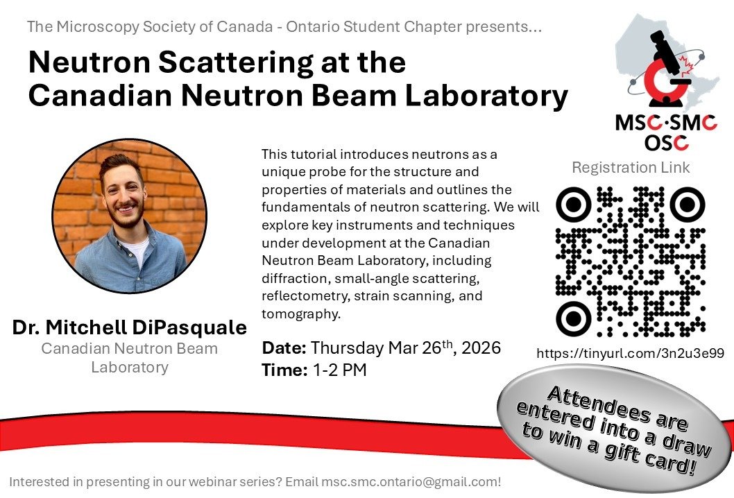
Dr. DiPasquale is the facility manager of the Canadian Neutron Beam Laboratory at @McMasterNuclear- home to Canada's largest nuclear research reactor. Join us and learn how neutron techniques enable advanced materials characterization ⚛️
English
0
0
0
7
MSC-OSC
MSC-OSC@msc_osc·
Do you know photons and electrons aren't the only particles used in microscopy and microanalysis? Neutrons can do that too! Join us on March 26 to explore neutron scattering with Dr. Mitchell DiPasquale 🔗Register: tinyurl.com/3n2u3e99
MSC-OSC tweet media
English
1
1
0
9
MSC-OSC
MSC-OSC@msc_osc·
📍 Image acquired on 𝐚 @zeiss_micro 𝐄𝐥𝐲𝐫𝐚 𝟕 𝐋𝐚𝐭𝐭𝐢𝐜𝐞 𝐒𝐈𝐌 (63×, 1.4 NA oil objective) © P. Ray & A. Ferrand, University of Base
English
1
0
0
5
MSC-OSC
MSC-OSC@msc_osc·
The image above displays a C. elegans larva imaged using Lattice Structured Illumination Microscopy. DNA appears in blue, apical junctions in yellow, and smFISH signal in magenta, revealing cellular organization beyond what conventional fluorescence microscopy can resolve.
English
1
0
0
15
MSC-OSC
MSC-OSC@msc_osc·
#ItsMicroscopyMonday What if we could not only see biological cells, but also resolve their structure beyond the classical limits of light? From the light sheet microscopy (2004) to Nobel Prize–recognized super-resolution (2014), and more recently, expansion microscopy...
MSC-OSC tweet media
English
1
0
0
22
MSC-OSC
MSC-OSC@msc_osc·
AFM measurements, collected at @icspicorp in Kitchener-Waterloo on a 𝗥𝗲𝗱𝘂𝘅 𝗔𝗙𝗠, highlight clear structural consequences of LytG inhibition. Want to learn more about AFM? Join us for our next webinar on 𝗙𝗲𝗯𝗿𝘂𝗮𝗿𝘆 𝟭𝟮,𝟭:𝟬𝟬 𝗽𝗺: tinyurl.com/ycru9shm
English
0
0
0
22
MSC-OSC
MSC-OSC@msc_osc·
Prof. Christopher Reid and his team at @BryantUniv investigated how antibiotic treatment alters cell-wall remodeling in Bacillus subtilis. In this study, the antibiotic masarimycin inhibits an autolysin involved in peptidoglycan remodeling during cell elongation and division.
English
1
0
0
13
MSC-OSC
MSC-OSC@msc_osc·
It’s #MicroscopyMonday What happens to bacterial cell walls under antibiotic stress? Atomic Force Microscopy (AFM) enables nanoscale characterization of biological surfaces, particularly for changes in surface morphology and roughness. Details in: doi.org/10.1039/D5CB00…
MSC-OSC tweet media
English
1
0
0
9
MSC-OSC
MSC-OSC@msc_osc·
Qianshu presentation titled: “𝗙𝗿𝗼𝗺 𝗕𝗮𝗰𝘁𝗲𝗿𝗶𝗮 𝘁𝗼 𝗣𝗼𝗹𝘆𝗺𝗲𝗿𝘀 𝗮𝗻𝗱 𝗦𝗶𝗹𝗶𝗰𝗼𝗻: 𝗜𝗻𝘁𝗿𝗼 𝘁𝗼 𝗔𝘂𝘁𝗼𝗺𝗮𝘁𝗲𝗱 𝗔𝘁𝗼𝗺𝗶𝗰 𝗙𝗼𝗿𝗰𝗲 𝗠𝗶𝗰𝗿𝗼𝘀𝗰𝗼𝗽𝘆” will happen on February 12, 2026 at 1 PM EST Attendees will be entered into a draw for a gift card
English
0
0
0
11
MSC-OSC
MSC-OSC@msc_osc·
Curious how automated AFM can make surface imaging easier and faster? Join us for our next webinar, featuring Qianshu W., an Application Engineer at @icspicorp, where he’ll share his expertise in automated AFM. Register here: tinyurl.com/ycru9shm
MSC-OSC tweet media
English
1
0
0
27
MSC-OSC
MSC-OSC@msc_osc·
It emerged unexpectedly during research aimed at extracting iron from ferritin, a common mammalian protein. The iron is used to grow carbon nanotubes for nano-biosensors for single-molecule detection —tools being explored for cancer screening and for studying molecular behaviour.
English
0
0
0
7
MSC-OSC
MSC-OSC@msc_osc·
“𝐀𝐥𝐥 𝐢𝐧 𝐚 𝐆𝐫𝐚𝐢𝐧 𝐨𝐟 𝐒𝐚𝐥𝐭” shows an ordinary grain of salt turned into a striking landscape of dendrites and crystallization islands, with a central crystal size of ~60 µm, all composed of salt. The image was captured using SEM at a magnification of 800x.
English
1
0
0
13
MSC-OSC
MSC-OSC@msc_osc·
Beautiful microscopy images don’t come from perfect experiments; they might come from unexpected lab errors! This image was captured by Jean-Nicolas Deraspe from the Université de Montréal and was selected as a finalist in the 2025 NSERC Science Exposed. 👇 See the image below
MSC-OSC tweet media
English
1
0
3
51
MSC-OSC
MSC-OSC@msc_osc·
Join us at the International Microscopy Student Mixer (ISM) on Wednesday, February 4, 2026, from 1 PM - 3 PM EST Register at the following link: tinyurl.com/yz4wpr77 Hosted by the yDGE, MSA StC, and the MSC-SMC Student Chapters
MSC-OSC tweet media
English
0
0
0
5
MSC-OSC
MSC-OSC@msc_osc·
These kinds of issues show up frequently in routine microscope use, and they can quietly affect data quality. Improving image quality doesn’t necessarily require new equipment, but a more deliberate look at how the microscope is set up!
MSC-OSC tweet media
English
1
0
2
5
MSC-OSC
MSC-OSC@msc_osc·
A useful tip from @zeiss_micro: when brightfield images don’t look right, the issue is often not the sample or the microscope itself, but small setup mistakes that remain unnoticed, especially those related to common objective lens setup. Sharing the document below.
MSC-OSC tweet mediaMSC-OSC tweet mediaMSC-OSC tweet mediaMSC-OSC tweet media
English
1
0
2
11
MSC-OSC
MSC-OSC@msc_osc·
Led by Arthur Blackburn, co-director of UVic’s Advanced Microscopy Facility, the team achieved this breakthrough using ptychography. This computational technique reconstructs high-resolution images from overlapping electron scattering patterns. Full paper: #Abs1" target="_blank" rel="nofollow noopener">nature.com/articles/s4146…
English
0
0
0
12
MSC-OSC
MSC-OSC@msc_osc·
Researchers at the University of Victoria have demonstrated a powerful new imaging approach that achieves sub-ångström resolution (0.67 Å) using a compact, low-energy SEM—a level of detail previously accessible only with large, high-cost TEMs.
MSC-OSC tweet media
English
1
0
0
18
MSC-OSC
MSC-OSC@msc_osc·
The Microscopy Society of Canada - Ontario Student Chapter is pleased to announce the 2026 Micrograph Contest! Participants can submit a micrograph for a chance to win a $50 Amazon gift card, plus custom stickers featuring the winning micrograph.
MSC-OSC tweet media
English
1
0
0
37