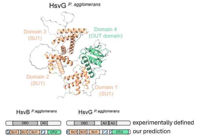
Sebastian Schornack

10.8K posts

Sebastian Schornack banner
Sebastian Schornack

Sebastian Schornack

@dromius

Plant cells and development, Plant-microbe research, Phytophthora, oomycetes & symbiotic fungi, TAL effectors (TAL DNA binding code codiscoverer) and Coleoptera

Cambridge, UK Katılım Temmuz 2009
1.3K Takip Edilen8K Takipçiler
Sabitlenmiş Tweet
Sebastian Schornack
Sebastian Schornack@dromius·
Be part of OMGN! Open to all researchers with an interest in oomycetes, from molecular genetics & genomics to biology, population biology, and ecology, at either an experimental or a computational level. Investigators new to the field are always welcome oomycetes.com
Sebastian Schornack tweet media
English
0
21
39
3K
Sebastian Schornack retweetledi
Elenor Kennedy
Elenor Kennedy@E_B_Kennedy·
Extremely excited to share the first pre-print from my PhD project in the Carella lab, co-first authored with @Khongsam_Chia 🎉 biorxiv.org/content/10.648… We investigated the EDS1-dependence of TIR domains from across the spectrum of land plant evolution, and found that TIR-NLR architecture itself can influence TIR domain activity, even for animal and bacterial TIRs. Huge thanks to all co-authors and the #CarellaCapybaras for all the support 😊 Thread below ⬇️
English
2
11
21
9.7K
Sebastian Schornack retweetledi
Philip Carella
Philip Carella@Phil_Carella·
Happy to share our latest pre-print on the evolution of TIR-mediated immunity in plants. Led by @E_B_Kennedy and @Khongsam_Chia from our lab @JohnInnesCentre with a big assist from @mhz1989 and @jonathandgjones @TheSainsburyLab. Check out El's twitter thread below:
Elenor Kennedy@E_B_Kennedy

Extremely excited to share the first pre-print from my PhD project in the Carella lab, co-first authored with @Khongsam_Chia 🎉 biorxiv.org/content/10.648… We investigated the EDS1-dependence of TIR domains from across the spectrum of land plant evolution, and found that TIR-NLR architecture itself can influence TIR domain activity, even for animal and bacterial TIRs. Huge thanks to all co-authors and the #CarellaCapybaras for all the support 😊 Thread below ⬇️

English
1
23
55
6.4K
Sebastian Schornack retweetledi
Sophien Kamoun
Sophien Kamoun@KamounLab·
Please consider supporting IS-MPMI and the Mishkind Travel Fund ismpmi.org 👇🏾
Sophien Kamoun tweet media
English
0
6
16
847
Sebastian Schornack retweetledi
IS-MPMI
IS-MPMI@ISMPMI·
We just passed the half-way point of our ambitious goal to raise $40,000 for the Mishkind travel fund for early career investigators to attend the IS-MPMI congress*. Please consider donating today! ismpmi.org
English
1
6
6
1.2K
Sebastian Schornack retweetledi
Mauricio Contreras
Mauricio Contreras@mpcontreras4·
Is this specific to the Rx-NRC2 pair, or a general rule? We predicted NRC sensor–helper complexes across asterid lineages spanning ~125 million years of evolution: 🫜 to 🥝 to 🥬. Same binding mode every time! A conserved structural logic, not conserved sequence.
Mauricio Contreras tweet media
English
1
5
26
1.2K
Sebastian Schornack retweetledi
Mauricio Contreras
Mauricio Contreras@mpcontreras4·
We made charge-reversal mutations across the predicted interface on both sensor and helper. The strongest test? A reciprocal charge-swap. Break one side of a salt bridge → dead. Break the other → dead. Flip BOTH → rescued.🧩
Mauricio Contreras tweet media
English
1
3
33
2.8K
Sebastian Schornack retweetledi
jean greenberg
jean greenberg@jeangre95682538·
@ISMPMI We passed the half-way point of our ambitious goal of raising $40,000 for the Mishkind Travel fund for early career researchers to attend our 2027 Congress. Please consider donating today! ismpmi.org
English
0
5
6
454
Sebastian Schornack retweetledi
Leibniz IPK
Leibniz IPK@LeibnizIPK·
Welcome to the International Symposium on the Nitrogen Nutrition of Plants - Nitrogen 2026. The event will take place at the @LeibnizIPK on August 23–27, 2026. The Early-bird registration deadline has been extended until 31 May! ➡️Further information: events.ipk-gatersleben.de/nitrogen2026/s…
Leibniz IPK tweet media
English
0
4
6
1.1K
Sebastian Schornack retweetledi
JJ
JJ@JosephJacks_·
Microtubules were discovered and named by looking at plants through an electron microscope. The pair behind it: Myron Ledbetter, a plant biologist, and Keith Porter, co-founder of cellular biology. 🌱 🔬 Porter was the giant. Harvard PhD 1938. From 1939 at the Rockefeller Institute, he and Albert Claude invented biological electron microscopy — first EM image of an intact cell in 1944, named the endoplasmic reticulum, co-invented the Porter-Blum ultramicrotome that made the whole field possible, co-founded the American Society for Cell Biology and the Journal of Cell Biology. Ledbetter was the plant person — Berkeley Master’s, Columbia PhD in botany, then came to Rockefeller to learn EM under Porter and followed him to Harvard. Their motivation was a long-standing botanical question. Plant cells lay down cellulose in highly patterned arrangements — circumferential, helical, and random — and these patterns determine cell shape. What in the cytoplasm tells the cellulose where to go? Porter argued in the 1950s the endoplasmic reticulum was the patterning agent, but too floppy and sparse for the job. Something else was orienting it — a structural element no one had yet resolved. The opportunity came from chemistry. In 1962, David Sabatini at Yale introduced glutaraldehyde as an EM fixative. Porter heard about it immediately and pushed Ledbetter to try it on plants. The plants… Three species, all with root tips full of wall-building cells. Phleum pratense (timothy grass) does most of the work and supplies every cortex image. Spirodela oligorrhiza (duckweed) catches a cell mid-division and gives them the spindle. Juniperus chinensis (Chinese juniper) provides the clinching image — tubes running parallel to cellulose fibers across the membrane, as if guiding their deposition. Two flowering plants and one conifer — enough phylogenetic spread to claim universality. The instrument… A standard 100 kV transmission electron microscope — fires electrons through tissue and builds an image from how they scatter off heavy-metal-stained structures. Far better resolution than light microscopy, but the sample is killed, dehydrated, embedded in plastic, and sliced thinner than a light wavelength. Whether your prep preserved anything real is the whole game. Osmium tetroxide alone shredded the delicate plant cortex. Glutaraldehyde locks proteins in place in seconds. They ran it on root tips, looked at the cortex, and the tubes were just there, waiting. What they saw… Hollow cylinders, ~250 Å across, with a clear central lumen and a wall suggesting smaller subunits packed side by side. Lengths indeterminate — traceable for several microns without finding an end. Each tube sat in a clear “halo” excluding ribosomes and everything else. In the cortex they ran parallel to the surface, wrapped like hoops on a barrel, up to three layers deep. In dividing cells, 500+ packed the mitotic spindle, oriented along its long axis, converging on the chromosomes. Before this paper, the cytoplasm was a messy soup with organelles floating around. A few people had glimpsed tubular profiles in odd places — Roth / Daniels in an amoeba a year earlier, Slautterback in Hydra in the JCB issue just prior. But Slautterback called them phospholipid membranes for ion transport… Wrong! Ledbetter and Porter predicted one kind of protein tube, of repeating subunits, present everywhere — plants and animals, cortices and spindles, cilia and flagella — doing the cell’s structural work. What’s amazing is how much they got right from so little! The subunit composition was correct — tubulin wouldn’t be isolated until 1968. The identity with cilia and flagella was also correct. The claim about spindle birefringence was correct. Plant cells forming spindles without centrioles anticipated decades of work on acentrosomal nucleation. And the alignment hypothesis, born from the juniper image, drove cell biology for the next fifty years.​​​​​​​​​​​​​​​​ pmc.ncbi.nlm.nih.gov/articles/PMC21…
JJ tweet mediaJJ tweet media
English
1
19
64
4K
Sebastian Schornack retweetledi
Yaowu
Yaowu@yaowu25·
Au🇦🇺
Yaowu tweet mediaYaowu tweet mediaYaowu tweet media
0
3
52
1.1K
Sebastian Schornack retweetledi
白水 貴
白水 貴@Takashirouzu·
テントウムシの食性進化に関する面白い論文が出ていた.アブラムシ食が祖先的で,そこから混合食を経て他の昆虫食に特化していくが,菌食への進化は混合食を経ずにダイレクトに起こっている.菌食への移行は顎の形態変化を伴うもので,一度だけの不可逆的な進化だったようだ. doi.org/10.1002/ece3.7…
白水 貴 tweet media
日本語
0
147
483
17.8K
Sebastian Schornack retweetledi
Keiko Sugimoto Lab.
Keiko Sugimoto Lab.@K_Sugimoto_Lab·
Fantastic 2 days to discuss plant cell plasticity and latest single cell & spatial technologies at our ASPIRE & GreenTE gathering 🌱🍃🌿! Many thanks to @PaszkowskiLab @JST_ASPIRE ❤️.
Keiko Sugimoto Lab. tweet mediaKeiko Sugimoto Lab. tweet media
English
0
4
38
2.5K
Sebastian Schornack retweetledi
Francis M. Martin
Francis M. Martin@fmartin1954·
Recent progress in genomics, phylogenetics & palaeontology yielded new insights into plant and fungal evolution since the 10th NPW. The 35th New Phytologist Workshop will highlight the progresses made since that time to move the field forward ... @CStrulluDerrien
Francis M. Martin tweet media
English
0
5
41
1.6K
Sebastian Schornack retweetledi
British Plant Galls
British Plant Galls@britgalls·
Gall of the week is caused by the fungus Protomyces macrosporus, an ascomycete, on many species of umbellifers, Apiaceae. Photo from BPGS website, photographer unknown.
British Plant Galls tweet media
English
0
7
11
862