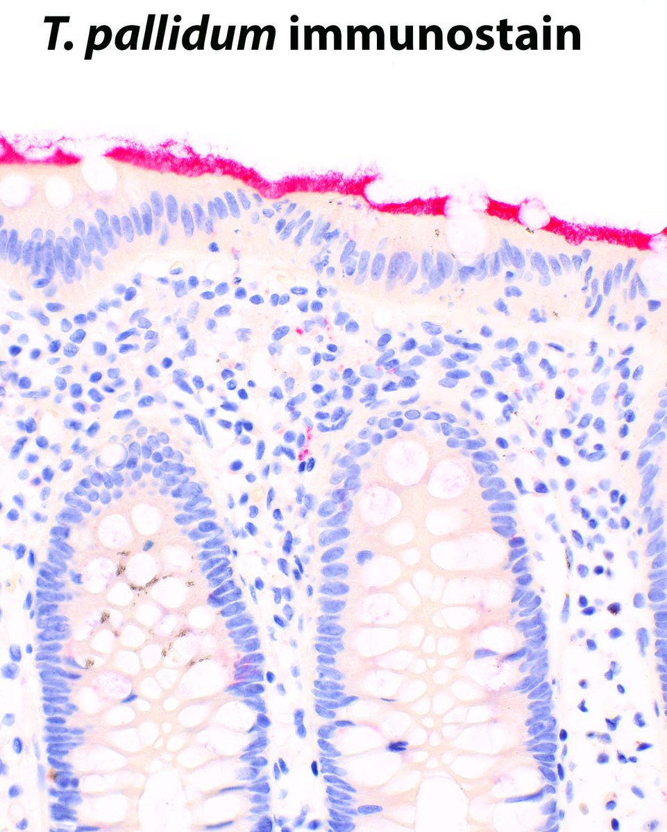
Intestinal spirochetosis, caused by Brachyspira spp, cross reacts with the T. pallidum immunohistochemical stain.
Pettit C, et al. Highlighting a Potential Pitfall: Positive Treponema pallidum Immunohistochemical Stain in a Patient Without Syphilis. PMID: 31389806.
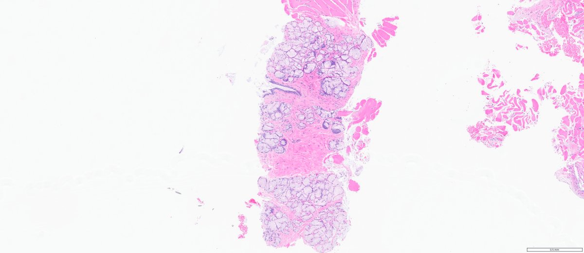
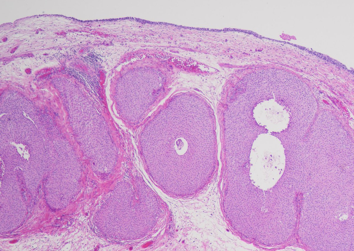
#GUpath prostate biopsy for elevated PSA
at first blush foamy gland pattern prostatic adenocarcinoma comes to mind...
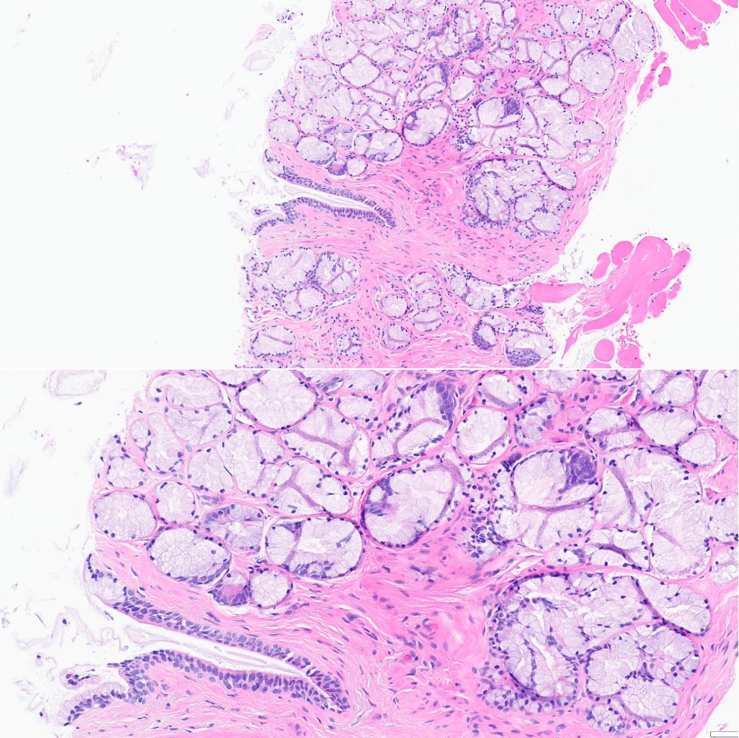
but other DDx include Cowper gland (Dx here) or mucinous metaplasia
⏬cont'd
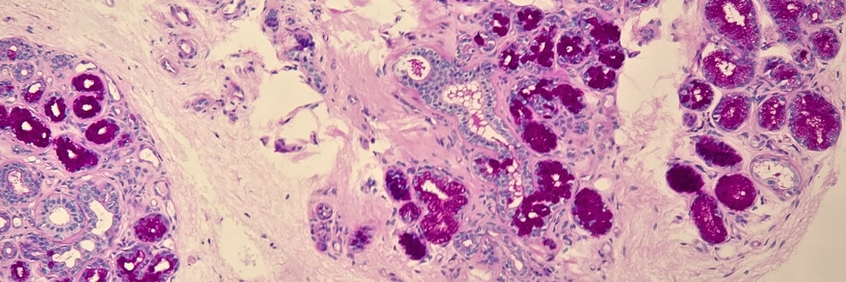
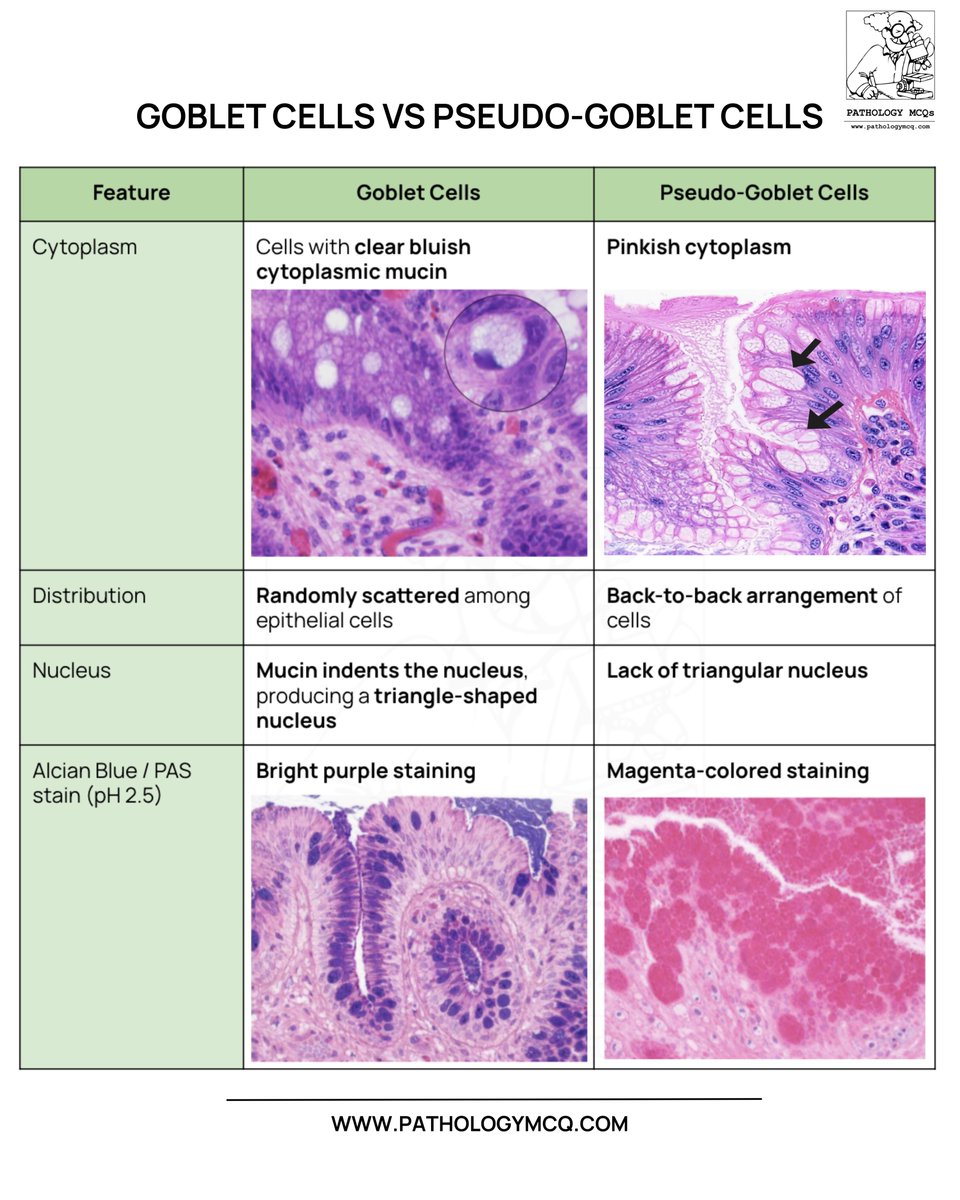
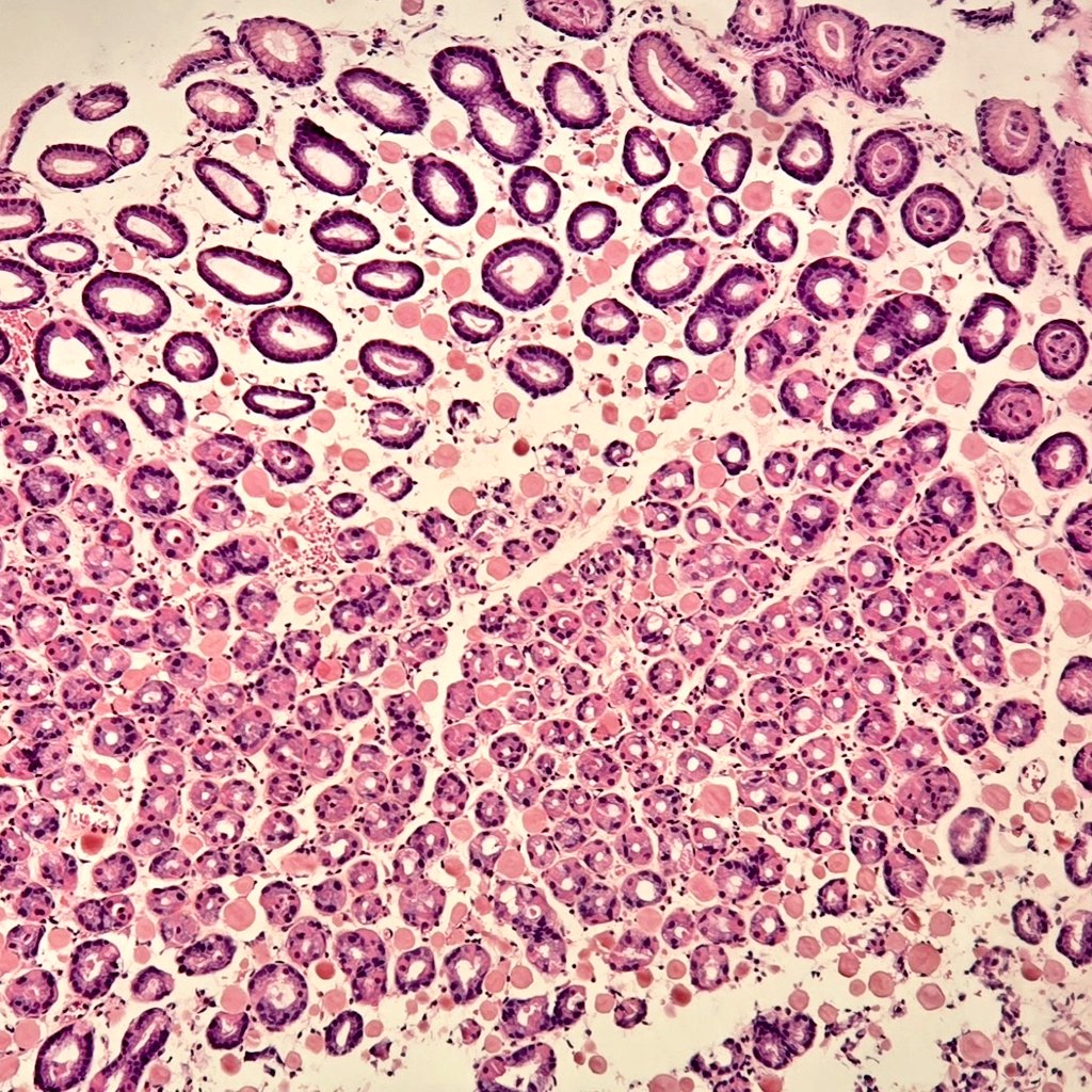
True goblet cells contain acid mucin,show a triangular nucleus, and stain Alcian blue positive🔵
Pseudo-goblet cells are actually gastric foveolar cells with neutral mucin, showing PAS magenta staining🟣
#Pathology#Histopathology#PathologyMCQ#GIPathology#BarrettEsophagus
#backtobasics
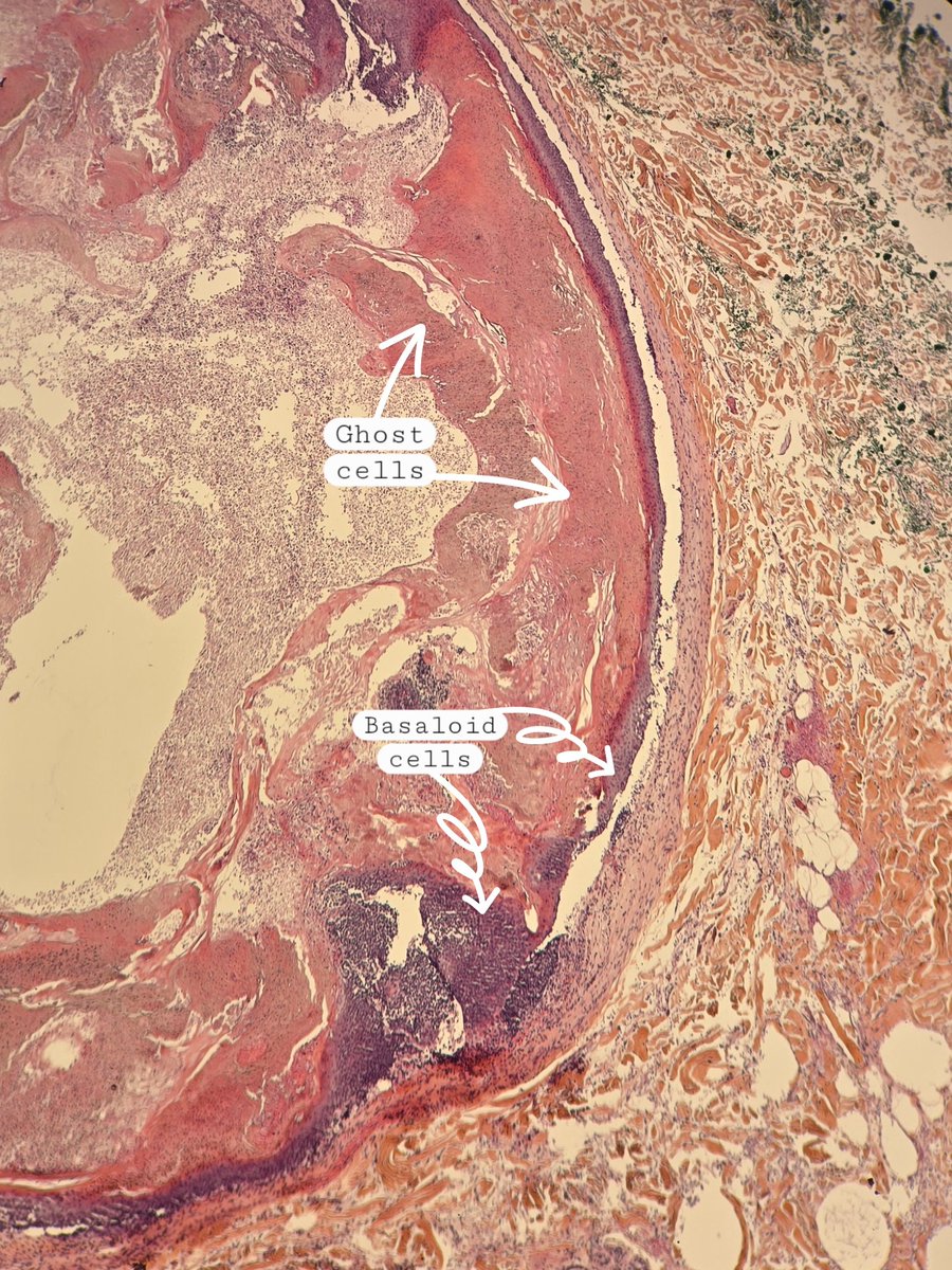
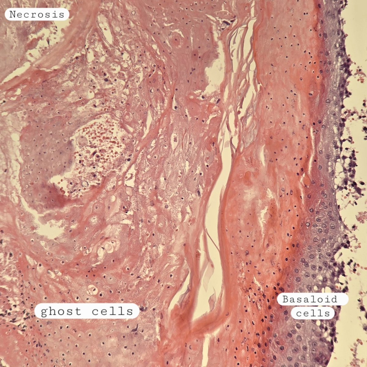
Pilomatrixoma 🔬
✔️Benign skin adnexal tumor
✔️basaloid proliferation resembling the hair matrix cells, which matures into structureless eosinophilic cells lacking nuclei called shadow/ghost cells
✔️Frequently there are areas of calcification
#xpath#dermpath
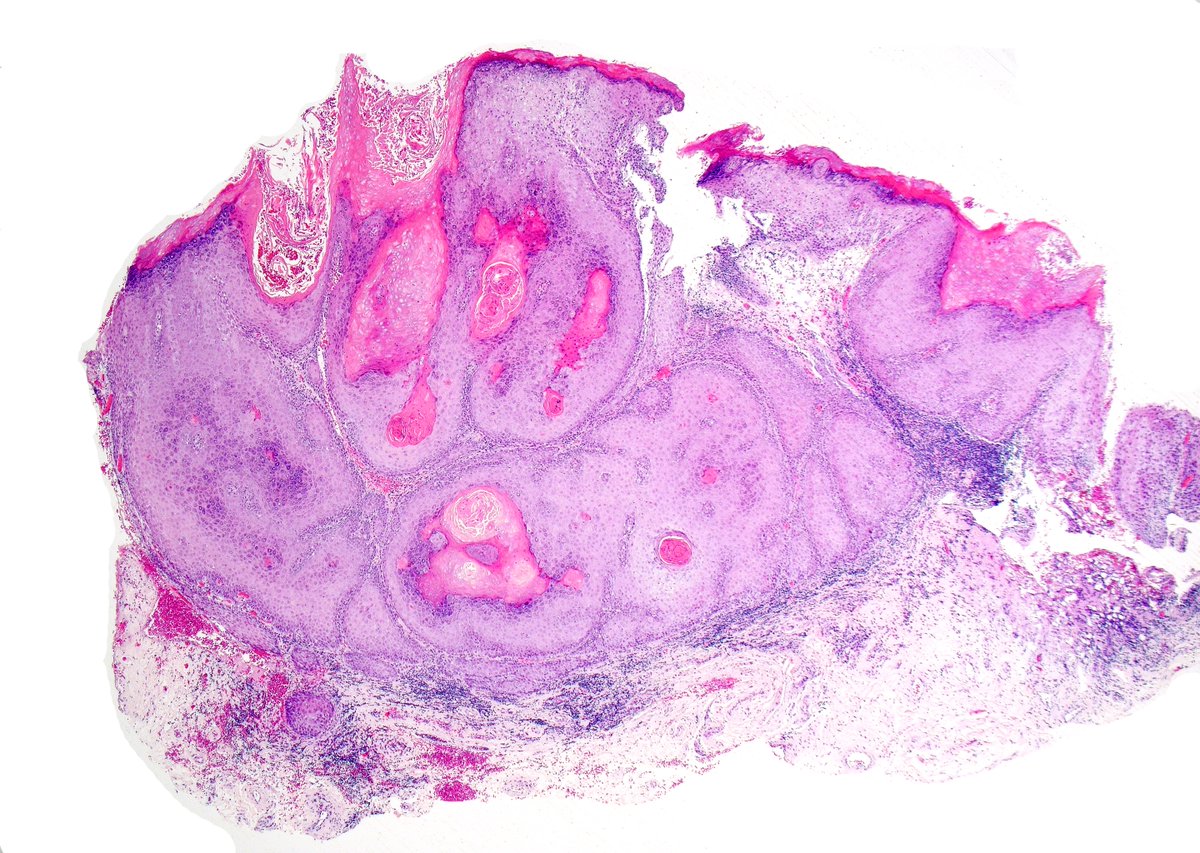
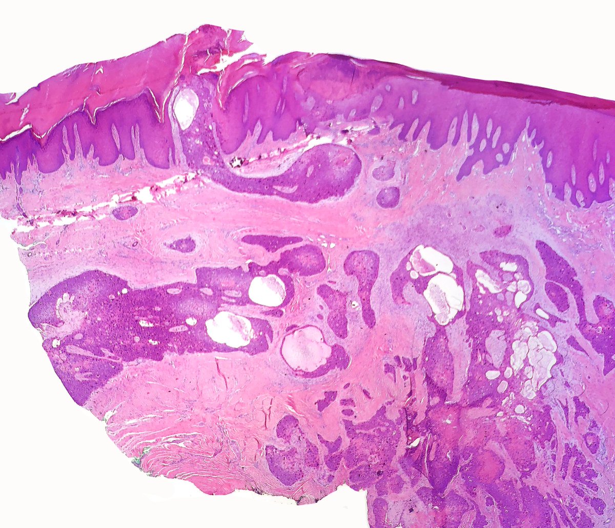
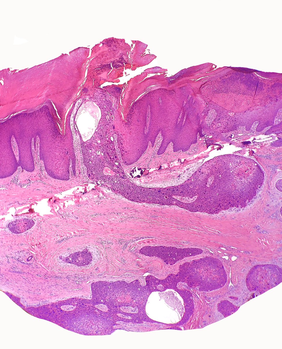
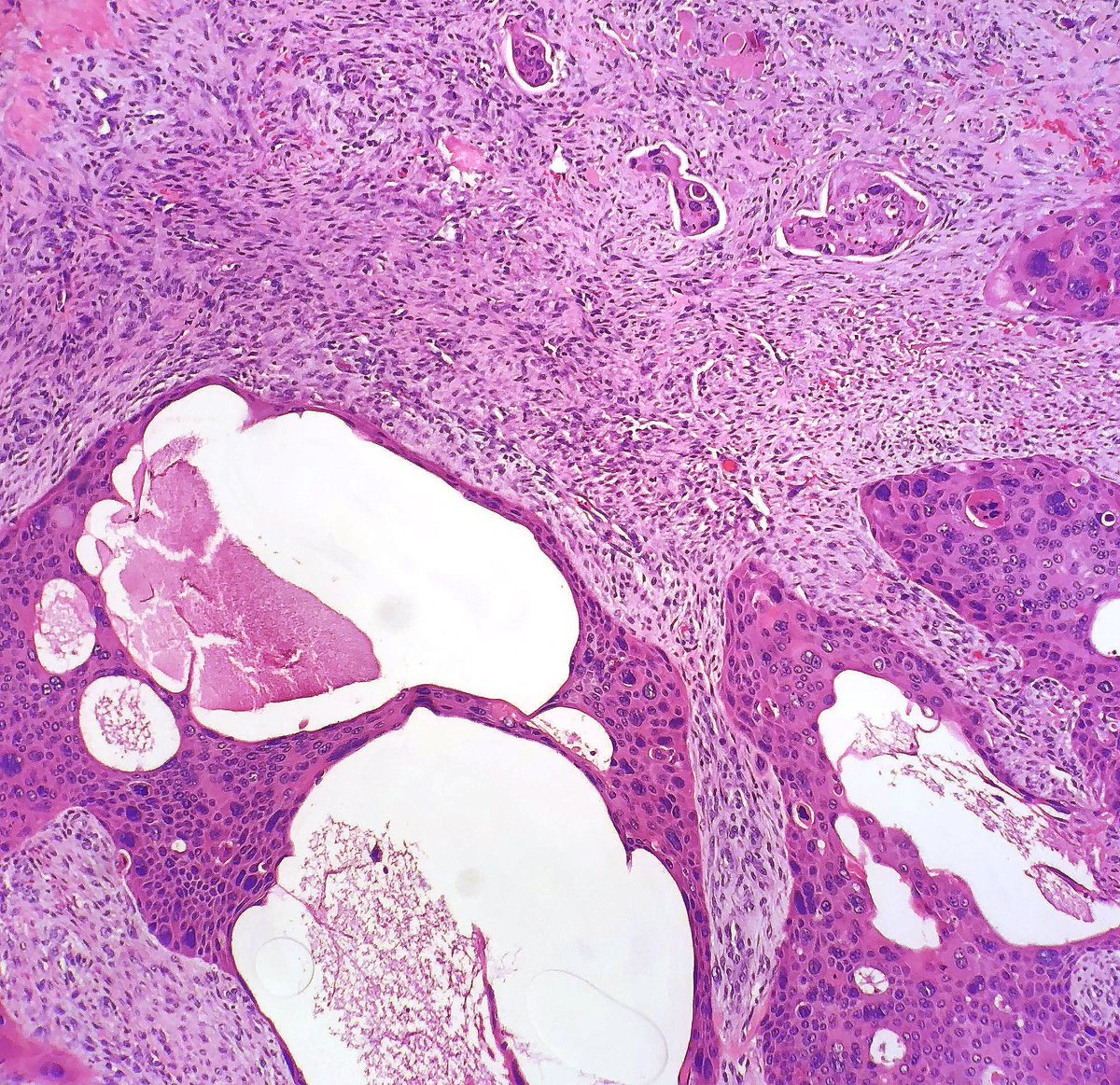
Verrucous carcinoma of the penis 🔬
Broad-based, exophytic lesion with pushing borders. Note the bulbous rete ridges and keratin-filled sinuses, classic for this low-grade variant. Despite its bland cytology, local invasion can be extensive. Early recognition is key.
#GUPath
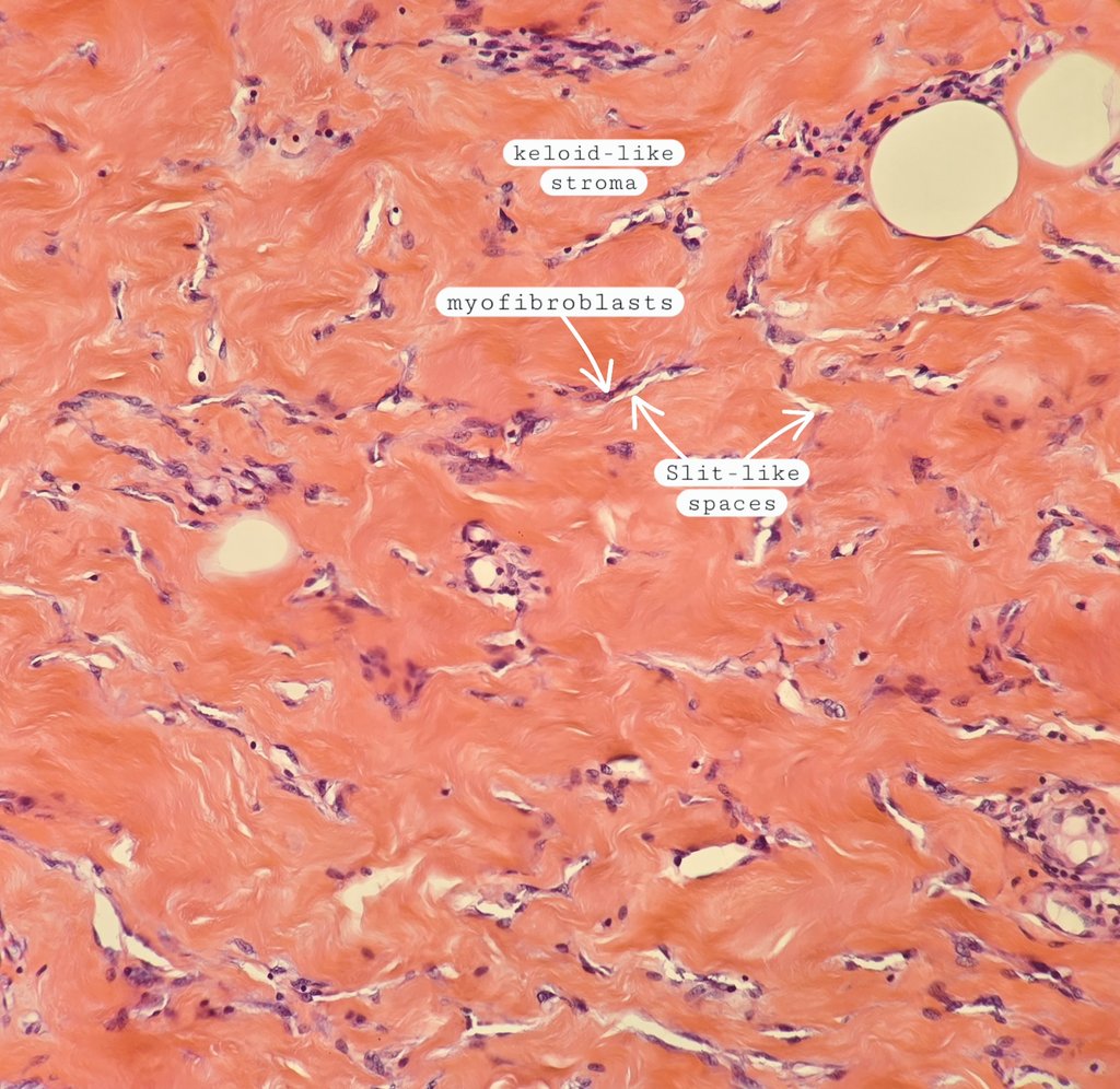
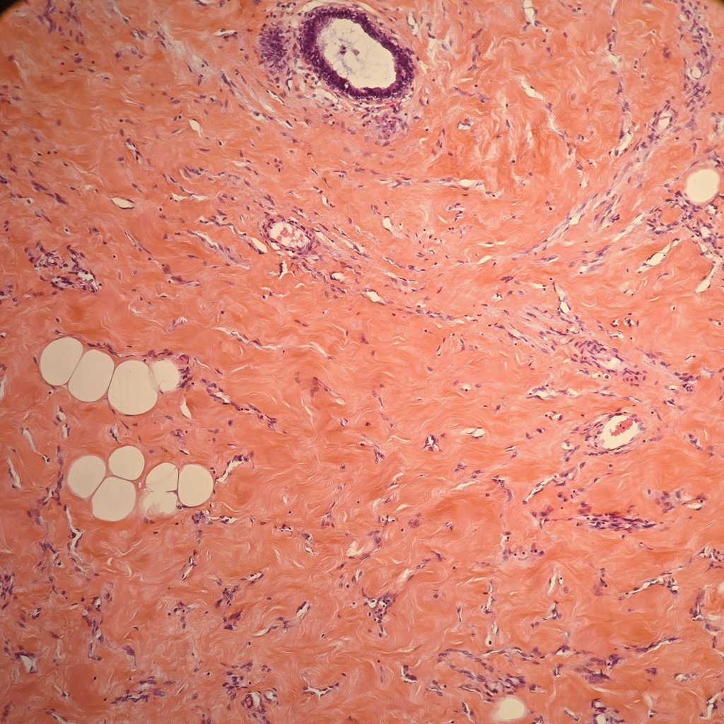
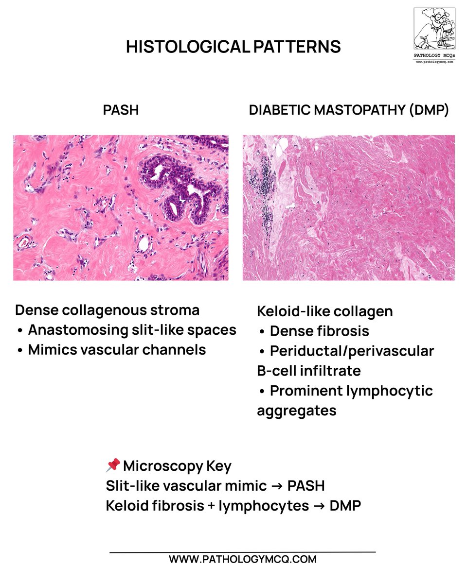
PseudoAngiomatous Stromal Hyperplasia ( PASH)
✔️Complex interanastomosing spaces in collagenous stroma resembling blood vessels
✔️Some of these spaces have spindle shaped myofibroblasts at their margins simulating endothelial cells
✔️- for vascular markers except CD34
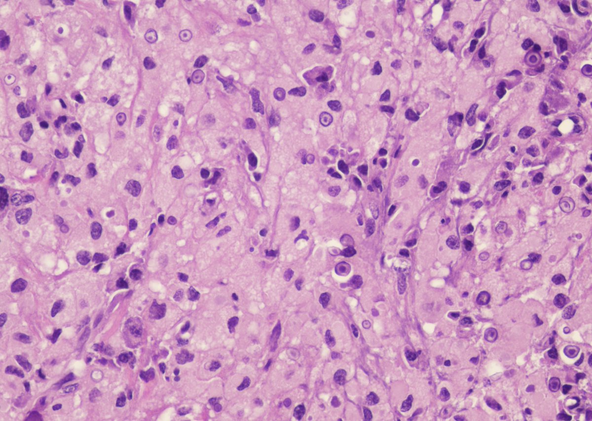
Malakoplakia of bladder: Sheets of histiocytes (von Hansemann cells) packed with granular cytoplasm and the pathognomonic Michaelis–Gutmann bodies—round, targetoid inclusions reflecting defective phagolysosomal activity and chronic E. coli infection.
#Pathology#UroPath#MedEd
Large Nested Urothelial Carcinoma on H&E 🔬
Deceptively bland cytology with expansile, well-circumscribed nests infiltrating the lamina propria—an architectural wolf in sheep’s clothing. Recognition is critical to avoid underdiagnosis of this aggressive variant.
#Pathology
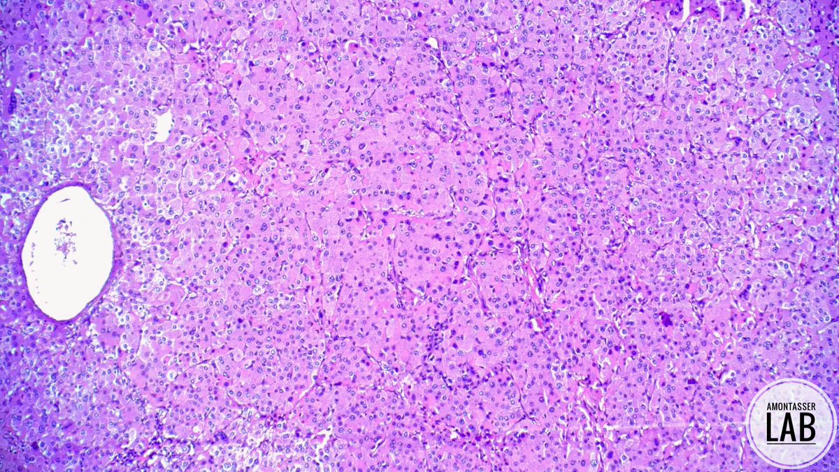
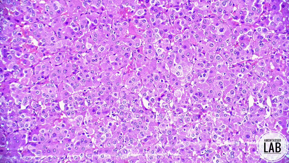
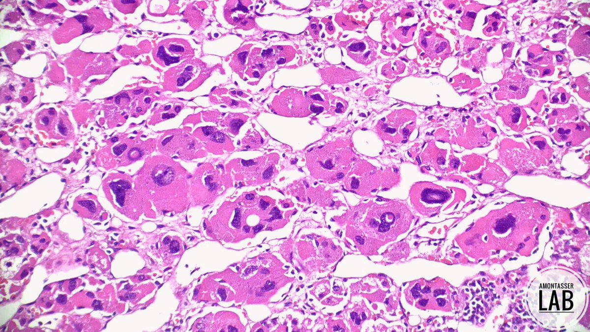
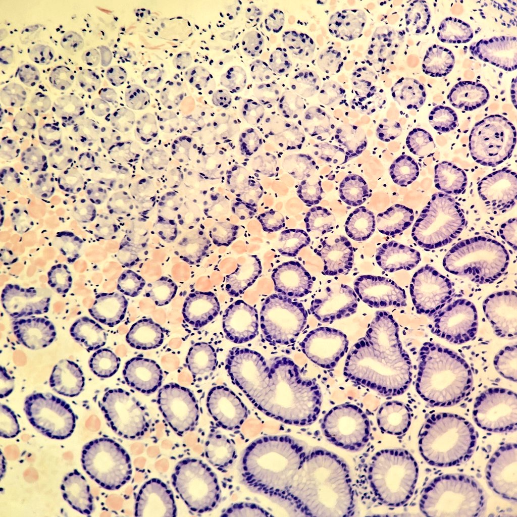
🔬Globular amyloid - gastric biopsy
✔️Amyloid doesn’t always appear as diffuse sheets
✔️“Globular” pattern = round, well-circumscribed eosinophilic deposits in lamina propia
✔️Congo red + → apple-green birefringence
✔️always confirm and type ( AL amyloidosis vs AA amyloidosis)