Moises Jimenez

4.6K posts

Moises Jimenez banner
Moises Jimenez

Moises Jimenez

@moisesjimenezs

Cardiologist. Cardiac CT and MR.

Mexico City. Katılım Nisan 2012
2.5K Takip Edilen2K Takipçiler
Moises Jimenez retweetledi
Victor Dayan
Victor Dayan@VictorDayan1·
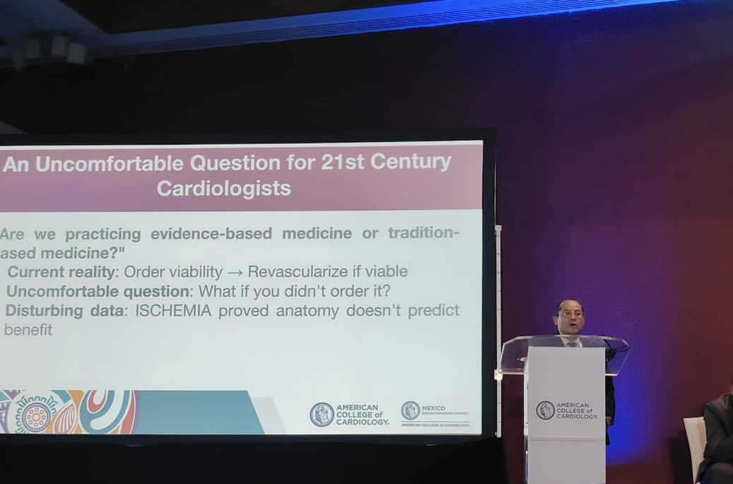
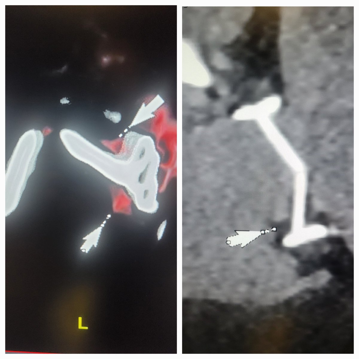
State of the Art Review of Coronary Revascularization without any surgeon as co-author ??? So much for Heart Team approach.
EHJ Editor-in-Chief@ehj_ed

A must-read paper in #EHJ! Coronary revascularization: a long-term perspective. A State-of-the-Art review forecasting advancements by 2040, drawing on historical trends and recent breakthroughs. @escardio @ESC_Journals #CardioTwitter academic.oup.com/eurheartj/adva…

English
7
11
64
25.3K
Moises Jimenez retweetledi
CardioTeca
CardioTeca@cardioteca·
🆕 Riesgo ≥7,5%: Cuándo iniciar fármacos en estadio 1 de hipertensión - Consenso AHA-ACC. 📕 Hypertension 🔓 Resumen del artículo en cardioteca.com/hipertension-a…
CardioTeca tweet media
Español
1
45
167
7.4K
Moises Jimenez retweetledi
Manlio Márquez Murillo
Manlio Márquez Murillo@ManlioMarquez·
For all ECG lovers, this book is for you!
Manlio Márquez Murillo tweet media
English
0
16
64
3.8K
Moises Jimenez retweetledi
Juan Lopez-Mattei, MD
Juan Lopez-Mattei, MD@onco_cardiology·
#ACCLATAM Powerful discussion panel of Women in Cardiology discussing breaking glass ceilings & increase representation of women in Cardiology 🫀 🇲🇽
Juan Lopez-Mattei, MD tweet media
English
1
7
34
3.7K
Moises Jimenez retweetledi
Juan Lopez-Mattei, MD
Juan Lopez-Mattei, MD@onco_cardiology·
#ACCLATAM Getting started with a special session with Dr. Valentin Fuster & Mexico’s Minister of Health discussing how we can do #cvPrev better.
Juan Lopez-Mattei, MD tweet media
English
0
4
22
2.8K
Moises Jimenez retweetledi
Gil Hildebrand
Gil Hildebrand@gilhildebrand·
MrBeast just went deep with Steven Bartlett. In a raw 2-hour conversation, he revealed: • Why he wants to quit YouTube • Why he's more unhappy than ever • Why you should never help anyone else These are his 7 insights you shouldn't miss🧵:
Gil Hildebrand tweet media
English
51
247
2.9K
977.1K
Moises Jimenez retweetledi
Dr. Filippo Cademartiri
Dr. Filippo Cademartiri@FCademartiri·
Myocardial extracellular volume measurement using cardiac computed tomography Rahul G Muthalaly et al. 2024 Oct 14. Int J Cardiovasc Imaging. DOI: 10.1007/s10554-024-03226-4 This paper reviews the utility of measuring myocardial extracellular volume (ECV) with cardiac computed tomography (CT) to assess myocardial fibrosis, a marker for several cardiac diseases and outcomes like heart failure, arrhythmias, and death. Key Points: • Background: Myocardial fibrosis, resulting from extracellular matrix expansion, impairs cardiac function. ECV measurement has traditionally been performed with cardiac magnetic resonance (CMR) using gadolinium, but recent research indicates CT-ECV, using iodine contrast, provides similar accuracy and is faster. • CT-ECV Methodology: CT-ECV quantifies fibrosis by comparing pre- and post-contrast myocardial images, adjusting for haematocrit. Dual-energy CT (DECT) can streamline this by creating iodine maps that remove the need for pre-contrast imaging. • Imaging Protocols: Protocols vary, but a delay of 5 minutes post-contrast appears optimal. High-pitch helical scans are not recommended due to image quality issues. Standardized parameters, including slice thickness and tube voltage, are crucial for accurate ECV measurements. • Clinical Applications: • Cardiac Amyloidosis: CT-ECV can distinguish amyloidosis from other conditions, with amyloid patients showing elevated ECV values. • Valvulopathies: CT-ECV predicts outcomes in conditions like aortic stenosis and can identify fibrosis before left ventricular dysfunction becomes apparent. • Non-Ischaemic Cardiomyopathy: Elevated ECV is indicative of myocardial damage in various cardiomyopathies. • Cardio-Oncology: CT-ECV could monitor cardiac fibrosis in patients undergoing cancer treatments known for cardiotoxicity. • Conclusions: CT-ECV is a promising tool for detecting and prognosticating myocardial fibrosis, with potential applications in multiple cardiac pathologies. Further research is needed to standardize CT-ECV protocols and explore its prognostic capabilities across a broader range of cardiac diseases. This advancement in CT extracellular volume (CT-ECV) measurement, particularly with photon-counting computed tomography (PCCT), represents a major step toward a “one-stop-shop” for cardiac diagnostics by addressing anatomy, function, tissue characteristics, and even molecular markers—all within a single modality. This integration positions cardiac CT to surpass traditional tools like cardiac magnetic resonance imaging (CMR), positron emission tomography (PET), and other modalities by providing a comprehensive, fast, and accessible solution for cardiac assessment. How PCCT-Enhanced Cardiac CT Addresses Key Diagnostic Areas 1. Anatomy Traditionally, cardiac CT has excelled in coronary anatomy visualization, helping detect coronary artery disease by offering high-resolution images of vessel structure. PCCT’s advanced resolution and photon sensitivity allow for more precise imaging of complex anatomical details, capturing even minor calcifications and plaque compositions, providing insight into coronary and vascular health without the need for multiple imaging modalities. 2. Function While functional imaging has been the realm of echocardiography, CMR, and nuclear imaging, PCCT is making strides here as well. Its ultra-fast scan capabilities and dynamic imaging provide valuable insights into cardiac function, including ventricular wall motion, ejection fraction, and flow dynamics. This functional assessment, combined with anatomical detail, supports comprehensive evaluations similar to those offered by echocardiography and CMR but with faster acquisition times and potentially greater accessibility. 3. Tissue Characterization With the addition of CT-ECV, cardiac CT can now quantify myocardial fibrosis, which was previously only accessible via CMR with gadolinium. PCCT improves this further by enhancing contrast differentiation at lower doses, making it possible to measure subtle differences in tissue composition with precision. This capability allows the identification of interstitial fibrosis, cardiomyopathies, and conditions like amyloidosis, providing tissue-level insights without requiring the extended protocols typical of CMR. The dual-energy capabilities of PCCT also support virtual non-contrast imaging, facilitating detailed tissue characterization with fewer scans. 4. Molecular Insights PCCT has the potential to extend into molecular imaging by differentiating between materials and contrasts at a molecular level. This energy-discriminating technology allows the detection of specific molecular markers (like calcium, iodine, and potentially other contrast agents) used in diagnostics. With future developments, it may enable visualization of molecular processes, such as inflammation or ischemic injury, offering similar insights to those currently provided by PET but in a faster, more accessible, and radiation-efficient manner. Benefits Beyond CMR, PET, and Other Modalities 1. Speed and Accessibility Cardiac CT, especially with PCCT, offers rapid imaging times compared to the longer protocols of CMR and PET. This speed not only enhances patient comfort but also allows CT to be more widely accessible for routine screening and early diagnosis. Cardiac CT can be performed without the need for dedicated cardiac MRI units, reducing logistical demands and increasing patient throughput. 2. Radiation Efficiency PCCT reduces radiation exposure by utilizing higher-efficiency photon detection and allowing lower-dose contrast agents, making CT safer for repeated evaluations. While PET also involves radiation exposure, its use of radioactive tracers can limit application frequency and accessibility. By reducing radiation doses, PCCT enables broader use for initial and follow-up assessments. 3. Single-Session, Comprehensive Data Collection Unlike the multi-step, multi-modality approach required with PET for molecular imaging, CMR for fibrosis, and echocardiography for functional imaging, PCCT can potentially capture all these elements in a single session. This integration into one test streamlines workflows, reduces patient burden, and lowers healthcare costs by eliminating the need for multiple appointments and tests. 4. Predictive and Preventative Potential By providing early insight into a range of cardiac conditions through anatomical, functional, tissue, and molecular imaging, PCCT-enhanced cardiac CT enables proactive patient management. This predictive capability allows clinicians to intervene earlier in disease progression and personalize treatments, improving outcomes and potentially reducing the need for future invasive tests. Conclusion The advancement of CT-ECV and the introduction of PCCT technology elevate cardiac CT to a multi-functional, comprehensive diagnostic tool that can address a wide range of cardiac evaluation needs. This approach surpasses the traditional roles of CMR, PET, and other modalities, offering a streamlined, efficient, and accessible path toward a true one-stop-shop in cardiac imaging. PCCT is more than a technological upgrade—it’s a paradigm shift that will change how we diagnose and treat patients for years to come. Investing in PCCT today means investing in the future of healthcare. PCCT is a NEW Imaging Modality. PCCT is changing the game, the field, the language, the priorities and in the end it will change the entire infrastructure of diagnostic medicine. Stay tuned and follow updates on the fine tuning and applications of Advanced Imaging within the "Expert Network of DECT, DSCT/Photon Counting CT and Advanced Imaging". #PCCT #photoncounting #QuantumHD #CT #computedtomography #yesCCT #coronaryarterydisease #ischemia #naeotomalpha Fulltext PDF at: link.springer.com/article/10.100…
Dr. Filippo Cademartiri tweet media
English
2
7
35
3.5K
Moises Jimenez retweetledi
Luis Gonzali
Luis Gonzali@LuisGonzali·
Si quieres entender mejor el mundo financiero y de inversiones, escucha y suscríbete a nuestro Podcast, Money Talks, el otro lado de la moneda. Salen 2 capítulos a la semana, uno el lunes y otro el miércoles. YouTube: @moneytalkselpodcast?si=9nxjONGC73GSRjZ1" target="_blank" rel="nofollow noopener">youtube.com/@moneytalkselp… Spotify: open.spotify.com/show/4zzqblCwY…
Luis Gonzali tweet media
Español
8
38
154
30.4K