
زَيد
1K posts

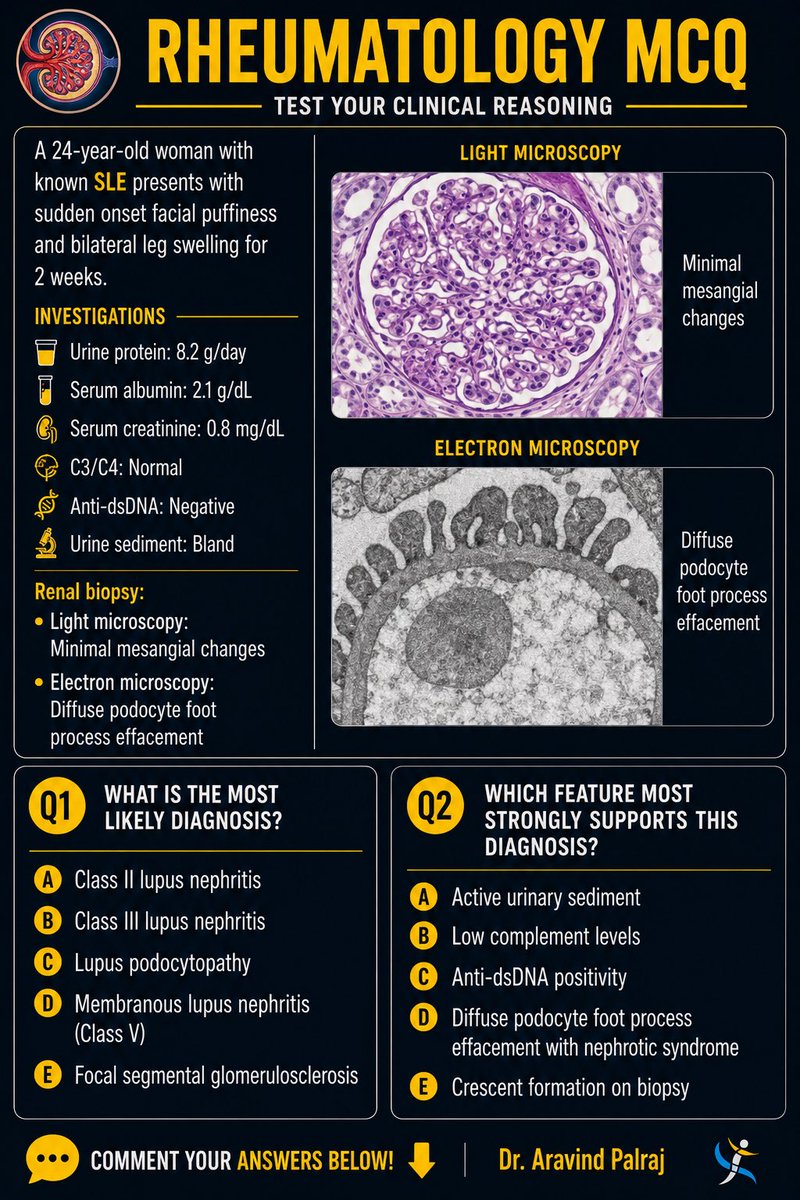
Not all nephrotic syndrome in SLE is proliferative LN 👀
24F with:
• 8.2 g/day proteinuria
• Normal creatinine
• Normal C3/C4
• dsDNA negative
• Bland urine sediment
Dx?
Which biopsy finding confirms it?
👇
— Dr. Aravind Palraj
#Rheumatology #SLE @docakx @IhabFathiSulima @DocPriyamMD @DrNikhilMD @Renalpathsoc #Nephrology #MedEd #ClinicalReasoning #RheumTwitter
English

A man in his 30s w/ AD on long-term upadacitinib presented w/ a 4-day hx of painful, mildly pruritic blistering & ulcerative papules developing 8 days after exposure to tear gas (CS gas) & pepper spray during police academy training. Initial symptoms included ocular burning, visual disturbance, & eyelid edema, followed several days later by vesiculobullous lesions beginning on the left posterior arm & progressing to the face, trunk, & extremities.
O/E: numerous scattered eroded vesicles & grouped papules on an erythematous base involving the face, trunk, & bilateral upper & lower extremities. Additional erythematous papulovesicles & eroded plaques extended in a dermatomal distribution from the right gluteal region to the suprapubic area.
What’s the diagnosis❔


English


#diagnosis? #dermatology #pathology #dermpath JAMA DermatologyCobblestoning Monomorphic Papules of the Lower Extremities jamanetwork.com/journals/jamad…

English
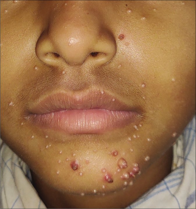
@dermatology Gianotti Crosti syndrome, aka papular acrodermatitis of childhood
English


كويز روماتولوجي:
- كم فيه كلاس لل Lupus nephritis ؟
- وش اخطر نوع ؟!

العربية
A man in his late 60s w/ longstanding well-controlled HIV (CD4 nadir 230 cells/mm³) presented w/ a 5-mo hx of a progressive generalized pruritic eruption that began on the head & neck and spread cephalocaudally.
O/E: erythroderma w/ ectropion, palmoplantar keratoderma, and diffuse orange-red scaly papules & plaques w/ islands of sparing.
What’s the diagnosis❓

English
زَيد retweetledi

This premature baby presented with maculopapular rash and blisters on the arms and legs with superficial desquamation particularly on the palms and soles.
Likely diagnosis?

English
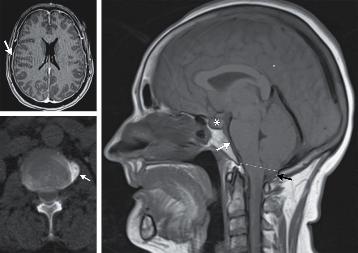
@drsthanus Spontaneous intracranial hypotension due to an acquired csf leak
English
This is a woman in her 50s presented with a 10-day hx of a throbbing headache that had suddenly started after she had felt a “pop” in her back.
The headache worsened with upright positioning and improved with lying flat.
PE, including fundoscopy, was normal.
Diagnosis?
English
A man in his late 50s, a former competitive bodybuilder w/ extensive tanning bed exposure, presented w/ a 25 × 25 cm nonhealing ulcerated plaque on the upper back.
O/E: a large pink ulcerated plaque w/ rolled borders involving most of the upper back, along w/ multiple scattered waxy plaques w/ telangiectasias on the torso.
He was afebrile & nontoxic, w/out palmoplantar pits, milia, skeletal abnormalities, lymphadenopathy, or hx of odontogenic cysts. CBC, CMP, & CK were w/in normal limits.
What’s the diagnosis?

English
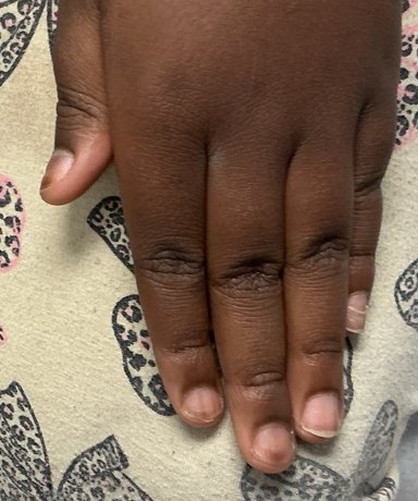
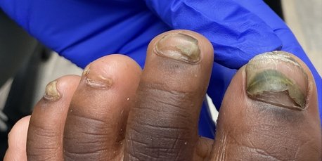
An otherwise healthy child presented w/ progressive proximal nail plate detachment & new green discoloration of several toenails.
One month earlier, she had experienced a self-limited viral exanthem w/ oral ulcers and acral vesicles. Shortly after resolution, painless loosening of several fingernails developed, followed by progressive proximal separation of multiple fingernail & toenail plates. Over subsequent weeks, green-yellow discoloration appeared on both great toenails. She denied fever, fatigue, pain, pruritus, recent aquatic exposure, or travel.
What are the diagnoses❔


English
🧪 Dermatology Case
Spot diagnosis challenge! What are we looking at? ______
Answer = go.dermrounds.com/r/QWhpp3
English
@schowardjd Pyoderma gangrenosum
Cutaneous leukocytoclastic vasculitis
Erythema nodosum
Metastatic Crohn's disease
Română
A woman in her late 20s w/ HS & untreated UC presented w/ a 2-yr hx of painful pruritic lesions on the lower extremities. She had previously been in UC remission on IFX for 8–9 yrs before d/c; ~5 mos later, she developed hematochezia w/ elevated CRP (86.2), ESR (51), & fecal calprotectin (511).
O/E: multiple 2–3 mm pink-to-violaceous papules, subcutaneous nodules, & ulcerated plaques involving the lower legs & feet.
What’s the diagnosis❔

English

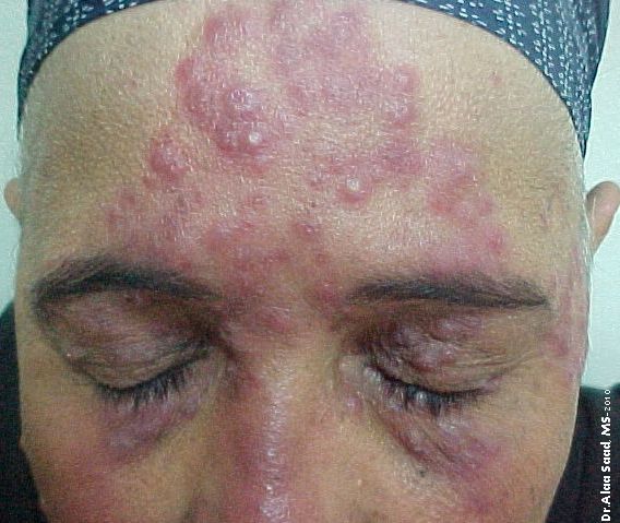
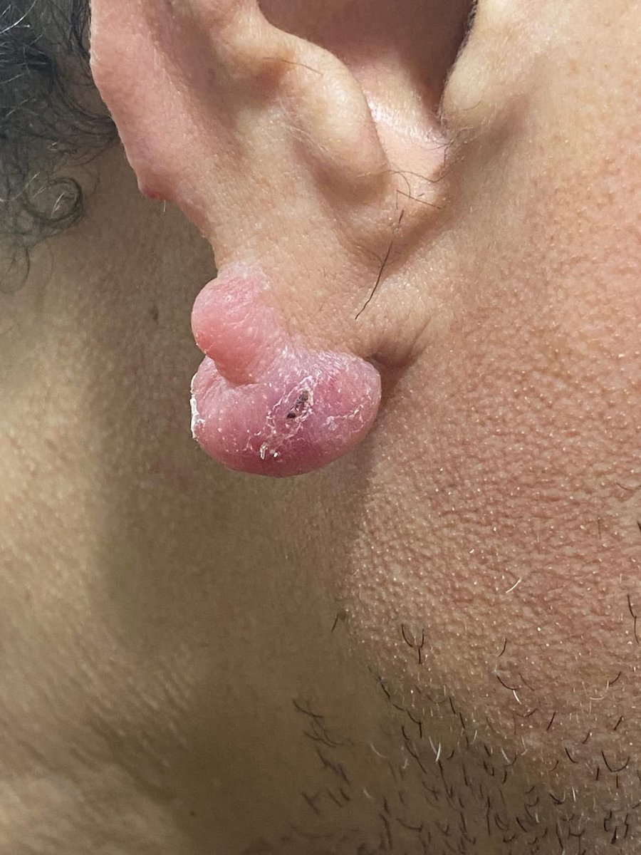
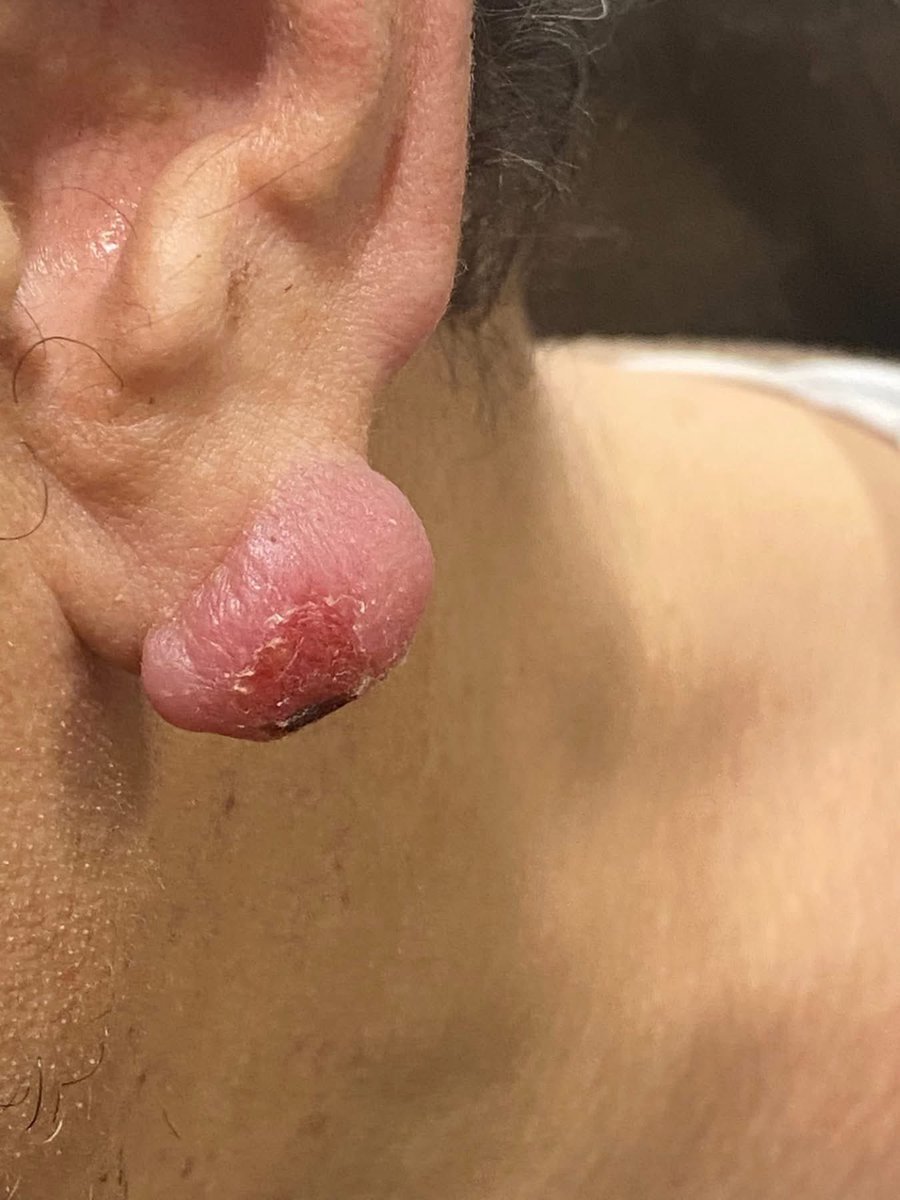
Clue: Head & neck nodules with eosinophils.
What is your diagnosis?
English

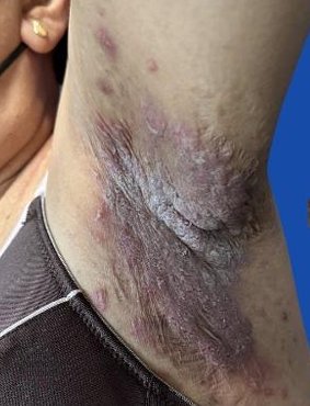
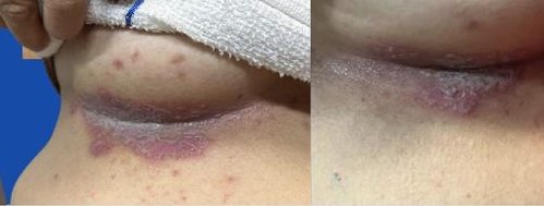
A middle-aged woman presented w/ a 4-yr hx of recurrent painful erosive plaques in intertriginous areas (axillae, inframammary folds, groin), associated w/ malodor, fissuring, & functional impairment. FHx notable for similar lesions in her father & two sisters.
What’s the diagnosis❔


English
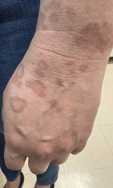
A woman in her 20s with a hx of SLE presented with a 2-week hx of an itchy, painful rash on her nose and hands. 👇
First appeared 1 day after the weather had turned cold.
Likely diagnosis?

English
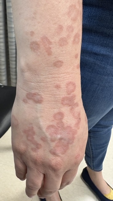
A middle-aged woman w/ hx of GERD presented w/ a >1-yr hx of a persistent eruption refractory to clobetasol & tacrolimus.
O/E: annular, dull erythematous plaques on the face, chest, & B/L upper extremities, most prominent on the dorsal hands.
What’s your diagnosis❔
English