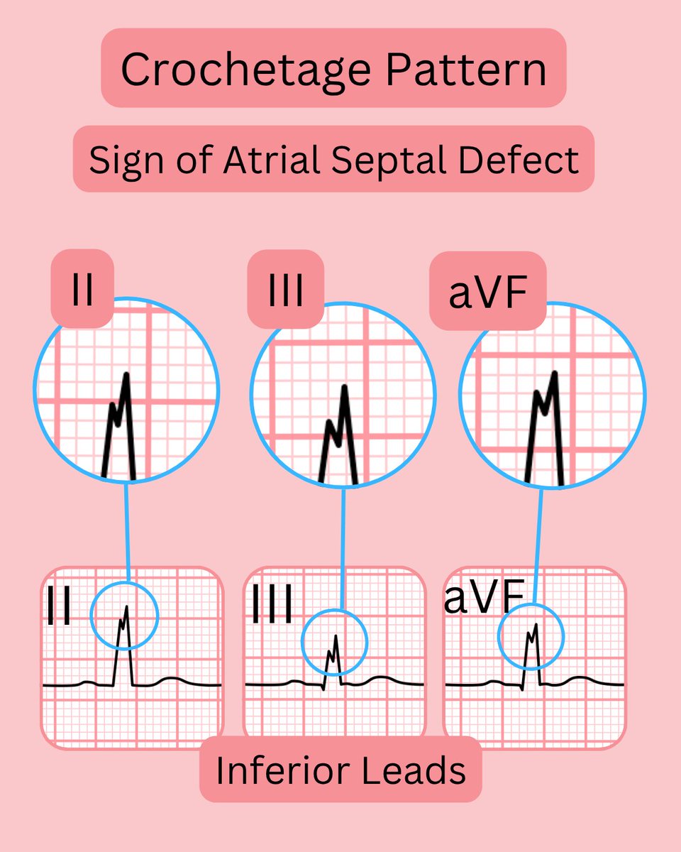
ECGsmart

22 posts

ECGsmart banner
ECGsmart

ECGsmart

@ECGsmart

🫀 Check your ECG online – from smart device or 12-lead ECG!

เข้าร่วม Mayıs 2024
25 กำลังติดตาม19 ผู้ติดตาม
ECGsmart
ECGsmart@ECGsmart·
S1Q3T3 pattern on #ECG – possible causes 🩺 Most classic association 🫁 Acute pulmonary embolism Other possible contexts 📉 Right ventricular strain 📈 Pulmonary hypertension 💔 Massive right-sided MI 🩸 Hypoxia or acute cor pulmonale 🧠 Neurological events (e.g. SAH) ⚠️ Severe COPD exacerbation 🏥 Post-operative states (e.g. after major surgery) Remember: #S1Q3T3 is suggestive, not specific. Always interpret in clinical context 🔍 #ECG #cardiology #medicaleducation #EKG #paramedic #MedstudentTwitter #CardioTwitter #FOAMed #MedEd #CardioEd #Cardiology #FOAMed #meded #MedEd
ECGsmart tweet media
English
0
0
0
93
ECGsmart
ECGsmart@ECGsmart·
Modified Sgarbossa Criteria – Simply Explained - Used to detect #STEMI in patients with #LBBB or Paced rhythm - Improves on original Sgarbossa by focusing on proportional ST elevation - Key point: Discordant ST elevation >25% of #QRS depth - Reduces false positives compared to original criteria 🔄 Concordant vs. Discordant – simple explanation: #Concordant - ST segment goes in the same direction as the QRS complex - (e.g., positive QRS and ST elevation) #Discordant - ST segment goes in the opposite direction to the QRS complex - (e.g., negative QRS and ST elevation) #ECG #cardiology #medicaleducation #EKG #paramedic #MedstudentTwitter #CardioTwitter #FOAMed #MedEd #CardioEd #Cardiology #FOAMed #meded #MedEd
ECGsmart tweet media
English
0
0
0
85
ECGsmart
ECGsmart@ECGsmart·
#ECG Fascicular Blocks Demystified #LAFB (Left Anterior Fascicular Block) – The most common fascicular block – Prevalence: ~1–4% in the general population – Left axis deviation – Often isolated but may coexist with #RBBB #LPFB (Left Posterior Fascicular Block) – Much rarer: <0.1% prevalence – Right axis deviation – Frequently occurs with RBBB → bifascicular block 🧠 Hemiblocks can signal early conduction disease — not always alarming on their own, but clinical context is key! #cardiology #medicaleducation #EKG #paramedic #MedstudentTwitter #CardioTwitter #FOAMed #MedEd #CardioEd #Cardiology #FOAMed #meded #MedEd
ECGsmart tweet media
English
0
0
0
75
ECGsmart
ECGsmart@ECGsmart·
Causes of pathological Q waves Most common ❤️ Myocardial infarction Other possible causes ⚡ Left bundle branch block ↔️ Left anterior hemiblock 🔄 Left posterior hemiblock 🫁 Pulmonary embolism (S1Q3T3 pattern) ⚡ Wolff-Parkinson-White syndrome 🦠 Myocarditis 💪 Hypertrophic cardiomyopathy 🧊 Infiltrative diseases (e.g. amyloidosis) 🤕 Cardiac contusion 🧍 Chest deformities 🎛️ Electrode misplacement 🔁 Positional Q waves (e.g. during deep expiration) Always interpret in clinical context #ECG #EKG #paramedic #MedstudentTwitter #CardioTwitter #FOAMed #MedEd #CardioEd #Cardiology #FOAMed #meded #MedEd
ECGsmart tweet media
English
0
0
0
81
ECGsmart
ECGsmart@ECGsmart·
ECG Color Codes & Lead Placement (IEC Standard) The IEC (International Electrotechnical Commission) ECG standard is commonly used in 🇩🇪 Germany, 🇦🇹 Austria, 🇨🇭 Switzerland, and in many parts of 🌏 Asia. Limb Leads 🔴 RA (Right Arm): Red 🟡 LA (Left Arm): Yellow ⚫ RL (Right Leg): Black 🟢 LL (Left Leg): Green Precordial Leads (C = Chest) 🔴 C1: Red – 4th ICS, right sternal border 🟡 C2: Yellow – 4th ICS, left sternal border 🟢 C3: Green – between C2 and C4 🟤 C4: Brown – 5th ICS, midclavicular line ⚫ C5: Black – same level as C4, anterior axillary line 🟣 C6: Purple – same level as C4, midaxillary line 📌 Some systems use ⚪ white for all precordial leads. Precordial Leads: "V" or "C"? 🇪🇺 IEC (used in Europe) → "C" = Chest lead (C1–C6) 🇺🇸 AHA (used in the USA) → "V" = Voltage lead (V1–V6) #ECG #ekg #paramedic #MedstudentTwitter #CardioTwitter #FOAMed #MedEd #CardioEd #Cardiology #FOAMed #meded #MedEd
ECGsmart tweet media
English
0
0
0
98
ECGsmart
ECGsmart@ECGsmart·
ECG Color Codes & Lead Placement (AHA Standard) The AHA (American Heart Association) ECG standard is widely used in the 🇺🇸 USA, 🇨🇦 Canada, and in most international medical textbooks and training courses. Limb Leads ⚪ RA (Right Arm): White ⚫ LA (Left Arm): Black 🟢 RL (Right Leg): Green 🔴 LL (Left Leg): Red Precordial Leads (V = Voltage) 🔴 V1: Red – 4th ICS, right sternal border 🟡 V2: Yellow – 4th ICS, left sternal border 🟢 V3: Green – between V2 and V4 🔵 V4: Blue – 5th ICS, midclavicular line 🟠 V5: Orange – same level as V4, anterior axillary line 🟣 V6: Purple – same level as V4, midaxillary line 📌 Some systems use brown for all precordial leads. 🔤 "V" or "C" for chest leads? 🇺🇸 AHA uses "V" = Voltage lead (V1–V6) 🇪🇺 IEC (used in Europe) uses "C" = Chest lead (C1–C6) #ECG #ekg #paramedic #MedstudentTwitter #CardioTwitter #FOAMed #MedEd #CardioEd #Cardiology #FOAMed #meded #MedEd
ECGsmart tweet media
English
0
0
0
75
ECGsmart
ECGsmart@ECGsmart·
🔴 Wellens Syndrome = LAD ticking time bomb 😮 Chest pain + normal troponin ≠ safe! 📊 3–5% of acute chest pain pts 📉 ECG: • Type I: Biphasic T in V2–V3 • Type II: Deeply inverted T in V2–V3 💣 Risk of massive anterior MI in days 🩺 Needs urgent PCI/CABG! #ECG #Cardiology
ECGsmart tweet media
English
0
0
0
68
ECGsmart
ECGsmart@ECGsmart·
#BrugadaPattern vs. #BrugadaSyndrome ⚡ Brugada Pattern - Asymptomatic ECG finding - Prevalence: 0.1–1% - Seen in #ECG leads V1–V2 (Type 1 or 2) 🚨 Brugada Syndrome - Brugada pattern with symptoms - Symptoms: Syncope, nocturnal agonal respiration, or sudden cardiac death
ECGsmart tweet media
English
0
0
0
88