Scott Haines, RCS, RDCS🫀 ‎

1.5K posts

Scott Haines, RCS, RDCS🫀 ‎ banner
Scott Haines, RCS, RDCS🫀 ‎

Scott Haines, RCS, RDCS🫀 ‎

@echogenics

Registered Cardiac Sonographer 🫀 | Member American Society of Echo | #ASE | #ARDMS |#CCI | #Echogenics | #EchoLab | #RCS | #RDCS

Simi Valley, CA Sumali Şubat 2018
129 Sinusundan91 Mga Tagasunod
Scott Haines, RCS, RDCS🫀 ‎ nag-retweet
CardiovascularCorner
CardiovascularCorner@TrackYourHeart·
A 21-year-old presented to the ER with fever, breathlessness, and palpitations. What's the most likely diagnosis ⁉️
English
12
7
41
12K
Scott Haines, RCS, RDCS🫀 ‎ nag-retweet
Jorge Parras
Jorge Parras@ParrasJorge2·
A 35-year-old woman with chronic liver failure.
English
5
8
27
3K
Scott Haines, RCS, RDCS🫀 ‎ nag-retweet
Dr G Rajesh (Gopalan Nair Rajesh).
Look beyond the heart in all cases of dyspnoea for evaluation, especially if X-ray chest findings is not entered in OP ticket/case sheet. Never do echo without seeing the patient clinically and without seeing the ECG& X-ray.
English
4
9
56
6.3K
Scott Haines, RCS, RDCS🫀 ‎ nag-retweet
Alexander Mladenow MD
Alexander Mladenow MD@alex1708ander·
Adult patient with normal segmental anatomy (no CHD). Which are the structures labelled with 🔢? #echofirst Learning point: 3D echo anatomy
English
3
3
22
5K
Scott Haines, RCS, RDCS🫀 ‎ nag-retweet
Dr Akhil Sharma
Dr Akhil Sharma@Drakhil_cardio·
#Sharing for learning Spotter for Fellows When the heart defies expectations…
and anatomy turns almost unrecognizable. A striking, ominous finding.
Exceptionally rare. Instantly unforgettable. Diagnosis and what else you are expecting on examination??
English
2
12
74
10.4K
Scott Haines, RCS, RDCS🫀 ‎ nag-retweet
Ross Prager
Ross Prager@ross_prager·
Patient with shock, what is driving their hemodynamic collapse more, the RV or the pericardial effusion? 👇
English
28
23
107
21.2K
Scott Haines, RCS, RDCS🫀 ‎ nag-retweet
Dr G Rajesh (Gopalan Nair Rajesh).
For fellows. Enumerate all findings this patient has. What warning will you give to the surgeon who is going to operate the patient?
English
11
8
57
10.9K
Scott Haines, RCS, RDCS🫀 ‎ nag-retweet
Lucy
Lucy@LucySafi·
Excited to share our latest work on T-TEER! We break down how to mirror M-TEER workflows for the TV—highlighting key anatomic parallels, imaging strategies, procedural steps to simplify a complex intervention. A practical step-by-step guide Link -> bit.ly/3Q0gURZ
Lucy tweet media
English
1
7
41
3.9K
Scott Haines, RCS, RDCS🫀 ‎ nag-retweet
CardiovascularCorner
CardiovascularCorner@TrackYourHeart·
Which intracardiac mass is shown being removed?
English
12
8
66
64.7K
Scott Haines, RCS, RDCS🫀 ‎ nag-retweet
Alexander Mladenow MD
Alexander Mladenow MD@alex1708ander·
Even if AV and LVOT are anatomically and functionally closely linked, SAVR mainly targets the aortic valve → the LVOT typically remains unchanged #SAM Pre & postop TEE in a patient undergoing SAVR for AS #echofirst
English
0
6
23
2K
Scott Haines, RCS, RDCS🫀 ‎ nag-retweet
CardiovascularCorner
CardiovascularCorner@TrackYourHeart·
Examine this specimen and make a diagnosis.
CardiovascularCorner tweet media
English
18
17
127
36.5K
Scott Haines, RCS, RDCS🫀 ‎ nag-retweet
Alexander Mladenow MD
Alexander Mladenow MD@alex1708ander·
3D ECHO literally means looking INSIDE the 🫀 Exactly right what @NMerke said: ✅ “The secret to great 3D is hidden into your 2D skills” ✅ “You should master 2D first” 📌 Using 3D #echofirst identifies in 2D (‼️) WHAT & WHY 🔍something is wrong IMMEDIATELY
Nicolas Merke@NMerke

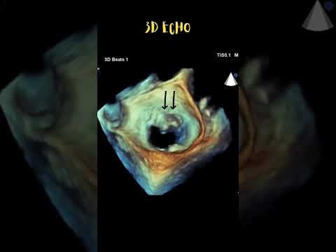
3D is an essential tool in echocardiography #echofirst 👉 Accurate LV volumes & function — beyond geometrical assumptions, minimizing foreshortening 👉 The only robust way to assess RV volumes & function without geometric limitations 👉 Key to understanding mechanisms of valvular regurgitation But here’s the catch: None of this works without mastering 2D first 👉 Reliable quantification starts with optimal 2D reference planes 👉 Full visualization of endocardial borders is non-negotiable 👉 Valve assessment always begins with precise 2D imaging 👉 #ALAMO Transthoracic echocardiography is the perfect training ground for 3D in valvular heart disease — every technique you refine here will elevate your TEE 3D performance The secret to great 3D? It’s hidden in your 2D skills 🎯 The better your 2D imaging → the better your 3D datasets → the more confident your clinical decisions

English
1
5
19
1.5K