Tweet ghimElectron Microscopy at uOttawaMed@AtElectron·23 OcaWhat’s your preferred organelle? We’d like to show it to you here 😇 #TEM #CellBiologyDịch English1010
Electron Microscopy at uOttawaMed@AtElectron·25 Oca@ramana_vaka Would this EV fit your taste? (preparation from Dr. Derrick Gibbings lab @uOttawaMed)Dịch English1010
Electron Microscopy at uOttawaMed@AtElectron·23 OcaWhat’s your preferred organelle? We’d like to show it to you here 😇 #TEM #CellBiologyDịch English1010
Electron Microscopy at uOttawaMed@AtElectron·24 Oca@NadyaMorrow As in those busy nascent endothelial cells? 🤔Dịch English0000
Electron Microscopy at uOttawaMed@AtElectron·23 Oca@adam_rudner from @uOttawaBMI visited us recently to image phages for @uOttawaTMM students 👇🏻👇🏻Dịch English0030
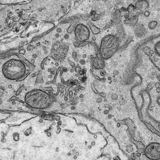
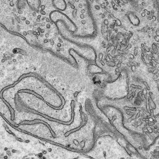
Electron Microscopy at uOttawaMed@AtElectron·23 OcaAin’t it good to navigate the 🧠?! Look at that beautiful synapse in the center at the end!🤩Dịch English1010
Electron Microscopy at uOttawaMed@AtElectron·23 OcaHow to calibrate TEM image magnification? Use tiny waffles (but better not be hungry, each square is only 500nm wide...)Dịch English1010
Electron Microscopy at uOttawaMed@AtElectron·22 OcaThe Gibbings lab @uOttawaMed was happy to image their beloved extracellular vesicles last week!Dịch English1040
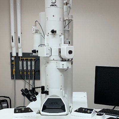
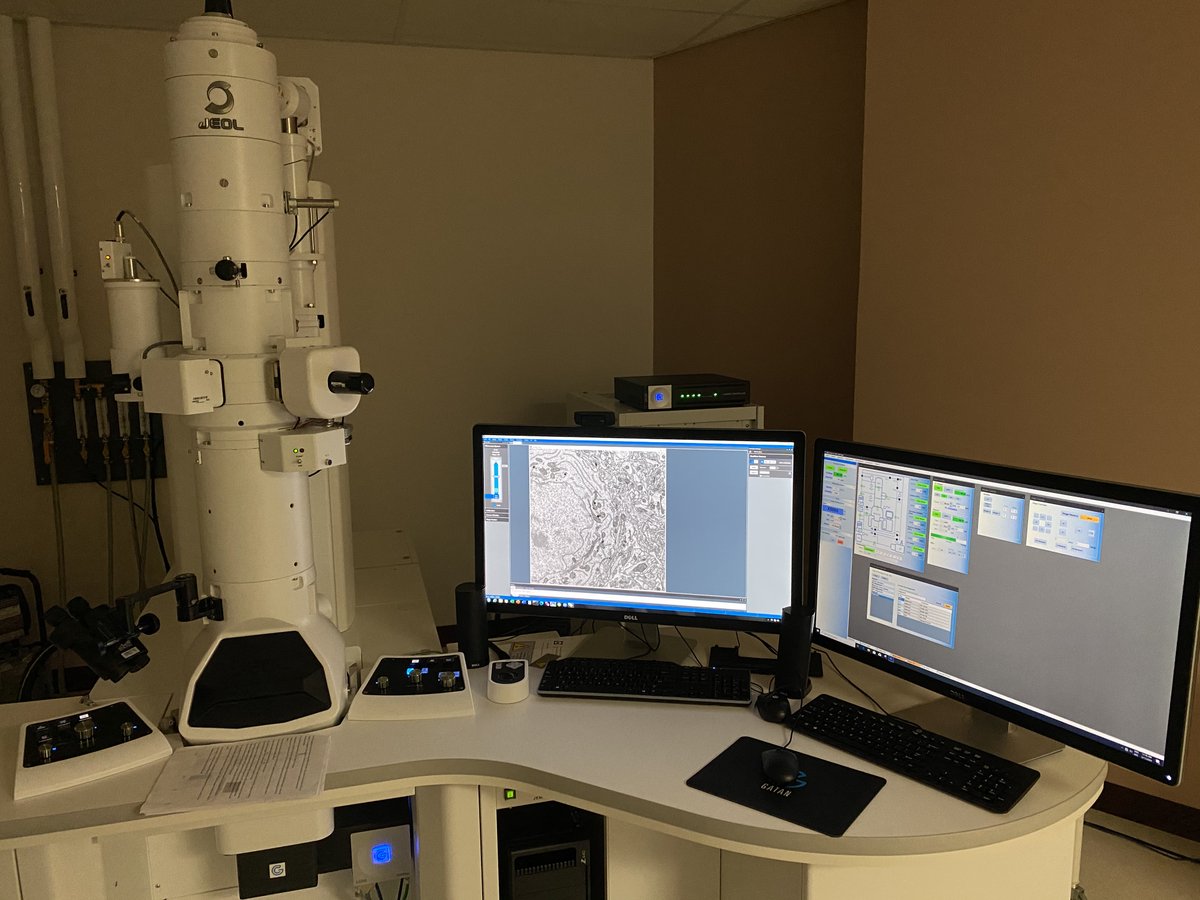
Electron Microscopy at uOttawaMed@AtElectron·22 OcaHere is our awesome set up! @JEOLUSA 120kv JEM-1400Flash TEM and @GatanMicroscopy OneView 4K high-speed camera!Dịch English0030
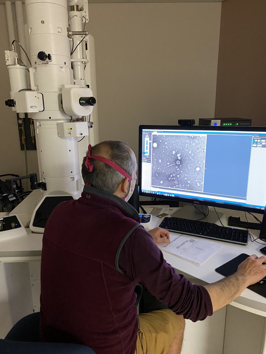
Electron Microscopy at uOttawaMed@AtElectron·22 OcaWelcome the our new TEM core! Stay tuned, we'll post images and news! More info here: med.uottawa.ca/core-facilitie…Dịch English0000