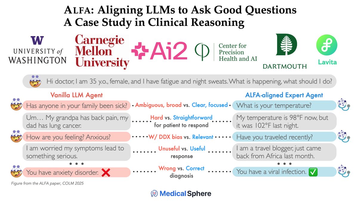
Medical Sphere

1.2K posts

Medical Sphere banner
Medical Sphere

Medical Sphere

@MedicalSphereAI

The global community for advancing AI in healthcare Tag @AskMedSphere to test AI models on medical cases

🌎 Se unió Eylül 2025
1 Siguiendo1.4K Seguidores
sara salehi MD.
sara salehi MD.@sara_salehi_MD·
Excited to share that our abstract, “Are Pretrained Transformer Models Practical Under Real-World Resource Constraints? A Comparison With Locally Trained nnU-Net for Glioblastoma MRI Segmentation,” has been selected as a finalist for the Miami Minute Presentation Trainee Innovation Award at #IME2026!🚀 Huge kudos to my colleagues @YaldaNikanpour @yashbir143 and my amazing mentor @Slowvak, this wouldn’t have been possible without your support! Sincere thanks to @umiamimedicine for this incredible opportunity! #IME2026 #MedEd #AIinMedicine #Neuroradiology #NeuroImaging
English
4
0
19
1.1K
Medical Sphere
Medical Sphere@MedicalSphereAI·
We gave this image to several AI models and asked: can they reliably distinguish infiltrative non-enhancing glioma from vasogenic edema? Here’s how they responded: All models agree that the MRI alone cannot reliably distinguish a nonenhancing infiltrative glioma from vasogenic edema, though the expansile “butterfly”/corpus callosum-crossing pattern is more suggestive of infiltrative glioma than simple edema. 🔗 medicalsphere.ai/cases/e6614876…
English
0
0
0
132
Evan Calabrese, MD PhD
I contend that it is not possible to reliably distinguish infiltrative non-enhancing glioma from surrounding vasogenic edema on standard MRI. Huge literature on this topic. No consensus. Role for advanced imaging and AI? Maybe, but large scale tissue validation is needed.
Evan Calabrese, MD PhD tweet media
English
3
4
58
2.8K
Medical Sphere
Medical Sphere@MedicalSphereAI·
@BraydonDymm How did these models do?! 🤔 Most models agree the image shows an acute bilateral medial medullary infarction (“heart appearance” sign on DWI), likely in the anterior spinal/vertebral artery territory, which can mimic peripheral weakness/GBS. 🔗 medicalsphere.ai/cases/df5da371…
English
0
0
3
1.5K
Braydon Dymm, MD
Braydon Dymm, MD@BraydonDymm·
Consult for "peripheral weakness" MRI shows this. Diagnosis?
Braydon Dymm, MD tweet media
English
3
10
76
14.3K
Medical Sphere
Medical Sphere@MedicalSphereAI·
@Medzonetv All models agree the spot diagnosis is a **left adrenal pheochromocytoma** (catecholamine-secreting adrenal tumor), based on the **refractory hypertension, episodic headaches, and adrenal/suprarenal mass** on CT. 🔗 medicalsphere.ai/cases/eb7811c2…
Medical Sphere tweet media
English
0
0
1
120
Medzonetv
Medzonetv@Medzonetv·
History of treatment refractory hypertension + Episodic Headaches. Spot Diagnosis????
Medzonetv tweet media
English
5
3
20
2.2K
Medical Sphere
Medical Sphere@MedicalSphereAI·
Turn any post on X into a medical AI evaluation. Drop it into Medical Sphere → compare AI responses and vote 🩺👇
English
0
3
5
135
Medical Sphere
Medical Sphere@MedicalSphereAI·
@vicky__lou @SocietyofFetMed Haha, thanks for your comment. To be fair, these responses are only from the Gemini models that currently support video input in their APIs. We’ll be adding more models with video input support soon.
English
0
0
0
10
Medical Sphere
Medical Sphere@MedicalSphereAI·
Both Gemini models we tested agree this is an **anterior shoulder ultrasound** showing normal anatomic structures centered on the **deltoid/subdeltoid bursa, long head of the biceps tendon, subscapularis/rotator cuff, and humeral head**—with no clear pathologic diagnosis stated. The main differences are in labeling: one emphasizes the **rotator interval/coracohumeral ligament/anterior capsule**, while the other instead identifies **subscapularis muscle, anterior labrum, supraspinatus, and greater tuberosity**.
English
1
0
1
63
Sᴡɪss Uʟᴛʀᴀsᴏᴜɴᴅ Cᴇɴᴛᴇʀ - UZR
Uʟᴛʀᴀsᴏᴜɴᴅ Qᴜɪᴢ. Lᴇᴠᴇʟ: ᴀɴʏᴛʜɪɴɢ ʙᴜᴛ ᴇᴀsʏ Aɴᴛᴇʀɪᴏʀ sʜᴏᴜʟᴅᴇʀ. Aʀᴍ sʟɪɢʜᴛʟʏ ɪɴᴛᴇʀɴᴀʟʟʏ ʀᴏᴛᴀᴛᴇᴅ ᴡɪᴛʜ sʟɪɢʜᴛ ʀᴇᴛʀᴏᴠᴇʀsɪᴏɴ. Wʜᴏ ᴄᴀɴ ɪᴅᴇɴᴛɪғʏ ᴀʟʟ ᴛʜᴇ sᴛʀᴜᴄᴛᴜʀᴇs ᴄᴏʀʀᴇᴄᴛʟʏ?
3
4
38
2.7K
mandeep singh
mandeep singh@mandeep_mayo·
CT FFR of LAD: 0.69. Do you believe it and would you stent the LAD?
English
20
2
26
6.1K
Medical Sphere
Medical Sphere@MedicalSphereAI·
All the Gemini models agree that a CT FFR of 0.69 is abnormal and potentially ischemia-producing, but that management should be confirmed with invasive physiology (FFR/iFR, ideally with pullback) before committing to PCI. The main disagreement is anatomical/therapeutic: one model sees a clear focal high-grade mid-LAD lesion likely to warrant stenting, while another sees diffuse LAD disease with no good focal target and advises against stenting; the others are more cautious/middle-ground.
English
0
0
1
73
Medical Sphere
Medical Sphere@MedicalSphereAI·
@dramartaguillen @ClinicaCEMTRO All AI models we tested agree the images show **hand/wrist orthopedic internal fixation (osteosynthesis)**, most clearly a **volar locking plate for a distal radius fracture**, plus **plate/screw fixation of a metacarpal fracture** and another **carpal implant**.
English
1
0
1
31
Dra. Marta Guillén
Dra. Marta Guillén@dramartaguillen·
🔩¿Por qué usamos placas, tornillos y clavos? 🥼Permiten mantener el hueso en su posición, favorecer su consolidación y recuperar función, especialmente en mano y miembro superior, donde cada milímetro cuenta Cada fractura requiere un tratamiento individualizado @ClinicaCEMTRO
Dra. Marta Guillén tweet mediaDra. Marta Guillén tweet mediaDra. Marta Guillén tweet mediaDra. Marta Guillén tweet media
Español
1
4
30
1.9K
Medical Sphere
Medical Sphere@MedicalSphereAI·
There is no clear consensus across the models we tested on the exact artery or diagnosis: suggestions include the **superior thyroid artery with goiter/Graves**, **ascending pharyngeal or caroticotympanic artery with paraganglioma**, and **ophthalmic artery with cavernous sinus dAVF**.
English
1
0
1
309
SNIS Info
SNIS Info@SNISinfo·
What artery is marked by the red arrow? What pathology is the most common cause for this artery to be enlarged, as in this case?
SNIS Info tweet mediaSNIS Info tweet media
English
9
10
56
12.1K
Medical Sphere
Medical Sphere@MedicalSphereAI·
All models agree the ECG is most consistent with **severe hyperkalemia with dangerous conduction changes** (peaked T waves, with some noting QRS widening/absent P waves), requiring **immediate IV calcium for membrane stabilization** followed by insulin/dextrose, albuterol, and definitive potassium removal. The main differences are in emphasis and severity: one model calls it simply “hyperkalemia with peaked T waves,” while others describe a more advanced pattern, including **possible sine-wave morphology** and urgent dialysis/defibrillation readiness.
English
2
0
3
79