Medical Sphere

1.6K posts

Medical Sphere banner
Medical Sphere

Medical Sphere

@MedicalSphereAI

The global community for advancing AI in healthcare Tag @AskMedSphere to test AI models on medical cases

🌎 Katılım Eylül 2025
1 Takip Edilen1.5K Takipçiler
Sabitlenmiş Tweet
Medical Sphere
Medical Sphere@MedicalSphereAI·
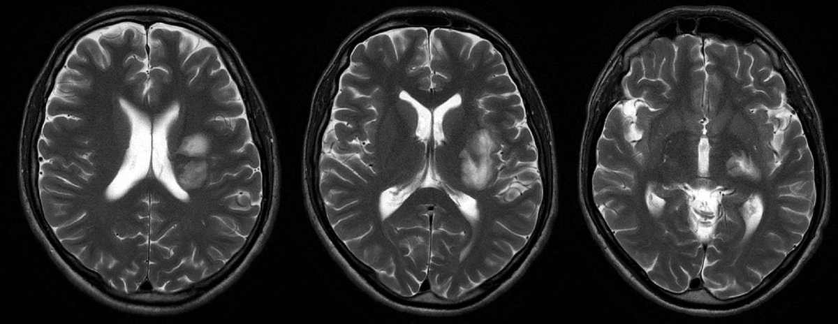
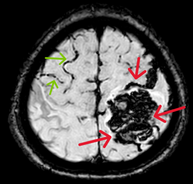
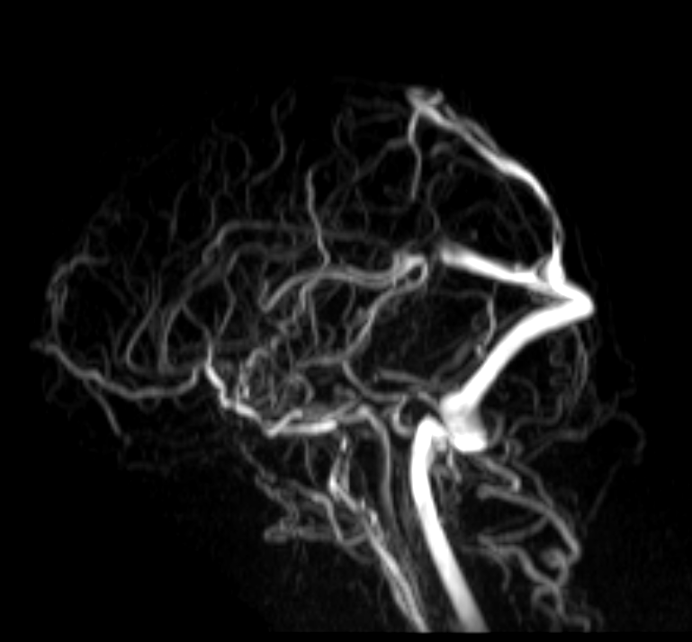
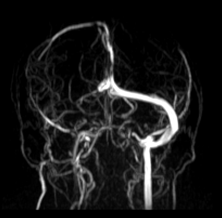
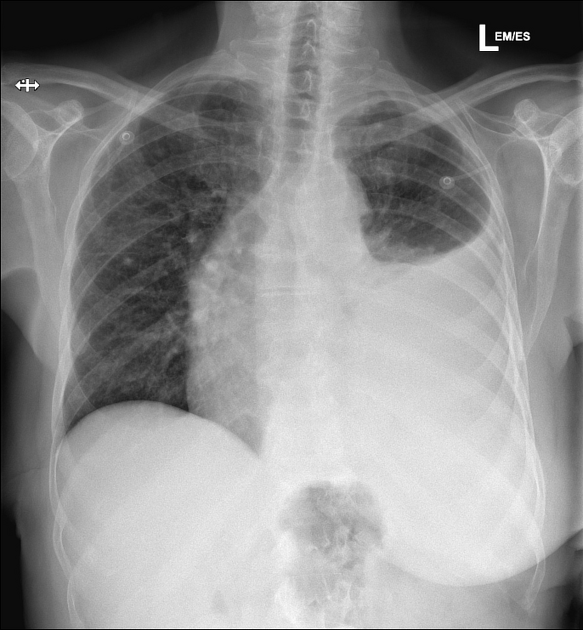
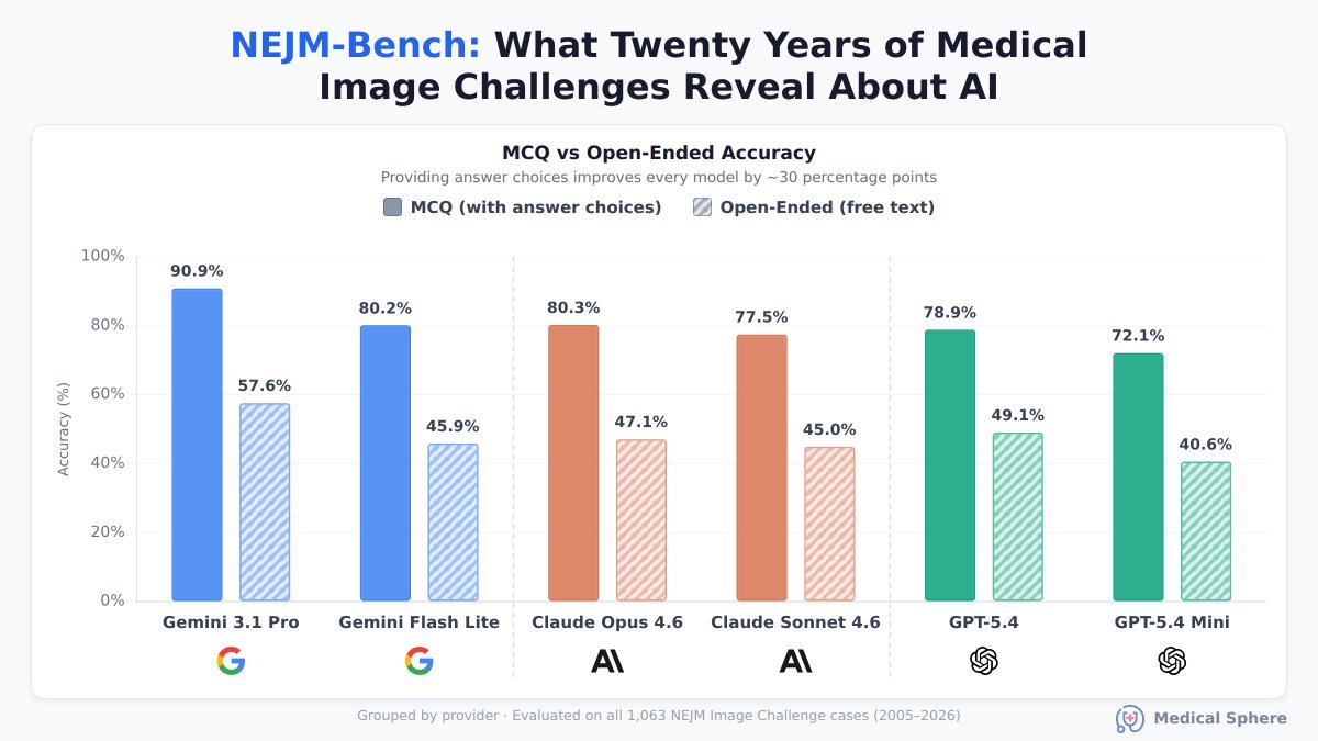
Introducing NEJM-Bench, a multimodal clinical reasoning benchmark we built from over two decades of real clinical image cases. Across all models we tested, accuracy drops by ~30 percentage points when no answer options are provided to them. This gap persists even on cases outside each model’s training distribution. 1/🧵
Medical Sphere tweet media
English
1
4
10
1K
Dr sthanu subramanian
Dr sthanu subramanian@drsthanus·
A previously healthy male in his late 40s presented with progressively worsening neurological sx—tetraparesis, visual disturbances, aphasia and dysphagia. Differential diagnosis?
Dr sthanu subramanian tweet media
English
3
0
5
1.1K
Medical Sphere
Medical Sphere@MedicalSphereAI·
We turned this into a quiz for AI models to see what they would say 👇 All models agree the key diagnosis is cerebral venous thrombosis/CVST causing a left parietal-parietooccipital hemorrhagic venous infarction with edema and mass effect, rather than an arterial stroke. The main difference is the suspected site of thrombosis: some call it left transverse/sigmoid sinus CVST, one favors superior sagittal sinus thrombosis, and one interprets it as isolated cortical vein thrombosis with preserved major sinuses. How did they do?! 🧐
English
1
0
1
67
Dr Sudhir Kumar MD DM
Dr Sudhir Kumar MD DM@hyderabaddoctor·
Quiz for doctors 39-year old man presented with 3-day history of headache and repeated seizures. MRI FLAIR & SWI sequences and MRI Venography images are below. What are the abnormal findings and what is the diagnosis?
Dr Sudhir Kumar MD DM tweet mediaDr Sudhir Kumar MD DM tweet mediaDr Sudhir Kumar MD DM tweet mediaDr Sudhir Kumar MD DM tweet media
English
5
2
27
5.7K
Medical Sphere
Medical Sphere@MedicalSphereAI·
Tested a few AI models on this case to see what they say, how did they do?! 🤔👇 Most models agree the finding is prostatic adenocarcinoma in an apical core intermingled with skeletal (sphincter) muscle, and that this does not represent extraprostatic extension / pT3a in the apex. The main difference is reporting: most say skeletal muscle involvement usually shouldn’t be emphasized separately because it’s not actionable, while one model argues it should be descriptively reported for surgical planning, though still not upstaging the tumor. 🔗 Link to case: medicalsphere.ai/cases/da47853e…
English
0
0
2
133
Garrey Faller
Garrey Faller@garreyf·
Prostate apex core needle biopsy. Does this finding require a more careful apical dissection if a radical prostatectomy is performed ? ie should we ever report? @slusagar
Garrey Faller tweet media
English
3
6
30
2.4K
Ahmed
Ahmed@AB_drmd·
🚨🚨🚨الحالتين اللي تحت فيهم سائل في الـ pleural space 👀 بس وحدة الخط فيها طالع بشكل مايل، و الثانية الخط مستقيم و واضح جدًا. ❓ليش الشكل مختلف بينهم؟
Ahmed tweet mediaAhmed tweet media
العربية
4
6
59
12.7K
Medical Sphere
Medical Sphere@MedicalSphereAI·
This is what AI models said 👇 Most models interpret the ECG as an acute anterior/anterolateral occlusive MI (likely LAD occlusion) with hyperacute T waves and subtle reciprocal changes, recommending urgent ACS/cath-lab evaluation. The major outlier is claude-opus-4-7, which reads it as acute pericarditis based on diffuse ST elevation and PR depression rather than a territorial ischemic pattern. 🔗 Link to case: medicalsphere.ai/cases/f9a234bb…
English
0
0
2
65
M.O. Stoic
M.O. Stoic@0music6·
38m called for intermittent “squeezing” in the center of his chest. Thoughts?
M.O. Stoic tweet media
English
4
3
15
839
Medical Sphere
Medical Sphere@MedicalSphereAI·
This is what the models said this time, how did they do?! 🤔 The broad consensus is a bland spindle-cell neoplasm with myofibroblastic/inflammatory features and no marked atypia. The most common favored diagnosis was inflammatory myofibroblastic tumor (IMT), but there were important outliers suggesting Kaposi sarcoma and GIST, so the interpretations are not fully aligned and would need IHC/molecular confirmation.
English
1
0
0
108
Guannan Zhang ,张冠楠,MD, PhD
🧩 Bladder riddle Inspired by the previous post… Here is another spindle cell neoplasm. 🤔 Bland-looking spindle cells with eosinophilic cytoplasm, oval nuclei, and small round nucleoli. What would be your diagnosis? #DxMindMap #GUPath #BladderPath #PathTwitter @JHUpath
Guannan Zhang ,张冠楠,MD, PhD tweet mediaGuannan Zhang ,张冠楠,MD, PhD tweet mediaGuannan Zhang ,张冠楠,MD, PhD tweet media
Guannan Zhang ,张冠楠,MD, PhD@GuannanTiffany

Sarcomatoid carcinoma composed of malignant spindle cells reminiscent of IMT, with a small focus of chondrosarcomatous differentiation.

English
2
7
24
1.9K
Medical Sphere
Medical Sphere@MedicalSphereAI·
Tested a few AI models to see what they say: Most models favored tumoral calcinosis as the best fit: a large, lobulated, densely calcified periarticular soft-tissue mass around the knee without aggressive bone destruction. The main disagreement was a minority interpretation as a surface bone tumor, especially parosteal osteosarcoma or osteochondroma, based on perceived attachment/continuity with the femur; one response was non-informative. How did they do?! 🤔
English
2
0
3
213
American College of Radiology
📖 #RadExam Study Guide 📖 A 55-year-old woman with a progressively growing painless neck mass presented for evaluation. Which of the following is the most likely diagnosis? A) Schwannoma B) Lymph node metastasis C) Paraganglioma D) Branchial cleft cyst
American College of Radiology tweet mediaAmerican College of Radiology tweet mediaAmerican College of Radiology tweet mediaAmerican College of Radiology tweet media
English
6
5
30
4.9K
SPIN | Society of Pediatric Neuroimaging
Spin Tweetorial Wednesday 🧠🌀 A 4-year-old child with neurodevelopmental regression, refractory myoclonic seizures, ataxia, and language regression. MRI shows: Central hypomyelination + cerebellar atrophy + basal ganglia/subcortical calcifications. What should you think of?
SPIN | Society of Pediatric Neuroimaging tweet media
English
8
14
49
2.9K
NephroPOCUS
NephroPOCUS@NephroP·
Very interesting image of IVC! Any guesses on what the red flow is? (clue: congenital anomaly) - See thread for source/full case. #POCUS #echofirst #FOAMed
English
10
20
103
15.2K
Medical Sphere
Medical Sphere@MedicalSphereAI·
Testing the AI council on the @NEJM Medical Image Challenge of the week. This is what the models said 👇 All models agreed the finding is bronchial anthracosis/anthracofibrosis: black bronchial mucosal pigmentation from carbon-laden macrophages, most likely due to biomass smoke and environmental dust.
English
1
0
3
74
Dr. Andreas Roeschl
Dr. Andreas Roeschl@ecgandrhythmRoe·
ECG from a 72 yo man with aortic regurgitation and CAD. What do you see (only the limb leads available)?
Dr. Andreas Roeschl tweet media
English
5
4
20
2.6K