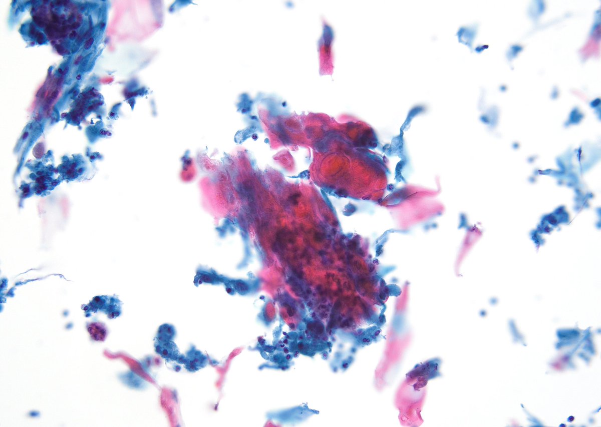
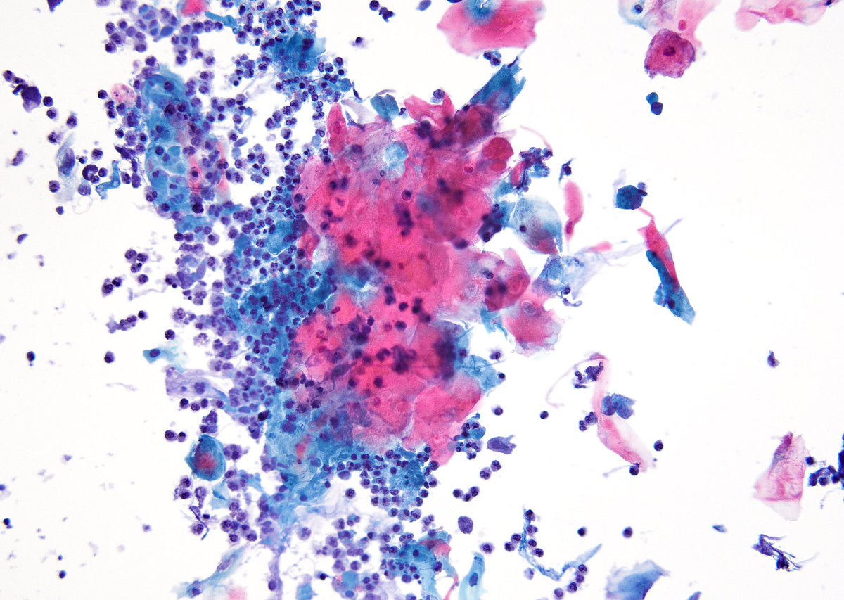
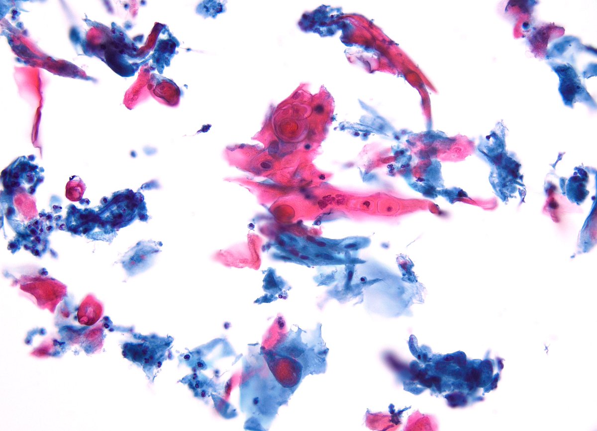
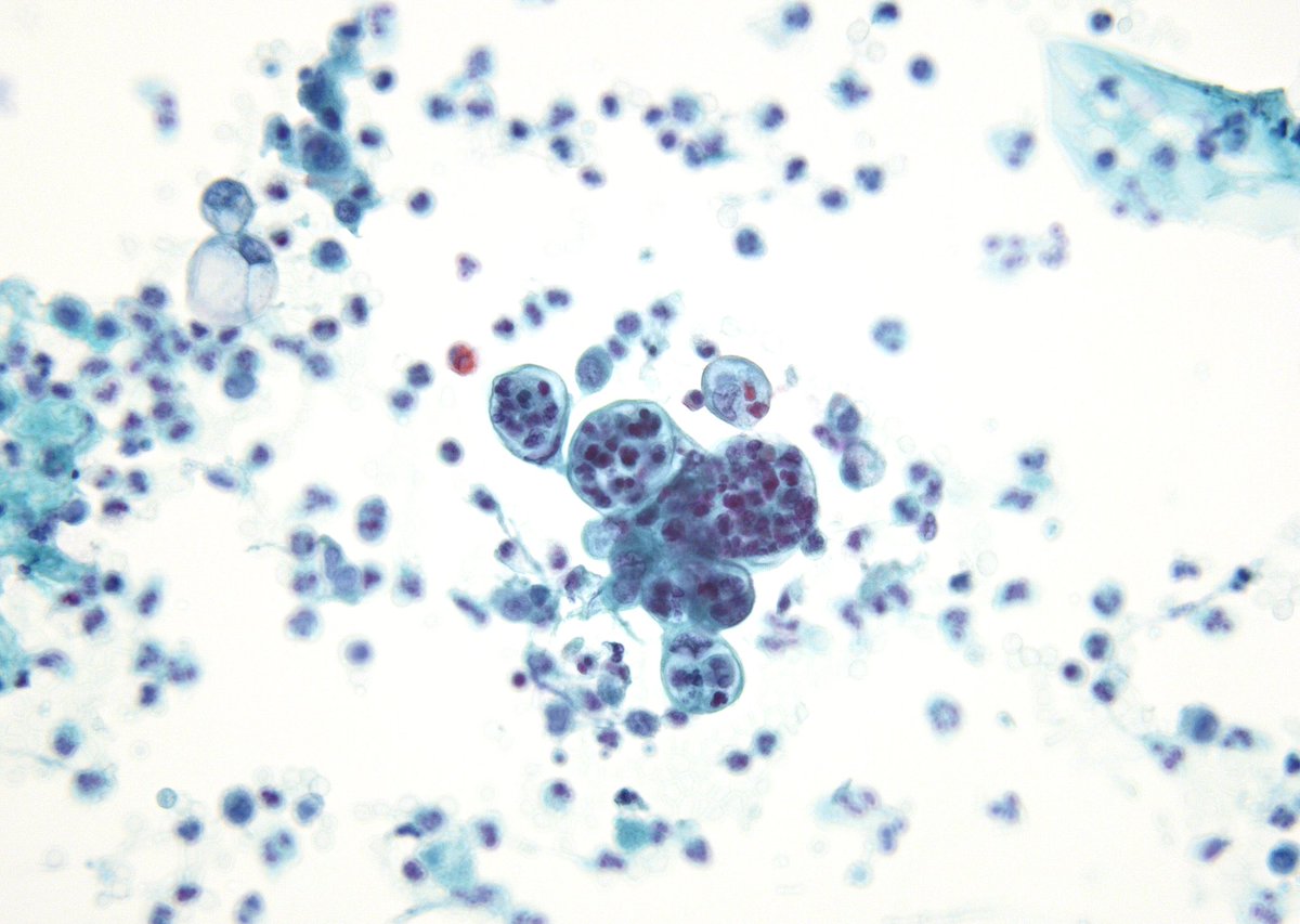
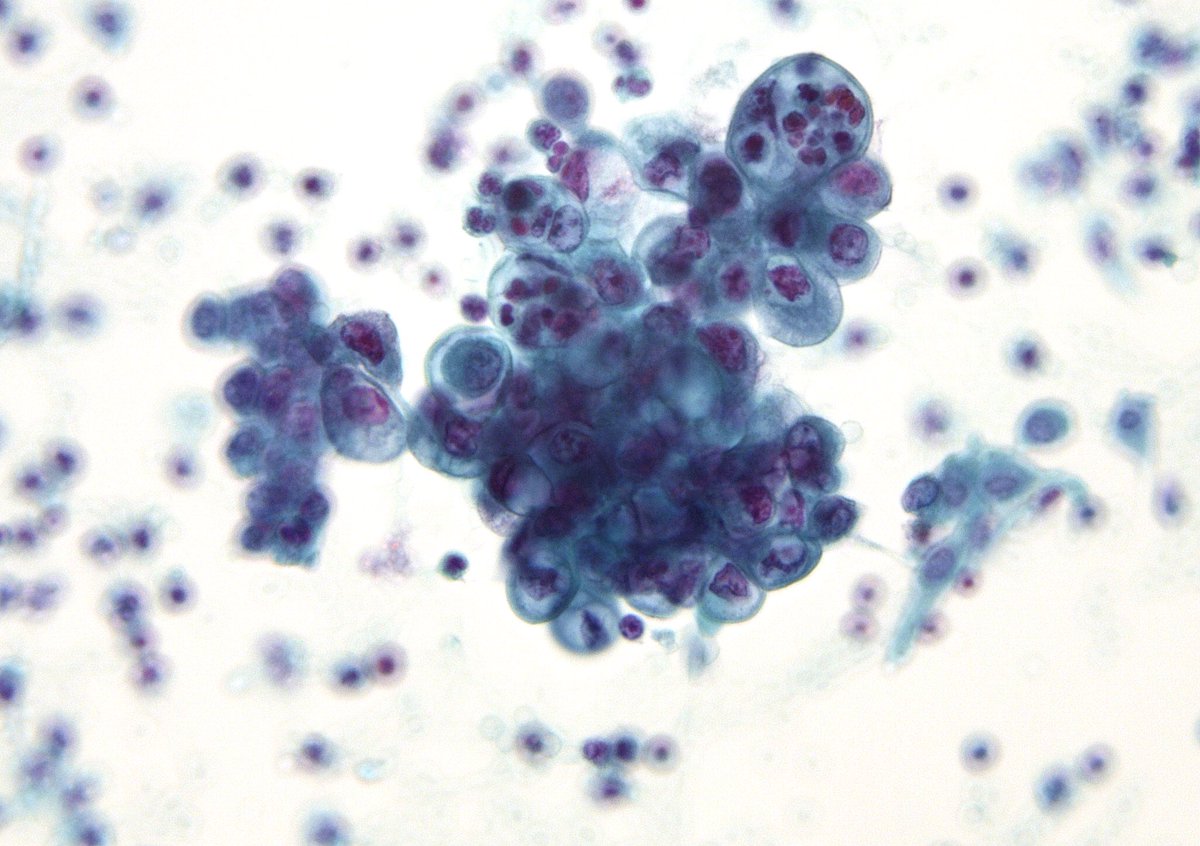
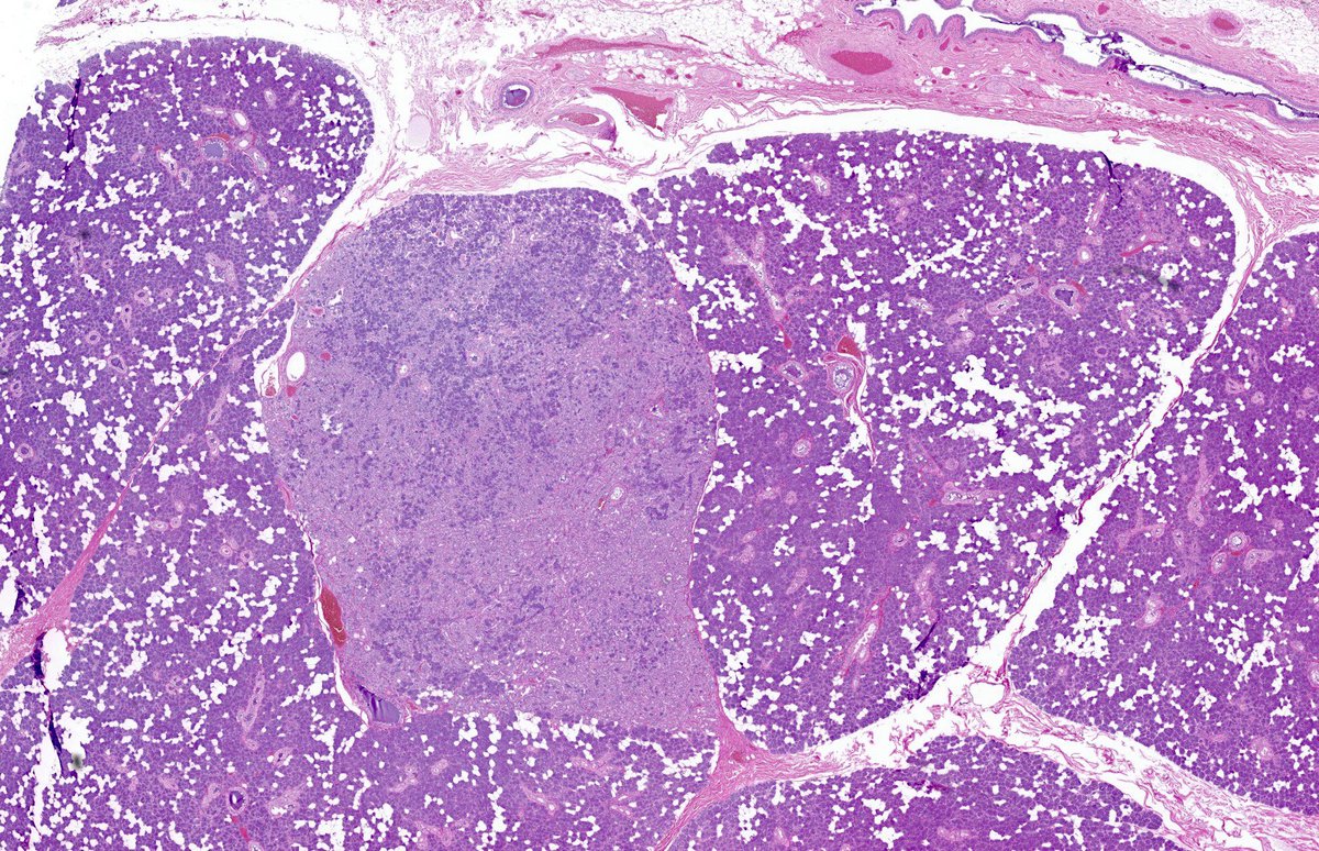
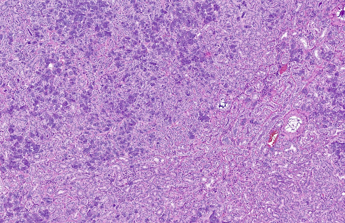
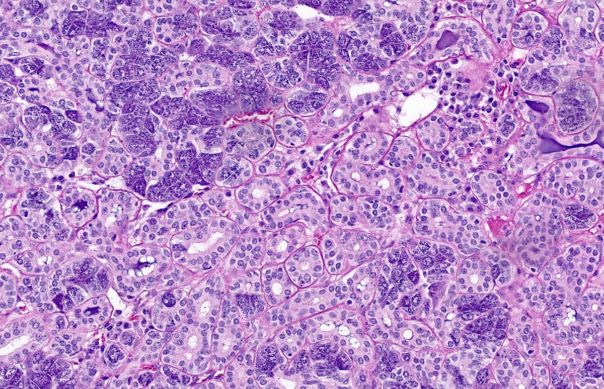
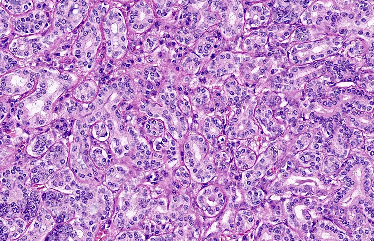
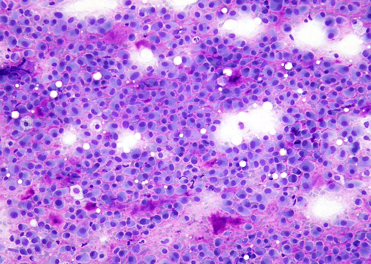
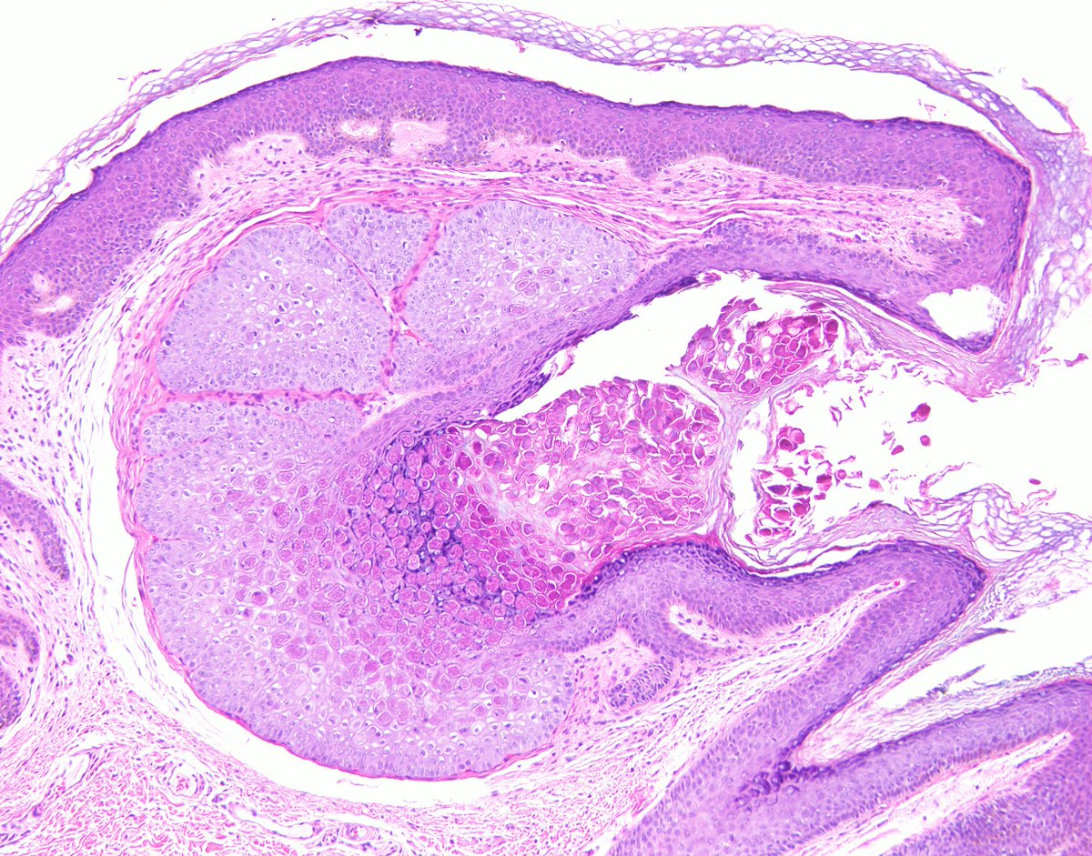
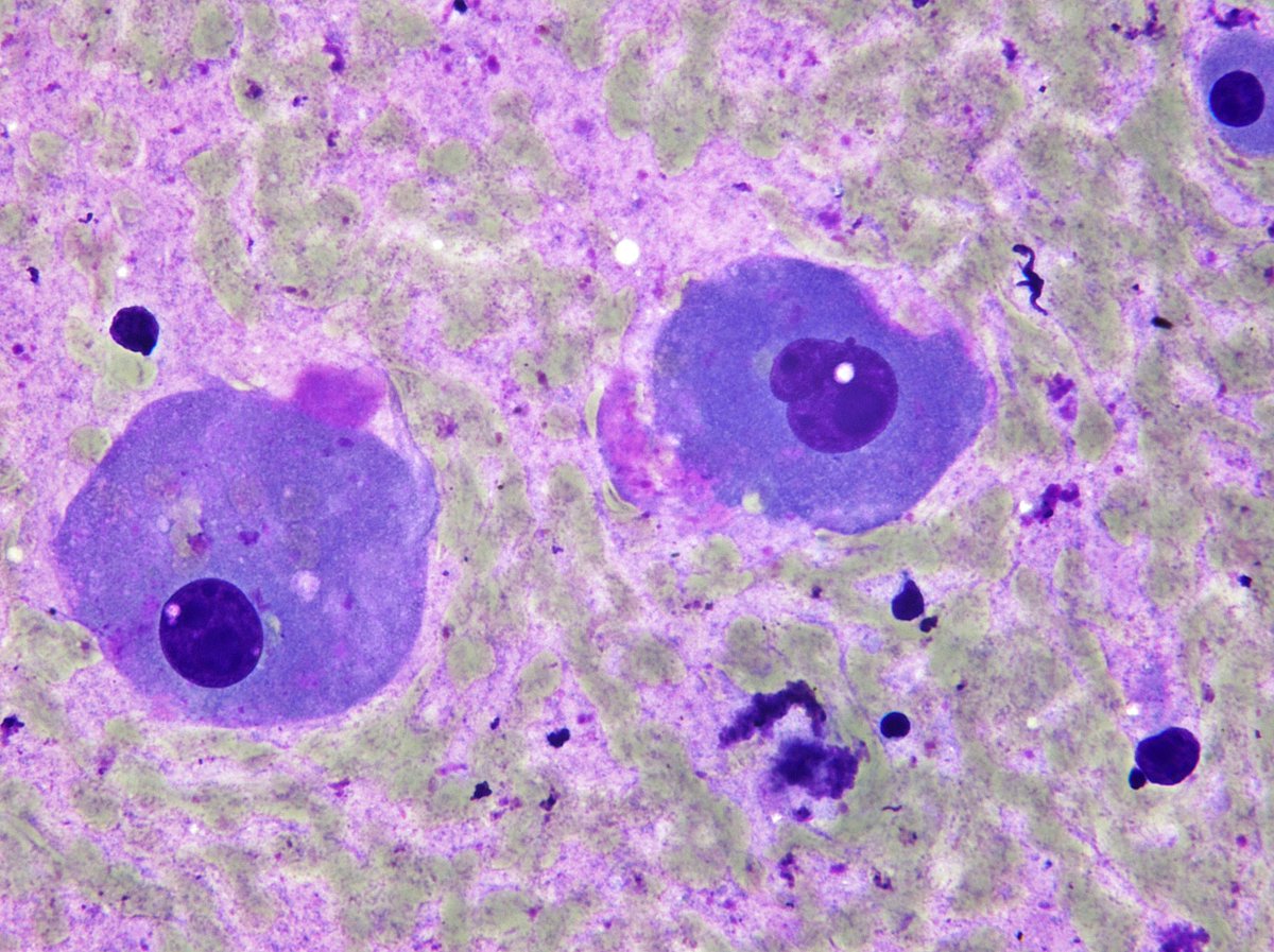
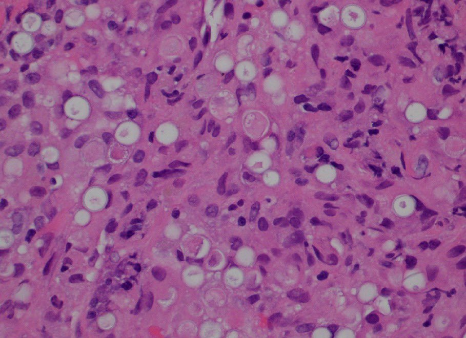
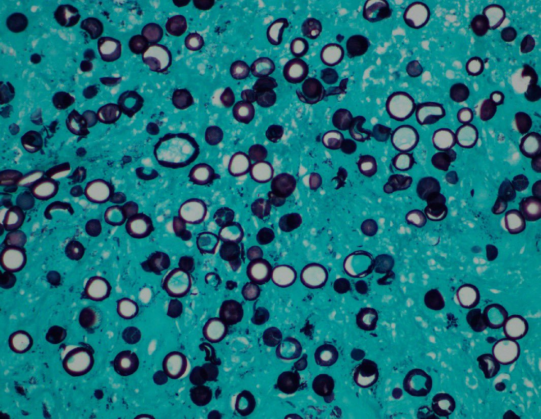
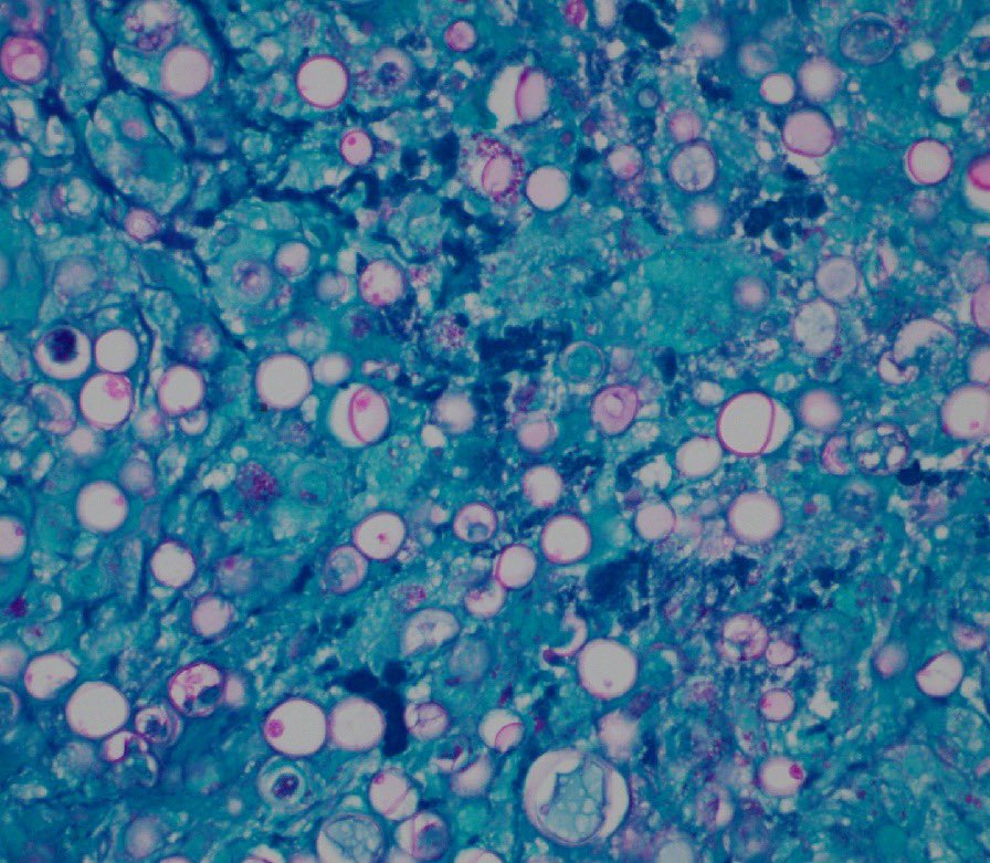
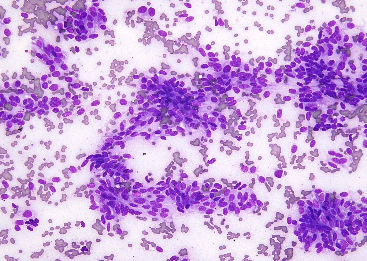
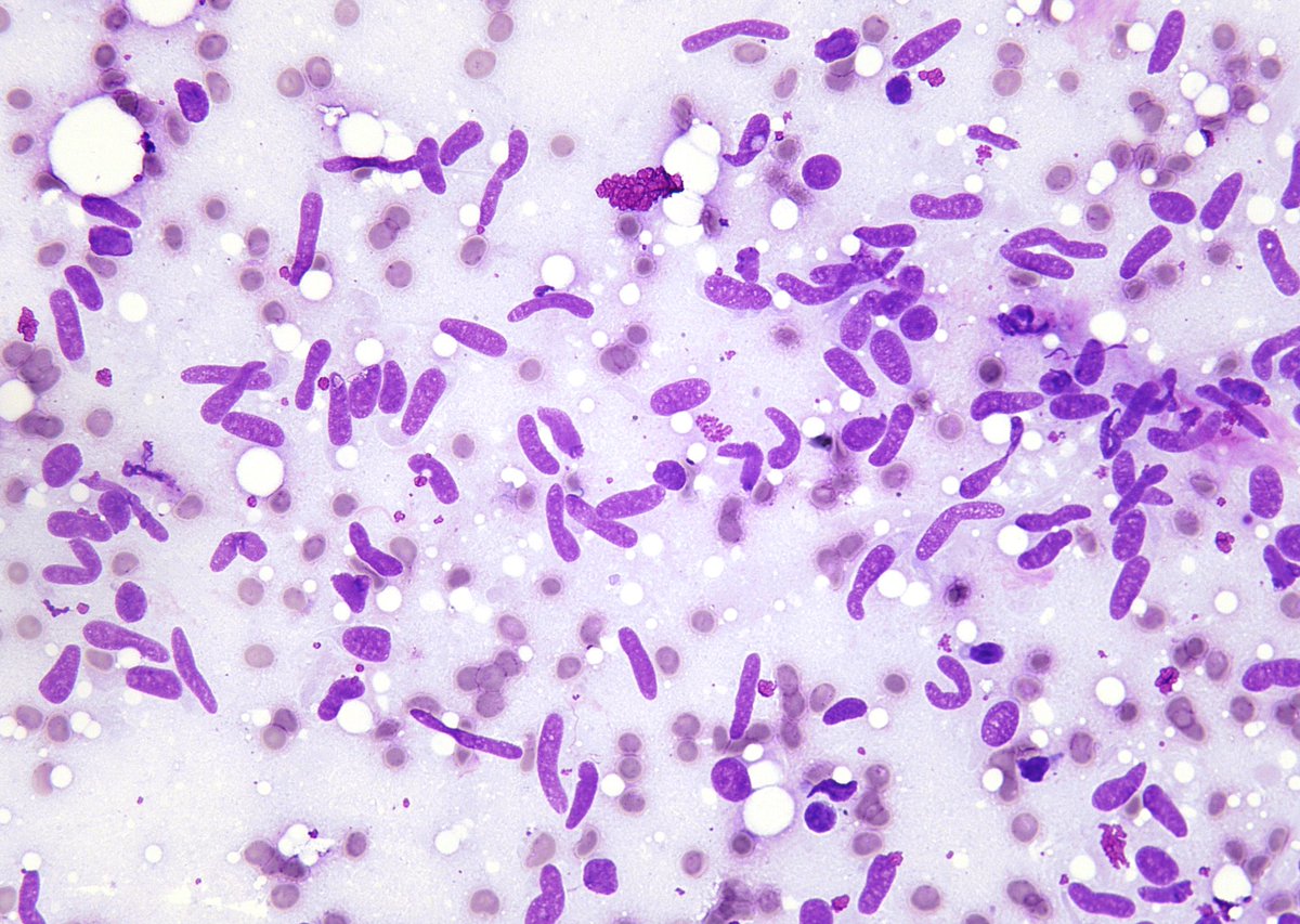
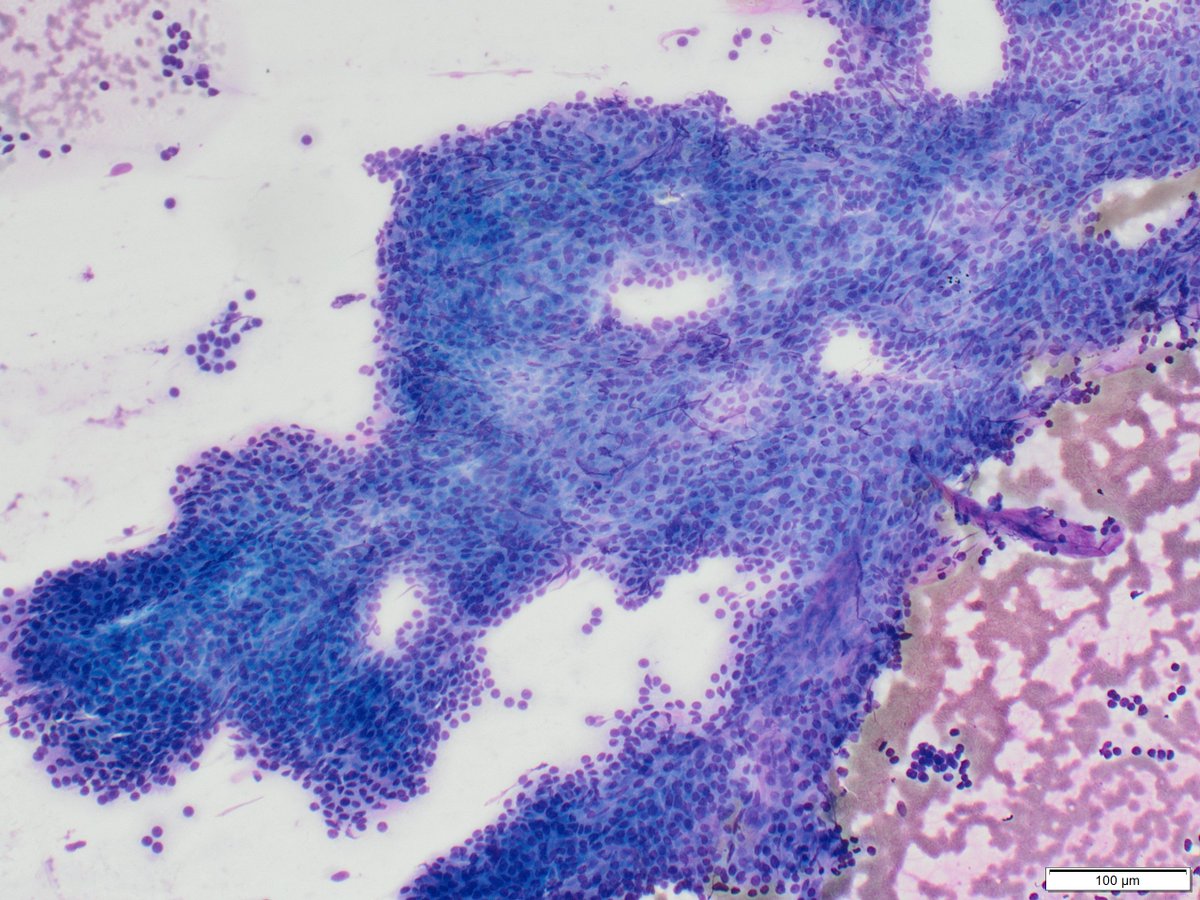
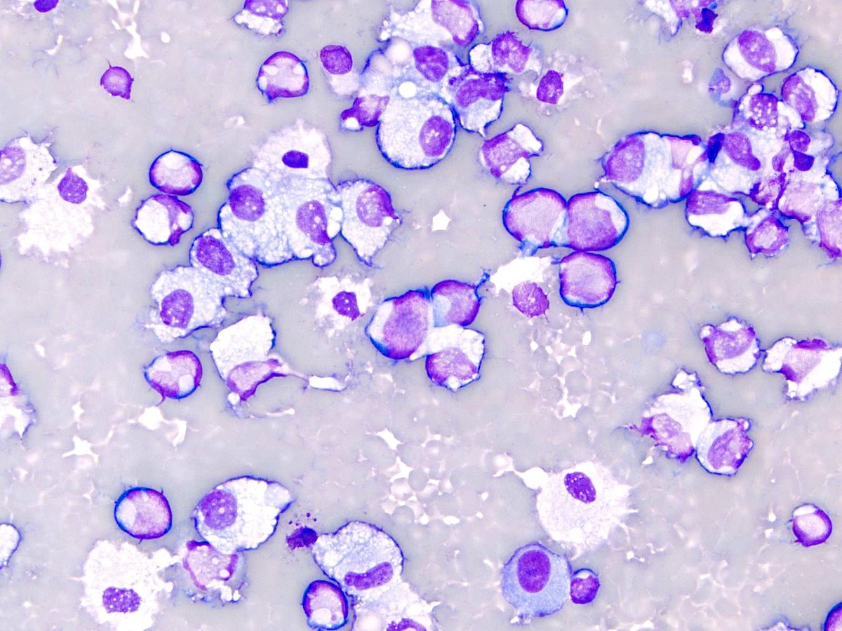
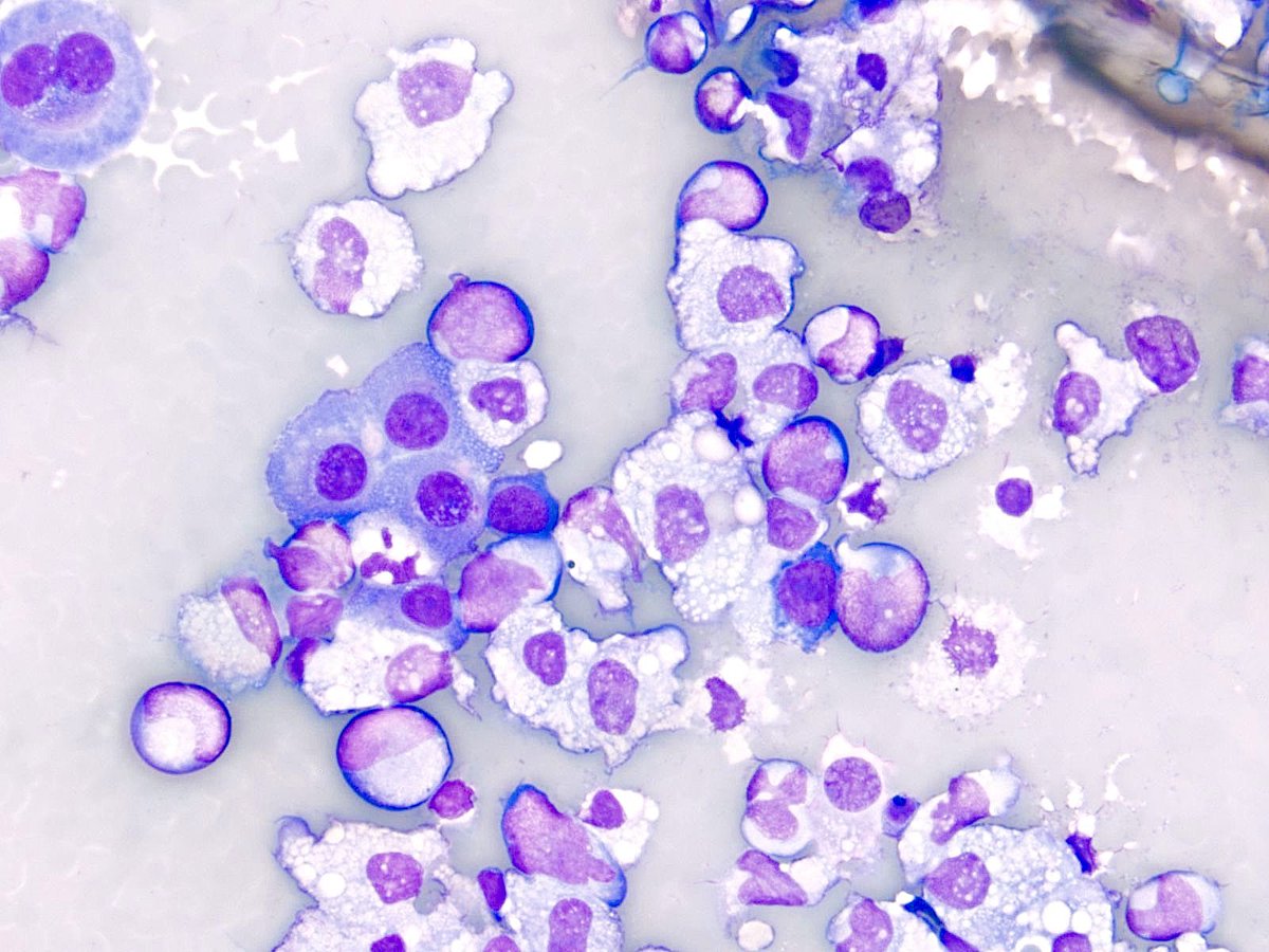
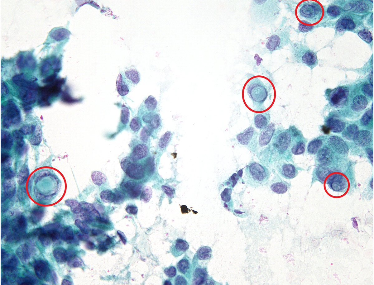
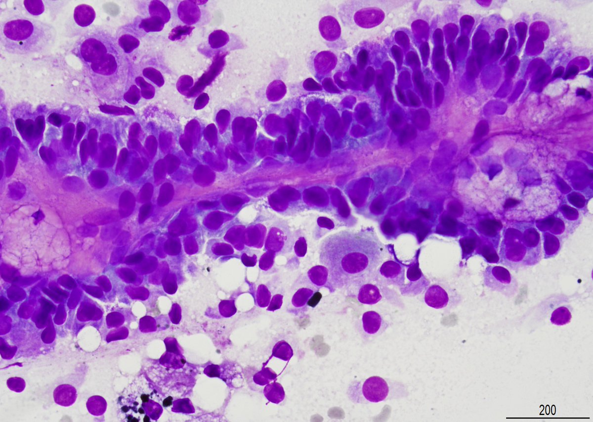
A Matloob, MD

744 posts

A Matloob, MD banner
A Matloob, MD

A Matloob, MD

@A_MatloobMD

Head & Neck Pathology #ENTPath. Cytopathology, #FNApath

Milwaukee, WI Katılım Mayıs 2016
347 Takip Edilen1.1K Takipçiler
A Matloob, MD retweetledi
Syed Z. Ali
Syed Z. Ali@sza_jhcyto·
“Vortex”, incipient tornado (or “onion skinning”), a subtle but helpful diagnostic feature of Papillary Thyroid Carcinoma. (FNA)
Syed Z. Ali tweet media
English
2
40
174
5.5K
A Matloob, MD retweetledi
Syed Z. Ali
Syed Z. Ali@sza_jhcyto·
“The Two Eyeballs”, located by my able fellow “John Sanchez” @JRS0298 in a case of Papillary Thyroid Carcinoma from today’s signout. (FNA)
Syed Z. Ali tweet media
English
3
20
87
2.9K
A Matloob, MD retweetledi
Syed Z. Ali
Syed Z. Ali@sza_jhcyto·
Textbook case, straight from the scope - “Squamous Cell Carcinoma of the Bladder” (Voided urine, ThinPrep)
Syed Z. Ali tweet mediaSyed Z. Ali tweet mediaSyed Z. Ali tweet media
English
1
27
102
3.7K
A Matloob, MD retweetledi
Syed Z. Ali
Syed Z. Ali@sza_jhcyto·
MELANOMA getting “spindlier”. (Lung, FNA)
Syed Z. Ali tweet media
Svenska
3
27
103
6.5K
A Matloob, MD retweetledi
Syed Z. Ali
Syed Z. Ali@sza_jhcyto·
My “CASE of the week” for inhouse cyto signout, illustrating the remarkable phenomenon of how the “bag of polys” are recruited by the “endometrioid endometrial adenocarcinoma”, even at metastatic sites. This case is an abdominal fluid involved by the pt’s known EM CA.
Syed Z. Ali tweet mediaSyed Z. Ali tweet mediaSyed Z. Ali tweet media
English
5
68
212
13.6K
A Matloob, MD retweetledi
Syed Z. Ali
Syed Z. Ali@sza_jhcyto·
TWO, often overlooked cytomorphologic features of Medullary Thyroid Carcinoma on FNA - Intranuclear inclusions and focal nuclear pleomorphism/anisonucleosis.
Syed Z. Ali tweet media
English
5
33
135
6.4K
A Matloob, MD retweetledi
Syed Z. Ali
Syed Z. Ali@sza_jhcyto·
“Squiggly Worms” - GIST. (FNA, Abdominal Wall)
Syed Z. Ali tweet media
English
6
74
269
14K
A Matloob, MD retweetledi
Syed Z. Ali
Syed Z. Ali@sza_jhcyto·
MYOEPITHELIAL cells stained with Giemsa on a parotid FNA are most photogenic. Although the DD here involves a “Myoepithelial Neoplasm”, other areas in the case had classic features of a “Pleomorphic Adenoma”.
Syed Z. Ali tweet media
English
0
34
144
6.4K
A Matloob, MD retweetledi
Sam Khader, MD
Sam Khader, MD@SamKhader·
DQ-stained cytospin preparation of pleural fluid with metastatic small cell ca. On the left you see a group of cells with ⬆️N:C ratio, finely granular chromatic, and molding. On the right you can appreciate the “single file”, “vertebral column”, or “stack of coins" arrangement.
Sam Khader, MD tweet mediaSam Khader, MD tweet media
English
4
22
59
4.4K
A Matloob, MD retweetledi
Tania Labiano
Tania Labiano@tlabiano·
77 yo M w/ biliary stenosis. Bush #cytology during ERCP. No dysplasia, Giardia Lambdia 👻
Tania Labiano tweet media
Indonesia
13
50
118
9.6K
A Matloob, MD retweetledi
Syed Z. Ali
Syed Z. Ali@sza_jhcyto·
Straight from the ultrasound room - Recurrent “ Fibrolamellar Carcinoma” (Fibrolamellar Hepatocellular Carcinoma). Liver FNA in a young boy.
Syed Z. Ali tweet mediaSyed Z. Ali tweet media
English
4
46
111
5.5K
A Matloob, MD retweetledi
Sam Khader, MD
Sam Khader, MD@SamKhader·
Pleural fluid in a patient with history of of #AML. It’s imperative to review the peripheral blood flow cytometry or smear in cases with abundant red blood cells. Some of these cells are suggestive of blasts; however, peripheral blood contamination cannot be excluded. #CytoPath
Sam Khader, MD tweet mediaSam Khader, MD tweet media
English
1
18
41
2.5K
A Matloob, MD retweetledi
Syed Z. Ali
Syed Z. Ali@sza_jhcyto·
Medullary Thyroid Carcinoma - "inclusion-rich". (FNA)
Syed Z. Ali tweet media
HT
4
49
163
7K
A Matloob, MD retweetledi
Sam Khader, MD
Sam Khader, MD@SamKhader·
EBUS-guided #FNA of a mediastinal lymph node demonstrating metastatic adenocarcinoma. #CytoPath
Sam Khader, MD tweet media
English
0
25
62
6.5K