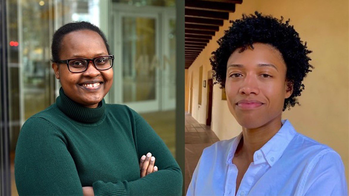
UCLA Bioengineering

553 posts

UCLA Bioengineering banner
UCLA Bioengineering

UCLA Bioengineering

@BioEngUCLA

Los Angeles, CA Katılım Kasım 2017
67 Takip Edilen1.5K Takipçiler
UCLA Bioengineering retweetledi
ABME
ABME@ABMEjournal·
When blood vessels are exposed to chronic inflammation, the cells lining them can quietly change identity in ways that drive heart disease and limit treatment options. Researchers examined whether cell chirality – how endothelial cells orient and twist in space – could serve as a new mechanobiological marker of endothelial-to-mesenchymal transition (EndMT). They built a simple, low-cost platform that uses 3D-printed protein stamps to guide how cells attach and align on a surface, making it easier to measure how their orientation changes. When exposed to inflammatory signals, human aortic endothelial cells not only showed the expected molecular signs of EndMT but also rotated by about 18°, indicating that chirality may shift alongside this transition. This work points toward earlier, more precise detection of harmful vessel remodeling, opening the door to interventions that could prevent long-term cardiovascular damage in patients. 𝗥𝗲𝗮𝗱 𝗺𝗼𝗿𝗲: link.springer.com/article/10.100… 𝗦𝘁𝘂𝗱𝘆: Development of a Stereolithography 3D Printing-Based Micropatterning Method to Study Endothelial-to-Mesenchymal Transition Mechanobiology 𝗔𝘂𝘁𝗵𝗼𝗿𝘀: Karina Bender, Sarah Chesley, Jay Lesny Drake, Megan Ho, Emily Lin, Kathryn Saxton, Ninava Sharma, Christina K. Tripsas, @QianLiMDPhD, and @JeffHsuMD @UCLA @UCLAengineering @BioEngUCLA @uclaibp @dgsomucla @DOM_UCLA @UCLAHealth 𝗦𝘂𝗯𝗺𝗶𝘁 𝘆𝗼𝘂𝗿 𝗺𝗮𝗻𝘂𝘀𝗰𝗿𝗶𝗽𝘁: link.springer.com/journal/10439
ABME tweet media
English
0
2
3
197
UCLA Bioengineering retweetledi
UCLA Samueli Engineering
UCLA Samueli Engineering@UCLAengineering·
New research led by @UCLA Prof. Gerard Wong of @BioEngUCLA shows calcium oxalate kidney stones—the most common type—can contain bacterial biofilms, challenging the idea they’re “noninfectious.” This may explain stone recurrence and infections triggered by stone fragmentation.
Science & Astronomy@sci_astronomy

For decades, doctors believed the most common kidney stones (calcium oxalate) were lifeless lumps formed purely by chemistry—minerals building up in the kidney. ​A groundbreaking study published this month (Jan 2026) by UCLA Health has proven this wrong. ​ Using high-tech fluorescence microscopy, researchers discovered that these stones actually contain live bacteria and fungal-like biofilms "entombed" inside them. The bacteria act as a scaffolding (nidus), allowing the minerals to crystalize and grow layer by layer. ​ This solves a long-standing medical mystery: Why do patients sometimes get severe infections (sepsis) after stone-breaking treatments (lithotripsy), even when their urine was sterile? The answer: breaking the stone releases the bacteria trapped inside. ​This could revolutionize treatment, shifting focus from just diet changes to targeting the hidden microbiome within the kidney. Journal Reference: Wong, Gerard C. L. et al, Intercalated bacterial biofilms are intrinsic internal components of calcium-based kidney stones, Proceedings of the National Academy of Sciences (2026). DOI: 10.1073/pnas.2517066123. ​#MedicalBreakthrough #Microbiology #KidneyHealth #UCLAHealth #NewDiscovery

English
0
2
2
646
UCLA Bioengineering retweetledi
Jun Chen
Jun Chen@JunChenLab·
Our research published in @NatureElectron is selected to be one of the UCLA Top 25 Bruinventions of the 21st Century! Thank you very much for your tremendous support @UCLAengineering @BioEngUCLA @NatureElectron over the years!
Julio Frenk@UCLAchancellor

Bruins continue to invent the future – from life-saving medical advances to world-changing tech. Our latest @UCLA Magazine features the top 25 Bruinventions of the 21st century. #ResearchPowersProgress newsroom.ucla.edu/magazine/top-2…

English
2
1
12
989
UCLA Bioengineering retweetledi
ABME
ABME@ABMEjournal·
When athletes suffer serious injuries to their knee ligaments, the road to recovery can be long, and some never return to the game. Understanding exactly how ligaments fail is essential to improving prevention and treatment, but studying these injuries in living people is difficult. Researchers have developed and validated a new robotic system that simulates natural knee movements with high precision using post-mortem human subject knees. By integrating advanced bone motion data from dynamic biplanar radiography, they were able to replicate motions like walking, running, jumping, and injury events to directly study ligament damage under controlled conditions. This advancement could unlock deeper insights into how and why knee injuries happen and bring us closer to better ways to keep athletes in the game. 𝗥𝗲𝗮𝗱 𝗺𝗼𝗿𝗲: lnkd.in/ePPncH8q 𝗦𝘁𝘂𝗱𝘆: A Robotic Clamped-Kinematic System to Study Knee Ligament Injury 𝗔𝘂𝘁𝗵𝗼𝗿𝘀: Ophelie Herve, Will Flanagan, Jake Kanetis, Bailey Mooney, Thomas Kremen, David R. McAllister, and Tyler Clites @UCLA @UCLAHealth @dgsomucla @UCLAMechAeroEng @BioEngUCLA @ThomasKremenMD @TylerClites 𝗦𝘂𝗯𝗺𝗶𝘁 𝘆𝗼𝘂𝗿 𝗺𝗮𝗻𝘂𝘀𝗰𝗿𝗶𝗽𝘁: lnkd.in/g6_6wwEu
ABME tweet media
English
0
2
2
184
UCLA Bioengineering retweetledi
UCLA Samueli Engineering
UCLA Samueli Engineering@UCLAengineering·
Join us at the first ever @UCLA Engineering & Medicine @dgsomucla Joint Research Symposium on Tuesday, April 22. This daylong event will bring together researchers from both schools to network, collaborate and hear from distinguished guest speakers. samueli.ucla.edu/upcoming-event…
English
0
1
2
318