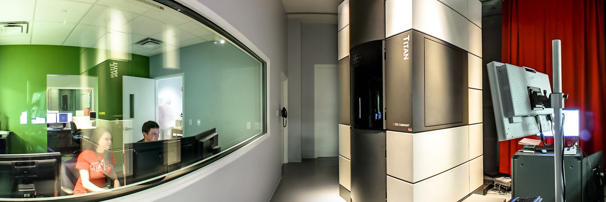
Center for Electron Microscopy and Analysis

827 posts

Center for Electron Microscopy and Analysis banner
Center for Electron Microscopy and Analysis

Center for Electron Microscopy and Analysis

@CEMAS_OSU

CEMAS is the center at The Ohio State University that breaks through the current limitations in medicine, environmental science, energy materials and beyond.

Columbus, OH Katılım Nisan 2013
130 Takip Edilen703 Takipçiler
Center for Electron Microscopy and Analysis
⏰ Early bird pricing ends today! Learn SEM fundamentals and advanced imaging techniques in CEMAS’ On the Scope course, featuring expert‑led online content and hands‑on training 🔬 Learn more & register: go.osu.edu/ots
English
0
0
1
84
Center for Electron Microscopy and Analysis
Happy #MicroscopyMonday 🔬 Here is a HRSTEM HAADF micrograph captured by Bryan Crossman, showing a low-angle grain boundary (LAGB) in a BCC Ta₅₅Re₄₅ (at.%) refractory alloy. This boundary is primarily a tilt boundary, formed by a ~12.6° rotation about the [001] zone axis.
Center for Electron Microscopy and Analysis tweet media
English
0
0
2
68
Center for Electron Microscopy and Analysis retweetledi
EDAX
EDAX@EDAX_LLC·
Our friends at @CEMAS_OSU are hosting the annual On The Scope – A Masterclass in Practical Scanning Electron Microscopy - April 1, 2026. If you would like more information or to register for the course, please visit: ow.ly/YSfF50U9wNy #Microscopy #Microanalysis
English
0
1
2
62
Center for Electron Microscopy and Analysis
These experiments were performed by graduate student Rogine Gomez in the Leonard research group in MSE with CEMAS Senior Research Associate Daniel Veghte. Recrystallization and growth are observed from the heavily deformed areas (initially non-indexable areas).
English
1
0
0
41
Center for Electron Microscopy and Analysis
What appear to be the “ears” are actually regions outlined by colloidal silica left behind during polishing and final cleaning. The “eye” is a void, and the rest of the “mouse” takes shape along the alloy’s grain boundaries.
English
0
0
0
27
Center for Electron Microscopy and Analysis
Check out the research SEM image that turned out unexpectedly artistic when a mouse appeared, taken by graduate student Nicole Hudak! Captured on Apreo 1/2 using standard conditions, it’s a BSE image of a titanium alloy. It takes a bit of imagination to see the tiny creature 🔬
Center for Electron Microscopy and Analysis tweet mediaCenter for Electron Microscopy and Analysis tweet media
English
1
0
0
79
Center for Electron Microscopy and Analysis retweetledi
Nature Biotechnology
Nature Biotechnology@NatureBiotech·
Antimicrobial peptide delivery to lung as peptibody mRNA in anti-inflammatory lipids treats multidrug-resistant bacterial pneumonia go.nature.com/3LZS6HD
Nature Biotechnology tweet media
English
1
29
119
29.9K
Center for Electron Microscopy and Analysis
Happy #MicroscopyMonday 🔬 This SEM backscattered electron micrograph, taken by PhD student Bryan Crossman, shows a three-dimensional, Christmas tree–like dendrite in a Ta₆₀Re₄₀ (at.%) refractory alloy, captured on the Apreo 1 at CEMAS.
Center for Electron Microscopy and Analysis tweet media
English
0
0
3
84
Center for Electron Microscopy and Analysis retweetledi
EDAX
EDAX@EDAX_LLC·
Our friends at @CEMAS_OSU are hosting the annual On The Scope – A Masterclass in Practical Scanning Electron Microscopy - April 1, 2026. If you would like more information or to register for the course, please visit: ow.ly/YSfF50U9wNy #Microscopy #Microanalysis
English
0
1
2
69
Center for Electron Microscopy and Analysis retweetledi
Ohio State Materials Science and Engineering
More spintronics applications are available thanks to current and former researchers of @CEMAS_OSU - Binbin Wang, Núria Bagués & David McComb, who helped identify how to control skyrmion states in metallic multilayers with broken inversion symmetry. doi.org/10.1002/adma.2…
English
0
4
4
303