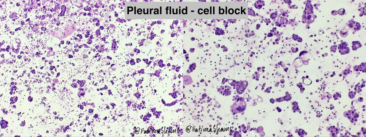
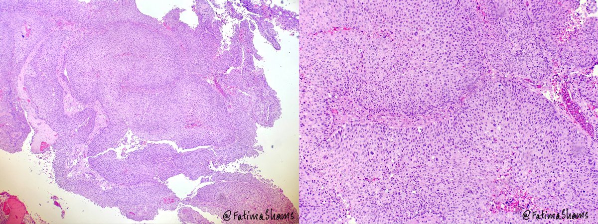
Fatima Shamsuddin

480 posts

Fatima Shamsuddin banner
Fatima Shamsuddin

Fatima Shamsuddin

@FatimaShamsPath

MD, FRCPath, FIAC

Kuwait Katılım Haziran 2023
114 Takip Edilen1.1K Takipçiler
Fatima Shamsuddin
Fatima Shamsuddin@FatimaShamsPath·
🔎 Histo-cyto correlation! 👉 74-year-old female with hematuria and bladder mass ⚓️ Urine cytology showed features suspicious for High-Grade Urothelial Carcinoma (HGUC) with: 🔬 Hyperchromatic crowded cell clusters 🔬 Single scattered atypical cells 🔬 Marked pleomorphism, high N:C ratio 🔬 Irregular nuclear membranes & coarse chromatin 💎 Biopsy confirmed High-Grade Papillary Urothelial Carcinoma showing fused/complex papillae, loss of maturation, diffuse cytologic atypia, and numerous mitoses. A nice histocytologic correlation case highlighting the importance of urine cytology in detecting HGUC. #PathTwitter #Cytopathology #Uropath #Surgpath #UrineCytology #HGUC #UrothelialCarcinoma #Histopathology #MedEd #Pathology
Fatima Shamsuddin tweet mediaFatima Shamsuddin tweet mediaFatima Shamsuddin tweet mediaFatima Shamsuddin tweet media
English
0
11
36
1.3K
Fatima Shamsuddin
Fatima Shamsuddin@FatimaShamsPath·
Indeed an inspiring journey and vision. It was truly wonderful attending the IAC meeting in Sweden this January and having the opportunity to meet you and so many distinguished faculty members from across the world. The exchange of knowledge, ideas, and experiences were invaluable. What stood out most was how approachable and encouraging all the office bearers and senior experts were — making all of us feel like equal members of one global cytology community. Looking forward to seeing IACytology and the cytology community grow even further.
English
0
0
4
136
Syed Z. Ali
Syed Z. Ali@sza_jhcyto·
“Dreams do come true.” During the recently concluded Asian Federation of Cytology Societies (AFCS) inaugural congress in Hong Kong, many people asked me how all of this happened - how we achieved such success so quickly. My answer was simple: it was neither quick nor easy. It took many years of vision, persistence, and collective effort. Over the last 25 years, I have made more than 75 trips to East Asia. During those visits, I repeatedly recognized the need for a consortium where national cytology societies across Asia could pool their talent, resources, and expertise, while also supporting smaller and resource-limited countries. The ultimate vision was to create a unified annual congress in which every Asian national society could participate, alongside distinguished faculty from other regions of the world, including the United States and Europe. About 10 years ago, I began discussing this idea with many Asian friends and colleagues. While everyone supported the concept enthusiastically, there was understandable skepticism about how such an ambitious vision could ever become reality. Years passed before I finally received a lucky break. About three years ago, I presented a rough plan to my close friends, Drs. Gary Tse and Bob Osamura, both of whom became instrumental in supporting the founding of AFCS. The name “AFCS” itself was finalized after I discussed the concept with Dr. Danijela Vrdoljak-Mozetic, secretary-general of the EFCS, as the mission statements of the two federations were remarkably aligned. It then took nearly a year of countless WhatsApp calls by me with many individuals across Asia, seeking support to enlist their national organizations as companion societies under the AFCS umbrella. Once our list grew to 20 countries, I felt the time was right to formally announce the launch of AFCS at the International Congress of Cytology in Florence, Italy in May 2025. That became a pivotal moment in our journey. Soon afterward, the executive council was established. Upon my strong recommendation, Bob Osamura was approved as the first President of AFCS, and Gary Tse as the Secretary-General, with Hong Kong selected as the federation headquarter and venue for the historic inaugural congress. Dr. Jamal Musayev from Baku designed the eye-catching insignia. The orange color symbolizes the warmth and optimism of Asia, with rays of sunlight reflecting hope, unity, and a bright future. Those who attended the recent congress witnessed firsthand the extraordinary work accomplished by Dr. Gary Tse and his remarkable team. Congratulations to them - and to everyone who contributed their time, effort, and expertise to this historic success. As the congress concluded, I finally allowed myself a quiet sigh of relief. A dream that once seemed distant had finally come true. As Ryūnosuke Satoro, a Japanese poet and philosopher so beautifully said: “Individually, we are one drop. Together, we are an ocean.” Today, with more than 20 national societies united together, we have created that mighty ocean - visible, collaborative, impactful, and full of promise for the future of cytopathology across Asia and beyond. @cytopathology @IACytology @CytologyEFCS @JM_CytoBox @MozeticV
English
14
20
86
4.6K
Fatima Shamsuddin
Fatima Shamsuddin@FatimaShamsPath·
Answer provided here
Fatima Shamsuddin@FatimaShamsPath

💎 Solitary Fibrous Tumor (SFT) involving the submandibular gland.   🔍  58-year-old male with a 5.5 cm left submandibular gland mass.   ⚓️ Grossly, the lesion was a well-circumscribed, fleshy, firm white-tan mass with a solid whorled cut surface.   🔬 Microscopy showed a bland spindle cell neoplasm arranged in a “patternless pattern” with alternating hypo- and hypercellular areas, dense collagenized stroma, and prominent branching/staghorn-like vessels. Tumor cells showed minimal atypia and low mitotic activity.   🧪 IHC profile: • STAT6 nuclear positive •.CD34: diffuse positive • PanCK & S100: negative • SMA / Desmin: highlighted only vessel walls, negative in tumor cells • Beta-catenin: cytoplasmic granular staining without nuclear positivity   💡 Key points: • SFT is a rare mesenchymal neoplasm in salivary glands, most commonly affecting the submandibular gland and parotid. • The characteristic molecular alteration is the NAB2::STAT6 fusion, resulting from inversion at chromosome 12q13 and leading to nuclear STAT6 expression which is highly sensitive and specific for SFT. • Most SFTs behave indolently, but risk stratification depends on factors such as size, mitotic activity, necrosis, and patient age. Rare cases may recur or metastasize.   📚 Important morphologic clues favoring SFT: ✔ Patternless architecture ✔ Ropey collagen ✔ Staghorn vasculature ✔ Bland spindle cells ✔ Diffuse CD34 positivity & nuclear STAT6 positivity Case courtesy @annsmiley78   #PathTwitter #SurgPath #HeadNeckPath #SalivaryGlandPathology #SoftTissuePathology #SolitaryFibrousTumor #SubmandibularGland #Histopathology #Pathology #MedEd

English
0
0
0
63
Fatima Shamsuddin
Fatima Shamsuddin@FatimaShamsPath·
What is your diagnosis and which IHC would support it?
English
2
0
0
189
Fatima Shamsuddin
Fatima Shamsuddin@FatimaShamsPath·
💎 Solitary Fibrous Tumor (SFT) involving the submandibular gland.   🔍  58-year-old male with a 5.5 cm left submandibular gland mass.   ⚓️ Grossly, the lesion was a well-circumscribed, fleshy, firm white-tan mass with a solid whorled cut surface.   🔬 Microscopy showed a bland spindle cell neoplasm arranged in a “patternless pattern” with alternating hypo- and hypercellular areas, dense collagenized stroma, and prominent branching/staghorn-like vessels. Tumor cells showed minimal atypia and low mitotic activity.   🧪 IHC profile: • STAT6 nuclear positive •.CD34: diffuse positive • PanCK & S100: negative • SMA / Desmin: highlighted only vessel walls, negative in tumor cells • Beta-catenin: cytoplasmic granular staining without nuclear positivity   💡 Key points: • SFT is a rare mesenchymal neoplasm in salivary glands, most commonly affecting the submandibular gland and parotid. • The characteristic molecular alteration is the NAB2::STAT6 fusion, resulting from inversion at chromosome 12q13 and leading to nuclear STAT6 expression which is highly sensitive and specific for SFT. • Most SFTs behave indolently, but risk stratification depends on factors such as size, mitotic activity, necrosis, and patient age. Rare cases may recur or metastasize.   📚 Important morphologic clues favoring SFT: ✔ Patternless architecture ✔ Ropey collagen ✔ Staghorn vasculature ✔ Bland spindle cells ✔ Diffuse CD34 positivity & nuclear STAT6 positivity Case courtesy @annsmiley78   #PathTwitter #SurgPath #HeadNeckPath #SalivaryGlandPathology #SoftTissuePathology #SolitaryFibrousTumor #SubmandibularGland #Histopathology #Pathology #MedEd
Fatima Shamsuddin tweet mediaFatima Shamsuddin tweet mediaFatima Shamsuddin tweet mediaFatima Shamsuddin tweet media
English
1
5
23
799
Fatima Shamsuddin
Fatima Shamsuddin@FatimaShamsPath·
Pleural fluid CYTOLOGY. 📌 Clinical: Male patient with known urothelial carcinoma of bladder with metastases to lung, chest wall, and bone, presenting with pleural effusion. 🔬 Cytology (Pap & DQ): •Moderate cellularity •Clusters and sheets of atypical epithelial cells in a hemorrhagic background •Cells show high N:C ratio, nuclear irregularity, coarse chromatin, and prominent nucleoli •No definite gland formation 🧫 Cell block: •Cohesive clusters of malignant epithelial cells with similar morphology 🧪 IHC: •BerEP4+, CK7+ •GATA3+, p63+ •CK20: focal weak+ •Calretinin– (excludes mesothelial origin)   🔹 Diagnosis: Malignant effusion – metastatic carcinoma consistent with urothelial origin   🔹 Teaching points 💡 ✔️ GATA3 + p63 co-expression → strong clue to urothelial carcinoma ✔️ In pleural effusion, always differentiate from lung adenocarcinoma & squamous carcinoma ✔️ BerEP4+ / Calretinin –ve → epithelial, not mesothelial ✔️ CK20 may be variable/focal in urothelial carcinoma   #PathTwitter #Cytopathology #PleuralFluid #EffusionCytology #UrothelialCarcinoma #MetastaticCarcinoma #DiagnosticPathology
Fatima Shamsuddin tweet mediaFatima Shamsuddin tweet mediaFatima Shamsuddin tweet mediaFatima Shamsuddin tweet media
English
1
6
20
831
Fatima Shamsuddin
Fatima Shamsuddin@FatimaShamsPath·
❄️ Frozen to Final: Diagnostic Correlation Case: 75M with massive compressive thyroid mass (9 cm) with tracheal invasion 🧊 Frozen: Malignant tumor (subtype deferred) 💎 Final diagnosis: Medullary Thyroid Carcinoma 🔬 Key features: • Nests, trabeculae, cords • Polygonal cells, granular cytoplasm • Salt & pepper chromatin • Lymphovascular invasion 🧪 IHC: Calcitonin+, CEA+, Synapto+, Chromogranin+, INSM1+ CK7+, CK20-, TTF1 (diffuse) Thyroglobulin–, PAX8–   🧬 Molecular: • Activating RET mutations in majority (germline in hereditary cases – MEN2A/MEN2B; somatic in sporadic) • RAS mutations (HRAS/KRAS) in RET–negative tumors • Routine: recommend RET mutation testing ± germline screening (important for family counseling)   📊 Prognosis: • Variable; worse with: Older age, large tumor, extrathyroidal extension (like tracheal invasion here) and distant metastasis • 10-year survival ~70–80% overall, but ↓ significantly in advanced disease • Calcitonin doubling time = key prognostic marker   💡 Pearl: When morphology & clinical picture mismatch on frozen → call “malignant” & defer typing. IHC + serum calcitonin clinches MTC. #PathTwitter #Surgpath #EndocrinePath #ThyroidPath #MedullaryThyroidCarcinoma #FrozenSection #Histopathology #Pathology Case courtesy @annsmiley78
Fatima Shamsuddin tweet mediaFatima Shamsuddin tweet mediaFatima Shamsuddin tweet mediaFatima Shamsuddin tweet media
English
3
10
35
1.7K