
Imaris 3D/4D Imaging
1.2K posts

Imaris 3D/4D Imaging
@ImarisSoftware
We enable the microscopy community with top performance Image Visualization, Analysis and Interpretation software.
Zurich, Switzerland Katılım Şubat 2013
628 Takip Edilen2.5K Takipçiler

𝟖 𝐑𝐞𝐚𝐬𝐨𝐧𝐬 𝐖𝐡𝐲 𝐈𝐦𝐚𝐫𝐢𝐬 𝐒𝐡𝐨𝐮𝐥𝐝 𝐛𝐞 𝐲𝐨𝐮𝐫 𝐈𝐦𝐚𝐠𝐞 𝐀𝐧𝐚𝐥𝐲𝐬𝐢𝐬 𝐒𝐨𝐟𝐭𝐰𝐚𝐫𝐞
Get your Imaris Free Trial today ⤵️
bit.ly/35I6gU2
#microscopy #microscopes #imageanalysis
English
Imaris 3D/4D Imaging retweetledi

Issue 13 is complete!
On the cover: zebrafish cardiac ECM labelled by Tg(ubb:ssNcan-GFP) & surface rendered in 3D using Imaris. Colors mark distinct cardiac regions: ventricle (purple), atrioventricular canal (yellow) & atrium (green). See Gentile et al.
journals.biologists.com/dev/article/15…

English
Imaris 3D/4D Imaging retweetledi

📢NEW IMARIS 10.2📷from @ImarisSoftware
The newest Imaris 10.2 #imageanalysissoftware brings faster data rendering for all users. All Imaris 10.2 functionality, including AI segmentation is now faster on Apple M3 processors.
#Imaris #Microscopy #ImageAnalysis #fluorescence
English
Imaris 3D/4D Imaging retweetledi

📣 NEW IMARIS 10.2 📣
The newest Imaris 3D image analysis software brings faster data rendering for all users. In addition, all Imaris 10.2 functionality, including AI segmentation is now faster on Apple M3 processors.
Take a 10 day Free Trial here ⤵️
imaris.oxinst.com/newrelease
English

📣 NEW IMARIS 10.2 📣
The newest Imaris 3D image analysis software brings faster data rendering for all users. In addition, all Imaris 10.2 functionality, including AI segmentation is now faster on Apple M3 processors.
Take a 10 day Free Trial here ⤵️
imaris.oxinst.com/newrelease
English
Imaris 3D/4D Imaging retweetledi

I'm giving a webinar next month! Want to learn how to do 3D high-resolution brain microscopy analysis?
Register using the link below; it is free! 🤓🔬🧠 selectscience.net/webinar/combin…
@ImarisSoftware

English

💥 𝐍𝐄𝐖 𝐈𝐌𝐀𝐑𝐈𝐒 𝟏𝟎.𝟐 💥
𝐃𝐨𝐮𝐛𝐥𝐞 𝟑𝐃 𝐑𝐞𝐧𝐝𝐞𝐫𝐢𝐧𝐠 𝐒𝐩𝐞𝐞𝐝 𝐟𝐨𝐫 𝐀𝐥𝐥 𝐔𝐬𝐞𝐫𝐬 𝐚𝐧𝐝 𝐌𝐚𝐜 𝐌𝟑 𝐕𝐞𝐫𝐬𝐢𝐨𝐧
Get Your Free Trial Now ⤵️
imaris.oxinst.com/newrelease
#Imaris #Microscopy #ImageAnalysis

English
Imaris 3D/4D Imaging retweetledi

🎞️ 𝟔 𝐑𝐞𝐚𝐬𝐨𝐧𝐬 𝐖𝐡𝐲 𝐁𝐂𝟒𝟑 𝐒𝐡𝐨𝐮𝐥𝐝 𝐁𝐞 𝐘𝐨𝐮𝐫 𝐍𝐞𝐱𝐭 𝐁𝐞𝐧𝐜𝐡𝐭𝐨𝐩 𝐌𝐢𝐜𝐫𝐨𝐬𝐜𝐨𝐩𝐞
💥 2 new models now available! Learn more about affordable and upgradable microscopy today ⤵️
andor.oxinst.com/products/bc43-…
#microscope #confocal #fluorescence #BC43
English

📷 𝐍𝐞𝐰 𝐁𝐞𝐧𝐜𝐡𝐭𝐨𝐩 𝐅𝐥𝐮𝐨𝐫𝐞𝐬𝐜𝐞𝐧𝐜𝐞 𝐌𝐢𝐜𝐫𝐨𝐬𝐜𝐨𝐩𝐞📷
𝑬𝒂𝒔𝒚. 𝑪𝒐𝒎𝒑𝒂𝒄𝒕. 𝑨𝒇𝒇𝒐𝒓𝒅𝒂𝒃𝒍𝒆. 𝑼𝒑𝒈𝒓𝒂𝒅𝒆𝒂𝒃𝒍𝒆.
Supercharge your Fluorescence Imaging today!
andor.oxinst.com/.../bc43-wf-be…...
#Microscopy #Fluorescence #BC43

English

𝐀 𝐁𝐞𝐧𝐜𝐡𝐭𝐨𝐩 𝐌𝐢𝐜𝐫𝐨𝐬𝐜𝐨𝐩𝐞 𝐭𝐡𝐚𝐭 𝐆𝐫𝐨𝐰𝐬 𝐰𝐢𝐭𝐡 𝐘𝐨𝐮𝐫 𝐍𝐞𝐞𝐝𝐬...
𝐹𝑎𝑠𝑡. 𝐸𝑎𝑠𝑦. 𝐶𝑜𝑚𝑝𝑎𝑐𝑡 𝐴𝑓𝑓𝑜𝑟𝑑𝑎𝑏𝑙𝑒. 𝑈𝑝𝑔𝑟𝑎𝑑𝑎𝑏𝑙𝑒.
andor.oxinst.com/products/bc43-…
#microscopy #fluorescence #confocal #superresolution #Microscope
English

💥𝟐 𝐍𝐞𝐰 𝐁𝐞𝐧𝐜𝐡𝐭𝐨𝐩 𝐌𝐢𝐜𝐫𝐨𝐬𝐜𝐨𝐩𝐞𝐬 𝐘𝐨𝐮 𝐂𝐚𝐧 𝐓𝐫𝐮𝐬𝐭!💥
𝑀𝑜𝑟𝑒 𝑀𝑜𝑑𝑎𝑙𝑖𝑡𝑖𝑒𝑠. 𝑀𝑜𝑟𝑒 𝑃𝑒𝑟𝑓𝑜𝑟𝑚𝑎𝑛𝑐𝑒. 𝑀𝑜𝑟𝑒 𝐶𝑒𝑟𝑡𝑎𝑖𝑛𝑡𝑦.
Discover today!
➡️ andor.oxinst.com/products/bc43-…
#Microscopy #Fluorescence #Confocal #Superresolution #BC43
GIF
English
Imaris 3D/4D Imaging retweetledi

💥𝟐 𝐍𝐞𝐰 𝐁𝐞𝐧𝐜𝐡𝐭𝐨𝐩 𝐌𝐢𝐜𝐫𝐨𝐬𝐜𝐨𝐩𝐞𝐬 𝐘𝐨𝐮 𝐂𝐚𝐧 𝐓𝐫𝐮𝐬𝐭!💥
𝑀𝑜𝑟𝑒 𝑀𝑜𝑑𝑎𝑙𝑖𝑡𝑖𝑒𝑠. 𝑀𝑜𝑟𝑒 𝑃𝑒𝑟𝑓𝑜𝑟𝑚𝑎𝑛𝑐𝑒. 𝑀𝑜𝑟𝑒 𝐶𝑒𝑟𝑡𝑎𝑖𝑛𝑡𝑦.
Discover today!
➡️ andor.oxinst.com/products/bc43-…
#Microscopy #Fluorescence #Confocal #Superresolution #BC43
GIF
English
Imaris 3D/4D Imaging retweetledi

Super happy with our brand new Imaris @ImarisSoftware station🖥️🔬🤩 at @ImperialNHLI
and @lloyd_lab with all the latest gadgets !
Thanks to @wellcometrust for funding !
English
Imaris 3D/4D Imaging retweetledi

Orthogonal (right) vs 45° perspective projection (left) of a #confocal z-stack of a #pollen grain, viewed with @ImarisSoftware 3D viewer - I love how the perspective adds to the 3D perception 😍
#microscopy #bioart #sciart #botany #plantscience #fluorescence #fluorescent #plants


English
Imaris 3D/4D Imaging retweetledi

A nice large airway for this week #FluorescenceFriday image🔬We had some fun preparing this image for an upcoming event👀 (stay tuned). Smooth muscle cells in blue and #mastcells in green with a @LeicaMicro and @ImarisSoftware at @ImperialNHLI and @lloyd_lab

English
Imaris 3D/4D Imaging retweetledi

Congrats @CHIRI_curtin image of the month winner Dr Gae Ellison!
Iron (ferroorange) in neuroblastoma cells, rendered using spots and surfaces in @ImarisSoftware
Captured using @AndorTechnology Dragonfly using @NikonInst 60x lens

English

Another awesome render using Imaris ❤️
Kevin Bishop@Kevin_W_Bishop
Our end-to-end workflow for 3D pathology is now published in @NatureProtocols! This includes all the steps to go from archived pathology tissues to 3D H&E-like datasets, with an emphasis on quality control for large studies. Full text at: rdcu.be/dwIYW
English

@guanggaoubc @RobertNabi @UBCcps @ubclifesciences @Hamarneh_MIA @LeicaMicro This is a beautiful image and a great achievement. Well done 👏
English

We are on the cover of JCB! Great work from @RobertNabi @UBCcps @ubclifesciences @Hamarneh_MIA. This cover image was collected on a @LeicaMicro STED and processed with @ImarisSoftware within the UBC LSI Imaging Core Facility.
Journal of Cell Biology@JCellBiol
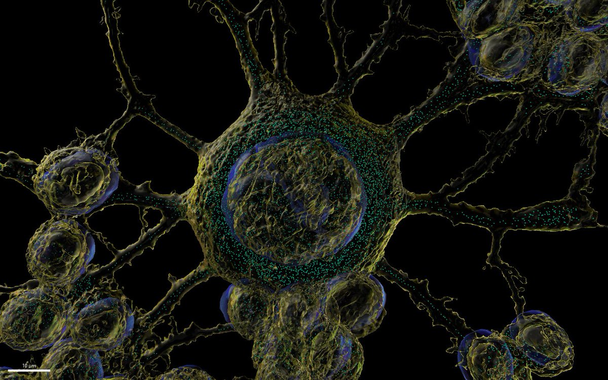
On our January cover: #mitochondria–ER contacts (white) in a COS-7 cell are speckled across the mitochondrial surface (purple) & are easily visualized in the absence of the ER (green). By @BenCardoen et al. hubs.ly/Q02dFDN50 JCB’s January issue ➡️ hubs.ly/Q02dFs_Q0
English
Imaris 3D/4D Imaging retweetledi
