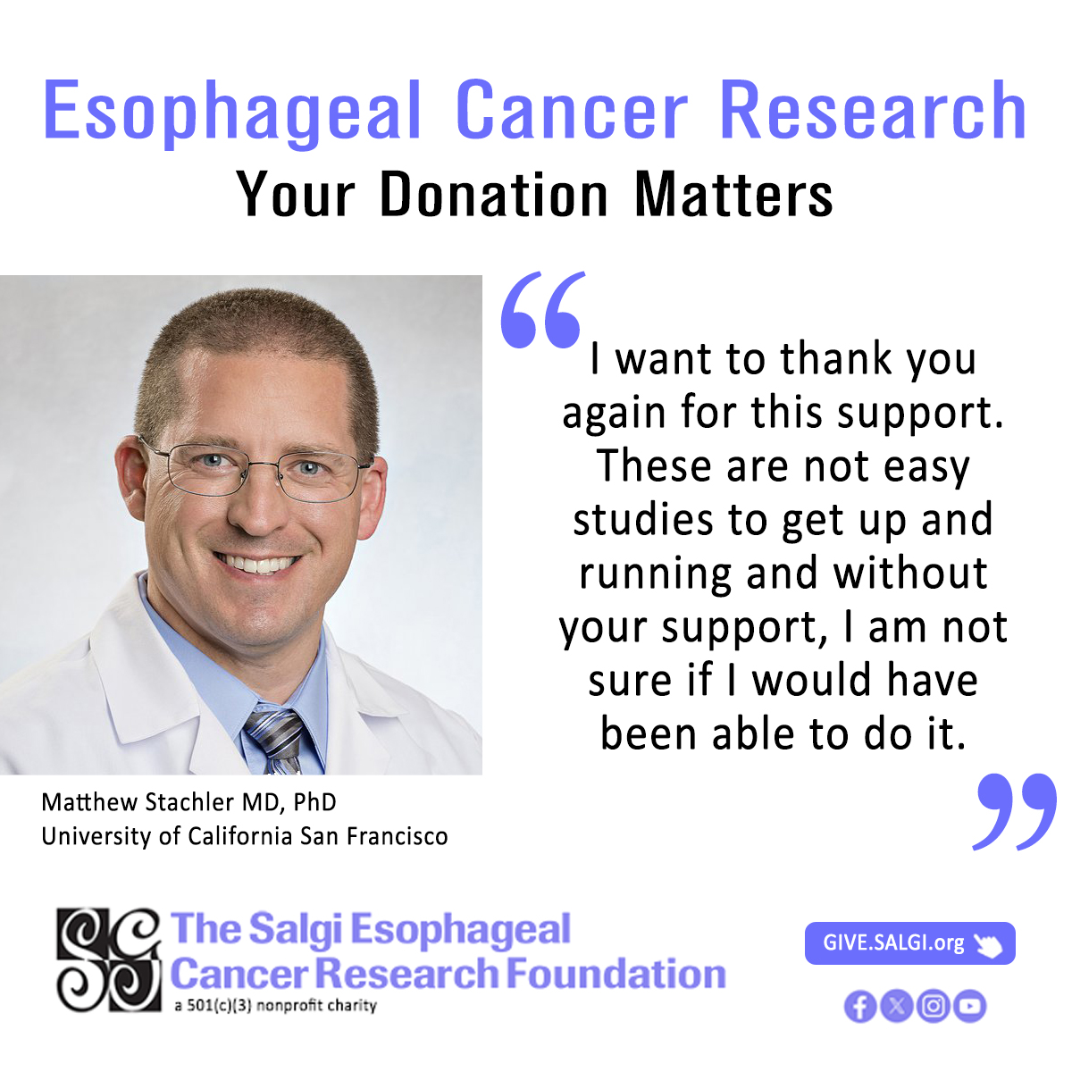
Matthew Stachler retweetledi

Your donation matters! Dr. Stachler of @UCSF expressed gratitude for the research funding he was awarded.
To donate in support of esophageal cancer research, visit: buff.ly/4dkI8tp
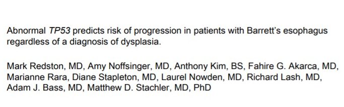
To read more about Dr. Stachler's research, please visit: buff.ly/3OOu1lm
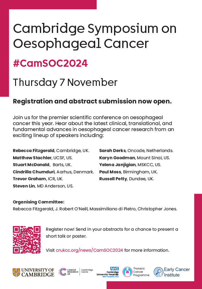
English