

Mohamed A H Ahmed, MD, PhD, FRCPath
1.7K posts
Mohamed A H Ahmed, MD, PhD, FRCPath
@MoAhmedMD
Consultant Histopathologist. NHS. UK: Uropathologist, lead of Uropathology NNUHFT#Norfolk and Norwich University hospital, UK
United Kingdom Katılım Şubat 2011
1.1K Takip Edilen823 Takipçiler
Histopathologic Flavors of Metastatic Prostate Carcinoma - Undifferentiated
🔷 High-grade carcinoma
🔷 Typically NKX3.1/AR negative. Neuroendocrine markers can be spotty/nonspecific.
NGS can be helpful in reaching the diagnosis of prostate origin.
Dr. Chan #USCAP2026 #pathology #PathX #PathTwitter


English

@garreyf great question!
while apical skel muscle does NOT constitute EPE as you well know, there is lit 👇🏼saying it should be reported (risk of + apical margin at RP)
in our group, we do include presence/absence of this in our synoptic summary table for prostate bx

English
Prostate apex core needle biopsy.
Does this finding require a more careful apical dissection if a radical prostatectomy is performed ?
ie should we ever report? @slusagar

English
Mohamed A H Ahmed, MD, PhD, FRCPath retweetledi
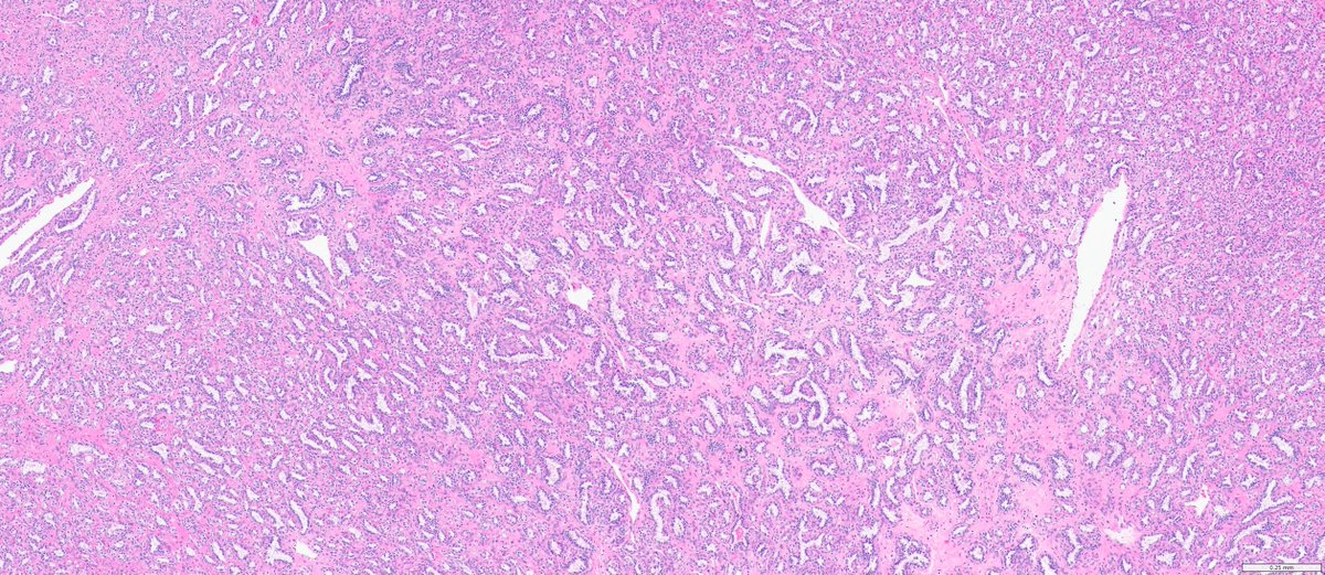
#GUpath nephrectomy:
Dx: clear cell papillary renal cell tumor
(I wish they'd named it clear cell TUBULOPAPILLARY renal tumor to emphasize morph!)
here,😍example of "shark smile" (tubulopapillary morph)🔬, as some cases lack prominent "reverse-apical" nuclei!

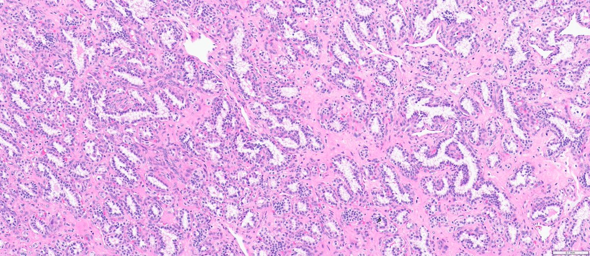
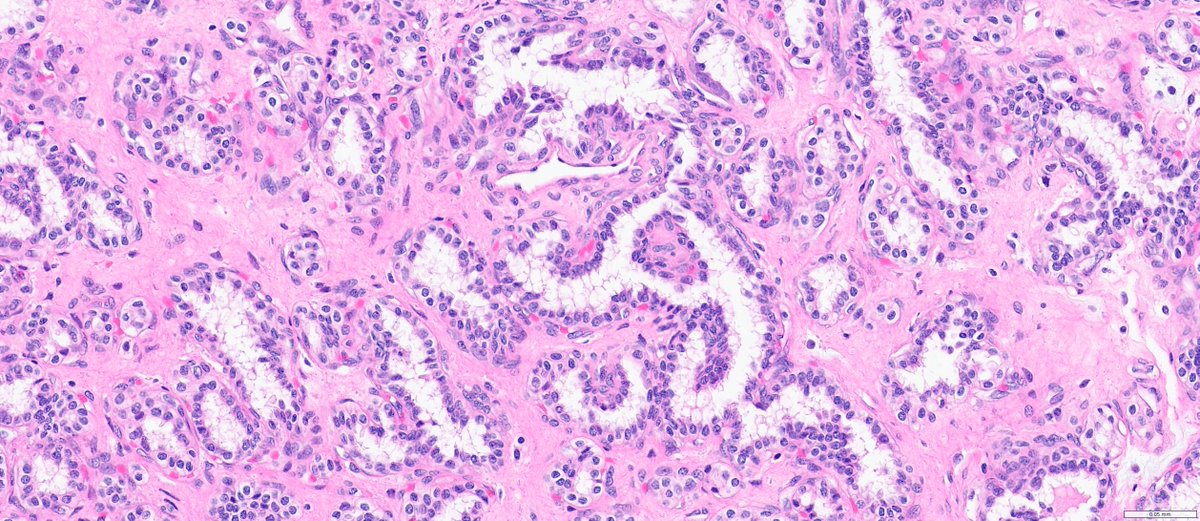
English
Mohamed A H Ahmed, MD, PhD, FRCPath retweetledi

Malignant phyllodes tumor with heterologous pleomorphic liposarcoma
#breastpathology #pathology
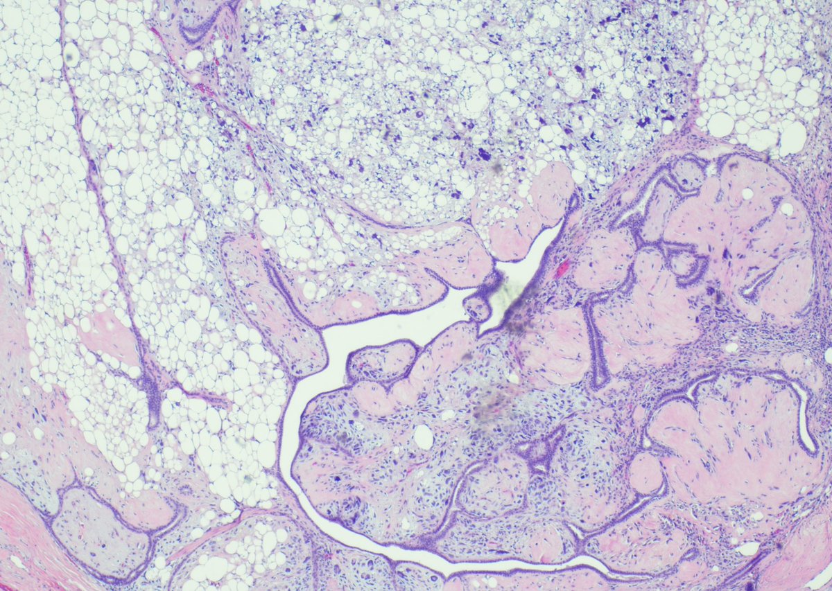


Filipino
Mohamed A H Ahmed, MD, PhD, FRCPath retweetledi

#GUPath #Surgpath #Pathtwitter #PathX
#Pathresidents: Let’s take a look at two biopsy cores (from the same patient) and decide what to call them.
Take a look at both cases (“A” in this post, “B” in reply). Cast your vote. Then, read on for some teaching points geared towards trainees and general fans of #GUPath.


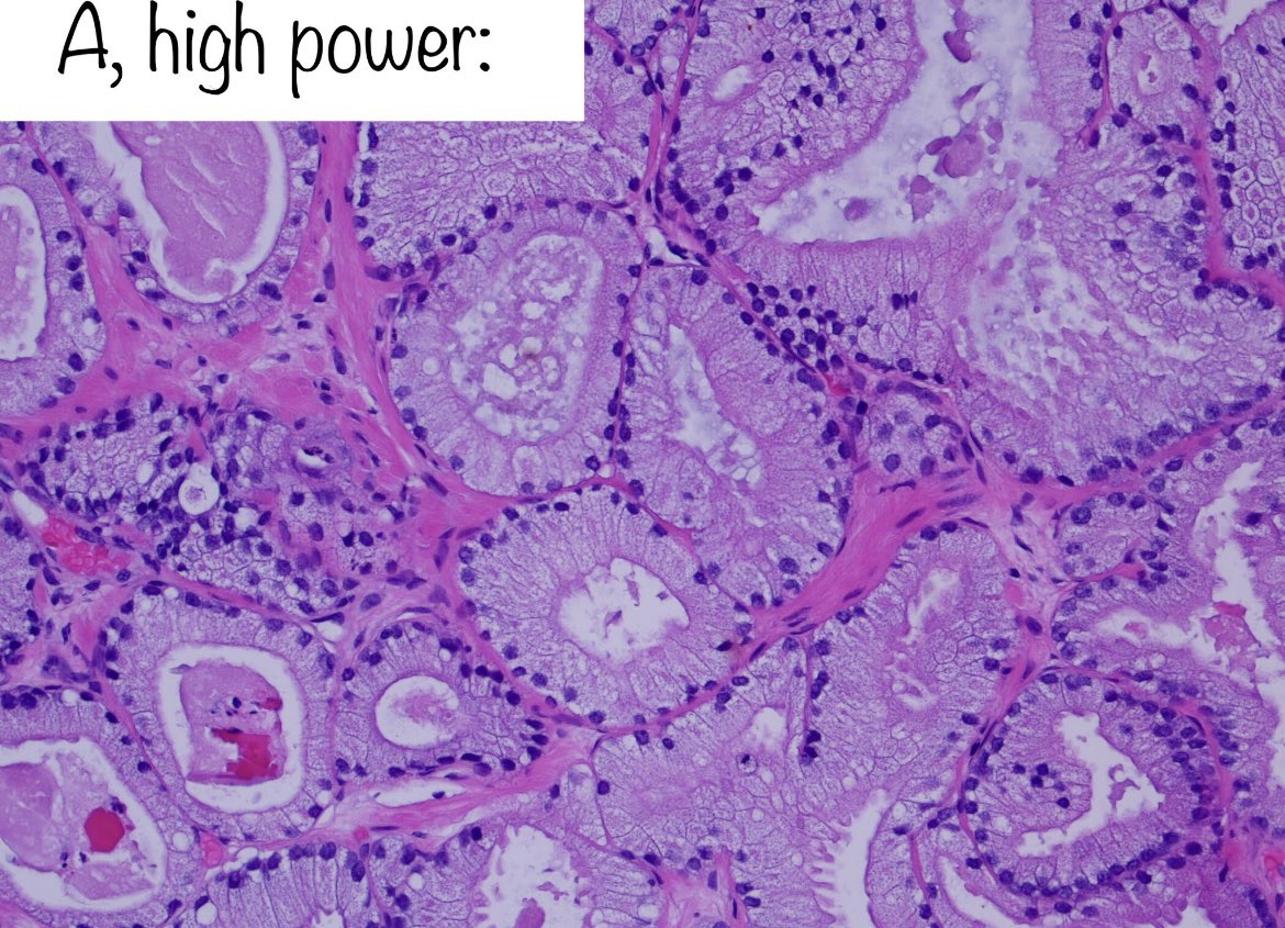
English
Mohamed A H Ahmed, MD, PhD, FRCPath retweetledi
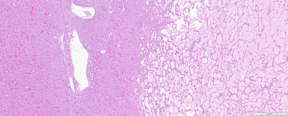
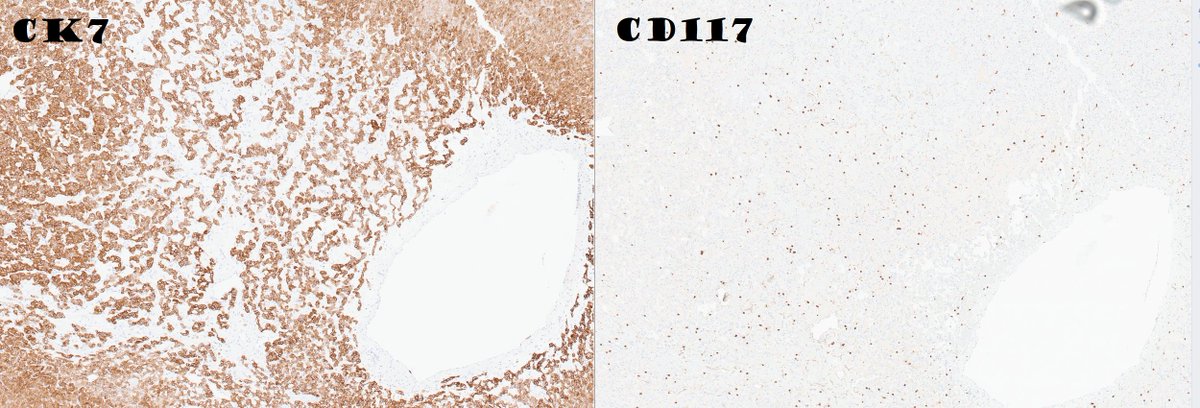
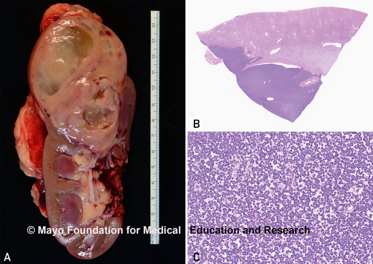
#GUpath #grosspath nephrectomy with central scar:
DDx: oncocytoma, chromophobe RCC...but also LOT!
🔬compact nests, loose cord-like nests floating in stroma, lymphocytic aggregates
🎨pos CK7/GATA3/L1CAM, neg CD117/CK20, retained SDHB
🧪another entity in TSC/MTOR family


English
Mohamed A H Ahmed, MD, PhD, FRCPath retweetledi

Mohamed A H Ahmed, MD, PhD, FRCPath retweetledi

A study in Virchows Archiv examines GPNMB immunohistochemistry in TFEB-altered renal cell carcinomas (RCCs). All 25 cases showed GPNMB positivity, indicating its potential as a sensitive screening marker for these tumors.
bit.ly/4sja0oG
English
Mohamed A H Ahmed, MD, PhD, FRCPath retweetledi
DCIS with clinging architecture vs flat epithelial atypia (FEA)
Dr Sciallis Cleveland Clinic MPS #Breastpath #pathtwitter #PathX #pathology #PathResident #NIPPathX

English
Mohamed A H Ahmed, MD, PhD, FRCPath retweetledi
Mohamed A H Ahmed, MD, PhD, FRCPath retweetledi

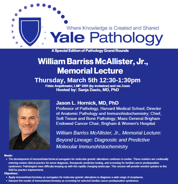
Join us for the annual William Barriss McAllister, Jr., Memorial Lecture @YaleMed on Thurs., March 5 at 12:30 PM ET as @JLHornick of @HarvardMed & @BrighamWomens, presents on, “Beyond Lineage: Diagnostic & Predictive Molecular Immunohistochemistry” shorturl.at/e5Aj9
English
Mohamed A H Ahmed, MD, PhD, FRCPath retweetledi

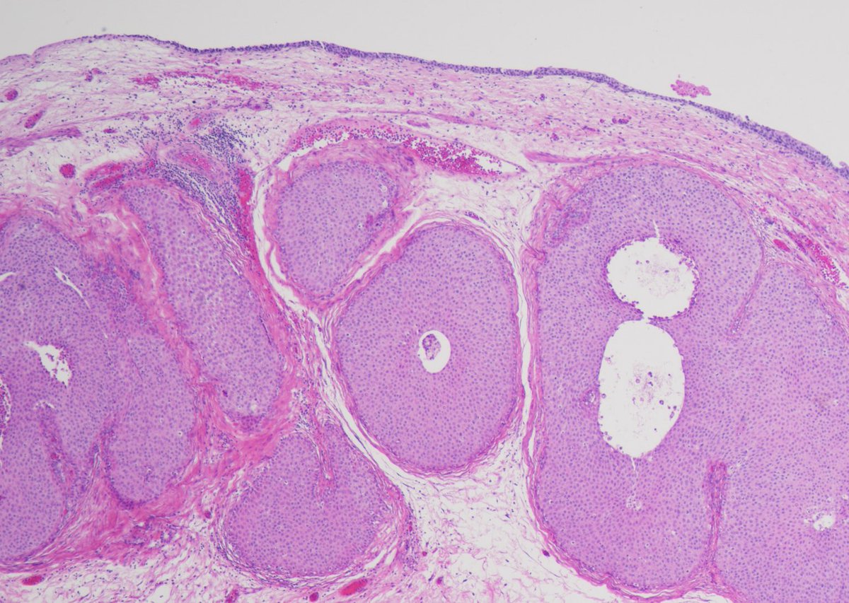
Large Nested Urothelial Carcinoma on H&E 🔬
Deceptively bland cytology with expansile, well-circumscribed nests infiltrating the lamina propria—an architectural wolf in sheep’s clothing. Recognition is critical to avoid underdiagnosis of this aggressive variant.
#Pathology
English
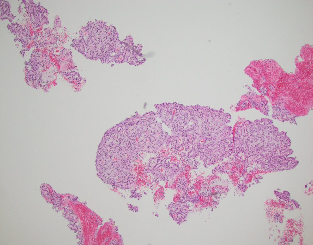
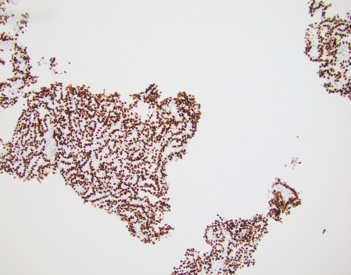
Can you make the diagnosis of TFE3 rearranged rcc on needle biopsy?
Yes!: 1) if you think about it and 2) are lucky to have enough tissue! (confirmed NONO:TFE3)
TFE3 IHC is not perfect but can be helpful as part of a screening panel
#GUPATH #pathology #pathres #kidneycancer


English
@LaraHarikMD @drmehrarohit @rovingatuscap @h_alahmadie @slusagar @priyaraomd @MiPathKunju @VarshaManuchaMD @lalpritu @Williamson_SR @MichelleDunno17 Nice case! I presume the intrachromosal inversion (inv(X)(p11.2;q12) was confirmed by RNA sequencing, because TFE3-NONO will give false negative or often subtle split signal on FISH if using Break-apart probes.
English
Mohamed A H Ahmed, MD, PhD, FRCPath retweetledi
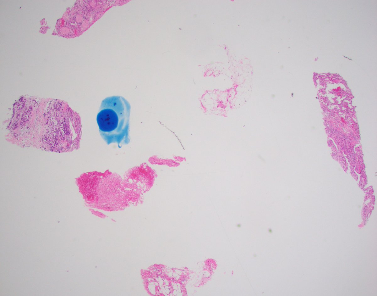
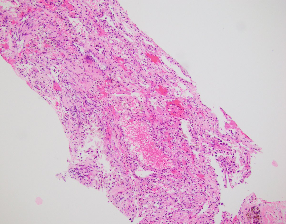
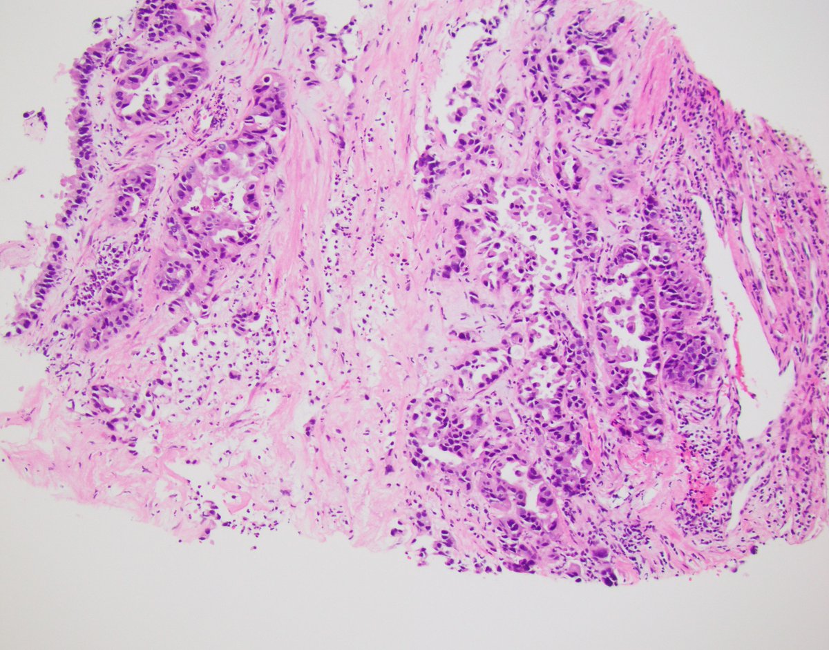
#Kidney biopsy, ⚠️unusual case (v rare)
70 yo male
⚡️notice 2 morphologically distinct areas
IHC and answer 👇
#rare #GUPATH #pathology #pathres

English
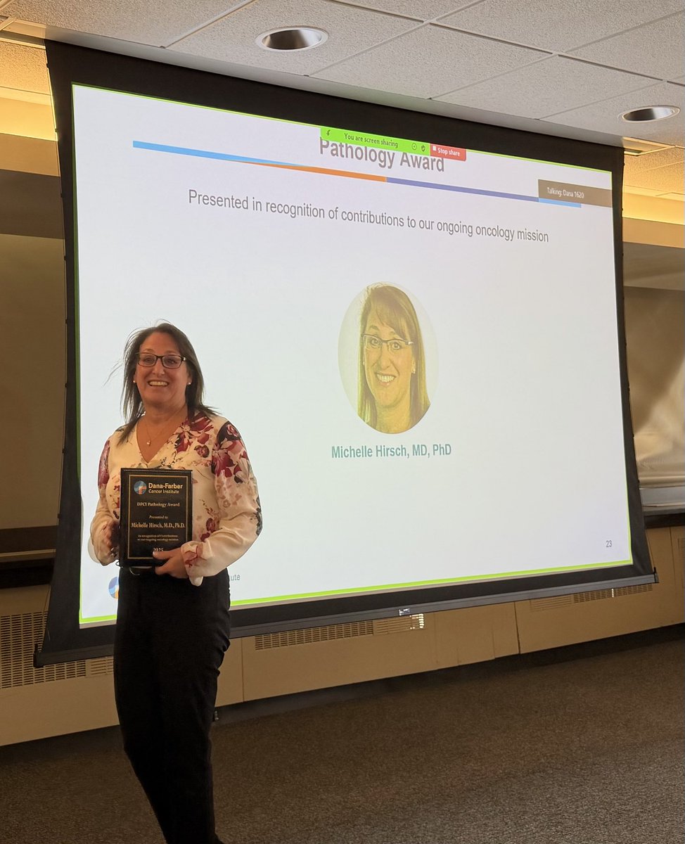
@DrChoueiri @GU_Path_Society @MichelleDunno17 @DanaFarber @DanaFarber_GU Well deserved… congratulations
English
Congratulations @MichelleDunno17 from all of us @DanaFarber and @DanaFarber_GU for being a role model of the best GU pathologist anyone can wish for !
Richly deserved !
English
Mohamed A H Ahmed, MD, PhD, FRCPath retweetledi
ALK-rearranged RCC are rare and can have variable morphologic features.
Here we describe some of these unusual tumors including an ALK-rearranged mesenchymal neoplasm:
nam12.safelinks.protection.outlook.com/?url=https%3A%…

English

Mohamed A H Ahmed, MD, PhD, FRCPath retweetledi

In primary aldosteronism, HISTALDO may overcall non-classical cases, missing curable resections. A simple “B2 ratio” (size of dominant vs 2nd-largest nodule) boosts accuracy and predicts surgical success. Better diagnosis = better outcomes. @EJEndo
academic.oup.com/ejendo/advance…

English
Mohamed A H Ahmed, MD, PhD, FRCPath retweetledi