Rafael Sirera

4.4K posts

Rafael Sirera banner
Rafael Sirera

Rafael Sirera

@ProfSirera

Professor of Cell Biology at Universitat Politècnica de València, with a particular passion for molecular medicine and a strong interest in natural philosophy.

Valencia Katılım Mart 2025
444 Takip Edilen2.3K Takipçiler
Sabitlenmiş Tweet
Rafael Sirera
Rafael Sirera@ProfSirera·
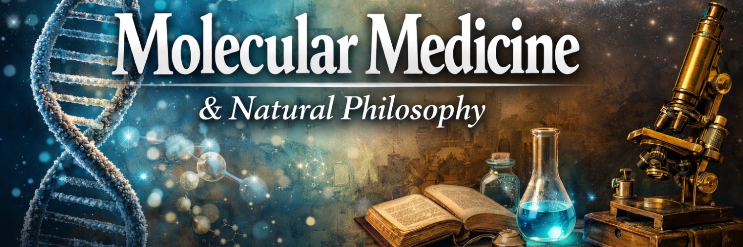
Molecular Medicine is not just a branch of science — it is the very language of life. It explains how cells communicate, how genes express identity, and how molecules decide between health and disease. Within its scope lies the most intimate narrative of our existence: the silent choreography of enzymes, receptors, and signals that sustain us every second. By following this account, you will explore the hidden logic that keeps us alive — and discover what happens when those delicate mechanisms fail, giving rise to illness. Here, we decode the molecular origins of disease and the rationale behind the action of drugs, tracing every therapeutic effect back to its biochemical root. Understanding Molecular Medicine is to see medicine itself under a microscope — where every cure begins as a molecular idea.
English
2
8
68
11.1K
Rafael Sirera
Rafael Sirera@ProfSirera·
Alzheimer’s disease cannot be understood only as a problem of amyloid or tau. It is also, at least in part, a disorder of lipid handling, repair and clearance. ApoE sits at the centre of this story because it is not just a cholesterol transporter; it is one of the brain’s lipid logisticians. ApoE is usually introduced as a “cholesterol protein”. But in the brain, that description is almost too small. Although the brain represents only about 2% of body weight, it contains nearly a quarter of all body cholesterol — a striking reminder that neuronal function is built not only on electrical signals, but on lipid architecture. The brain is a lipid-rich organ, yet it is largely isolated from circulating cholesterol by the blood–brain barrier. Most cerebral cholesterol is therefore produced, redistributed, remodelled and cleared locally. Neurons need lipids for synapses, membrane repair and plasticity, but they are not the main lipid managers. That role belongs largely to astrocytes and microglia, which package cholesterol and phospholipids into ApoE-containing particles and deliver them to neurons through receptors such as LDL receptor family members. This is where ApoE becomes biologically fascinating. There are three common ApoE isoforms: ApoE2, ApoE3 and ApoE4, encoded by the ε2, ε3 and ε4 alleles. They differ by only two amino acid positions, yet those small structural differences alter lipid binding, receptor interaction, protein stability and inflammatory behaviour. ✳️ ApoE3 is the most common variant and is considered the neutral reference. ✳️ ApoE2 is relatively protective against Alzheimer’s disease, although not absolute. ✳️ ApoE4 is the strongest common genetic risk factor for late-onset Alzheimer’s disease. But ApoE4 is not a diagnostic sentence. It is a risk modifier, not a deterministic mutation. A person carrying one ε4 allele has increased risk; carrying two increases it further. Yet many ApoE4 carriers never develop Alzheimer’s, and many patients with Alzheimer’s do not carry ApoE4. That is why ApoE genotyping is better understood as a predictive and stratification marker, not as a standalone diagnostic test. Mechanistically, ApoE4 appears to disturb several converging processes. It is less efficient in supporting lipid redistribution and neuronal repair. It favours amyloid-β aggregation and impairs its clearance. It also influences tau pathology, neuroinflammation, mitochondrial stress and blood–brain barrier integrity. The brain does not simply dump cholesterol directly into the blood. Excess cholesterol is converted mainly into 24S-hydroxycholesterol, an oxysterol able to cross the blood–brain barrier. Once outside the brain, it reaches the liver, where it can be metabolised and eliminated through bile acid pathways. So ApoE sits at a crossroads: lipid trafficking, amyloid clearance, immune tone and vascular interface biology. In Alzheimer’s disease, ApoE4 does not “cause” degeneration in a simple linear way; it shifts the brain towards a less resilient lipid–immune state, where damage is harder to clear and repair is less efficient.
Rafael Sirera tweet media
English
2
7
25
1K
Rafael Sirera retweetledi
Louisa Nicola
Louisa Nicola@louisanicola_·
ApoE4 is best understood as a systems level vulnerability, not an isolated amyloid trigger, because it simultaneously disrupts lipid trafficking, impairs synaptic repair, weakens blood-brain barrier integrity, and amplifies neuroinflammatory signaling. Alzheimer’s increasingly looks less like a single proteinopathy and more like a chronic failure of cerebral resilience and clearance biology.
English
0
1
1
199
Rafael Sirera retweetledi
Jordi Sabaté Pons
Jordi Sabaté Pons@pons_sabate·
ESTOY DESESPERADO, COMPARTIR POR FAVOR Busco profesional sanitario para que me asista en Barcelona ciudad indefinidamente. Tengo ELA con traqueo y PEG. Imprescindible experiencia y conocimientos. HORAS Y TURNOS: Una media de 26h semanales de mañanas y noches. SUELDO: 1284,10€ BRUTOS en 14 pagas. HORAS EXTRAS NETOS: 15€/hora laboral diurno 20€/hora, noches, fines de semana y festivos. Interesad@s: Enviar currículum WhatsApp 630324954 y entrevista con la empresa @Aiudo_es
Español
121
6.6K
5.1K
1.2M
Rafael Sirera
Rafael Sirera@ProfSirera·
The colour of urine is, biochemically, the visible end of haemoglobin turnover. Every day, red blood cells age and are dismantled mainly by macrophages in the spleen, liver and bone marrow. Their haemoglobin is split into globin and haem. Globin is recycled as amino acids. Haem, however, is chemically dangerous because its iron-containing porphyrin ring can promote oxidative reactions, so the body rapidly converts it into safer products. The pathway begins with haem oxygenase, which opens the porphyrin ring and produces biliverdin, carbon monoxide and free iron. Biliverdin is green. Then biliverdin reductase converts biliverdin into bilirubin, which is yellow-orange and hydrophobic. At this stage, bilirubin cannot simply dissolve in plasma. It travels bound to albumin towards the liver, where hepatocytes conjugate it with glucuronic acid via UDP-glucuronosyltransferase. This makes bilirubin water-soluble enough to be secreted into bile. Most conjugated bilirubin enters the intestine. There, bacterial enzymes convert it into urobilinogen. Part of this urobilinogen remains in the gut and is oxidised to stercobilin, which gives faeces their brown colour. But a fraction is reabsorbed into the portal circulation. Some returns to the liver; a small amount escapes into the systemic circulation and reaches the kidney. In urine, urobilinogen is oxidised to urobilin, historically called urochrome. This yellow pigment is the main reason normal urine has its characteristic colour. So the colour of urine is not simply “waste colour”. It is the chemical signature of red blood cell renewal, hepatic processing, intestinal bacterial metabolism and renal excretion. Hydration then modifies what we see. When urine is diluted, the same pigments are dispersed in more water, so urine appears pale yellow. When urine is concentrated, urobilin becomes more visually intense, giving amber or dark yellow urine. Other colours add diagnostic clues. Very dark brown urine may reflect bilirubin, haemoglobin, myoglobin or some drugs. Red urine may come from blood, beetroot pigments or medications. Orange urine may appear with dehydration, rifampicin or phenazopyridine. Green or blue urine is rare but can occur with dyes, drugs or unusual infections. Urine colour is haem biochemistry made visible: the body turning potentially toxic blood pigment into soluble molecular traces that the kidney can remove.
Rafael Sirera tweet mediaRafael Sirera tweet media
English
2
17
41
1.3K
Rafael Sirera
Rafael Sirera@ProfSirera·
Why can someone with advanced chronic kidney disease still pass a lot of urine? At first, it seems paradoxical. We tend to imagine kidney failure as a state in which urine simply disappears. And sometimes, in very advanced disease, that is exactly what happens. But often the biology is subtler: the kidney may continue to produce urine while progressively losing the ability to produce the right urine. In advanced chronic kidney disease (CKD), excluding diabetes, high diuresis is often largely more diluted urine, but not only that. The key concept is that the damaged kidney may still produce a lot of urine while progressively losing the ability to concentrate, dilute, and regulate solute excretion efficiently. In other words: ✳️ High urine volume ≠ preserved renal function. ✳️ Diuresis is not the same as renal function. A healthy kidney does not merely eliminate water. It decides how much water should accompany sodium, urea, potassium, phosphate, acid load and thousands of soluble molecules. It concentrates urine when water must be conserved, dilutes it when water is excessive, and adjusts this balance continuously. In CKD, the number of functioning nephrons falls. The remaining nephrons are forced to excrete a larger solute load per unit. Sodium, urea, phosphate and other retained solutes still need to leave the body, and water follows solute. This can generate a form of osmotic diuresis, even without diabetes. But there is a second, crucial layer. The damaged kidney loses its concentrating machinery. Tubulointerstitial fibrosis, medullary disruption, impaired countercurrent multiplication and reduced responsiveness to vasopressin mean that the collecting duct can no longer reclaim water efficiently. The urine becomes relatively fixed around plasma osmolality: neither very concentrated nor properly diluted. This is why nocturia is often one of the early clinical clues. So yes, the urine may be abundant — but often it is more diluted, less regulated, and metabolically inefficient. A patient may pass two or three litres per day and still retain potassium, acid, phosphate or uraemic toxins. This is the central distinction: urine volume measures output, not precision. Advanced CKD is not simply a kidney that “makes less urine”. It is a kidney that progressively loses discrimination — still moving water, but no longer controlling chemistry with the elegance that health requires.
Rafael Sirera tweet media
English
3
18
50
1.9K
Rafael Sirera
Rafael Sirera@ProfSirera·
Clinicians typically use 24-hour urine collections to calculate creatinine clearance and measure urinary potassium excretion, often combined with serum levels or spot urine potassium-to-creatinine ratios to evaluate glomerular filtration and tubular handling. These measurements help distinguish mere urine volume from actual solute clearance efficiency, aligning with the thread's emphasis that high diuresis in CKD often reflects poorly regulated, osmotically driven urine rather than healthy kidney performance.
English
0
0
2
28
CYN
CYN@cstrader66·
@ProfSirera What test is used to determine if the urine is removing creatine and potassium et al?
English
1
0
1
40
Rafael Sirera
Rafael Sirera@ProfSirera·
Entiendo perfectamente la escena. Mi hija va a un colegio británico y tiene esos tres modos dependiendo del contexto, Español, Inglés y Spanglish. El Spanglish lo usa con las compañeras del colegio de origen español y con los padres (que sabemos inglés) pues ni se plantea traducirnos al español cosas que dicen en el colegio/amigas o de sus asignaturas. Eso sí, las matemáticas le son más naturales en inglés.
Español
0
0
3
549
Pablo Grueso
Pablo Grueso@PabloGrueso·
Me resulta fascinante cómo un gibraltareño simultanea el inglés y el español.
Español
98
215
1.9K
158.6K
Rafael Sirera retweetledi
Ward Reflections
Ward Reflections@WardReflections·
This is one of the clearest explanations of CKD physiology I have seen written for a general audience, a really good one👍 The central distinction between urine volume and renal precision is exactly what gets missed in clinical conversations with patients. Families watching a loved one with advanced CKD pass normal volumes of urine genuinely believe the kidneys are coping. The nuance that quantity and quality of excretion are entirely different things is almost never communicated. The osmotic diuresis from solute loading in reduced nephron mass is underappreciated even among clinicians outside nephrology. Each surviving nephron carrying a proportionally larger excretory burden generates exactly the urine volumes that create false reassurance. The nocturia as early clinical clue point is particularly valuable. Loss of concentrating ability before overt GFR decline means the tubular damage precedes what standard creatinine based markers detect. A patient reporting new nocturia deserves renal evaluation earlier than the bloodwork might suggest. "Still moving water but no longer controlling chemistry with the elegance that health requires" is a genuinely beautiful clinical summary. Excellent post.
English
1
1
1
138
Rafael Sirera
Rafael Sirera@ProfSirera·
Dear @docakx, your query about the lacrimal puncta — the primary entry points of the lacrimal drainage system — gives me the opportunity to take your audience one step upstream: to the tear film itself. Because tears are not just salty water. They are a finely organised biological interface, designed to keep the ocular surface transparent, lubricated, protected, and optically smooth. And one of the most overlooked actors in this system is something we usually associate with the lung: surfactant. In the lung, surfactant reduces surface tension at the air–liquid interface, preventing alveolar collapse during expiration. In the eye, the logic is different but the physical principle is similar. The tear film must spread rapidly over the cornea with every blink, remain stable between blinks, and resist premature break-up. That stability depends on the outermost lipid layer, mainly secreted by the meibomian glands. This layer contains polar and non-polar lipids that behave, in functional terms, as ocular surfactants. They help lower surface tension, allowing the aqueous tear layer to distribute evenly over the hydrophobic epithelial surface. Without this surfactant-like behaviour, tears would not form a smooth optical sheet. They would retract, fragment, and evaporate too quickly. This is why tear film dysfunction is not simply a matter of “not producing enough tears”. In many patients with dry eye disease, the problem is qualitative rather than quantitative: the lipid layer is altered, meibomian gland secretion becomes abnormal, evaporation increases, and the tear film loses its stability. The consequence is deceptively simple: the eye feels dry. But mechanistically, what has failed is a delicate biophysical system. The ocular surface becomes exposed to friction, hyperosmolarity, inflammation, and epithelial stress. Blinking, which should renew the tear film, becomes a mechanical reminder that the interface is no longer properly protected. So yes, the lacrimal puncta drain tears. But before tears are drained, they must first behave as a living film — and that film depends on surfactant-like molecules quietly doing in the eye what surfactant famously does in the lung: making an air–liquid interface biologically possible. By the way, many thanks to @birloque for shedding light on this fascinating topic in @rtve.
Rafael Sirera tweet media
English
0
2
14
725
Dr. AK 🇮🇳
Dr. AK 🇮🇳@docakx·
What are these pits? What is their purpose?
Dr. AK 🇮🇳 tweet mediaDr. AK 🇮🇳 tweet media
English
16
9
85
28.8K
Rafael Sirera retweetledi
Abdelhamed Hamdy
Abdelhamed Hamdy@Abdelhamed6261·
Complement inhibitors for kidney disease
Abdelhamed Hamdy tweet media
English
0
48
112
3K
Rafael Sirera retweetledi
Rafael Sirera
Rafael Sirera@ProfSirera·
Scientific recognition often follows a predictable hierarchy. Prizes such as the Nobel Prize or the Lasker Award tend to crystallise visibility, projecting certain discoveries into collective awareness. Yet some of the most structurally transformative contributions in biology remain, paradoxically, less visible. 🌟 The work of Carl Woese sits precisely in that category. ✳️ The decisive transition in biological classification—from morphology to molecules—begins with him. In the 1970s, Woese recognised that ribosomal RNA offered something unprecedented: a record of evolutionary history embedded within the cell. Specifically, 16S ribosomal RNA in prokaryotes (and its eukaryotic counterpart, 18S rRNA) provided an ideal phylogenetic marker: universally conserved, functionally constrained, yet punctuated by variable regions that accumulate mutations at informative rates. ✳️ This was not simply a methodological refinement. It redefined taxonomy as a molecular science. Today, 16S rRNA sequencing underpins microbial classification, ecological profiling, and metagenomics. Entire fields—from microbiome research to environmental microbiology—depend on this principle: that evolutionary relationships can be inferred from sequence divergence in a conserved molecular scaffold. ✳️ What emerged from Woese’s analyses was unexpected. The apparent homogeneity of prokaryotes dissolved into deep evolutionary divisions, culminating in the identification of a new domain of life: Archaea. ✳️ This was not an incremental addition, but a fundamental restructuring. Life was no longer divided into prokaryotes and eukaryotes, but organised into three domains: Bacteria, Archaea, and Eukarya. ✳️ Interestingly, this framework also reshaped how we understand cellular complexity. Eukaryotic cells are now interpreted as chimeric systems, emerging through processes consistent with endosymbiosis—a merger of distinct prokaryotic lineages rather than a linear progression. ✳️ At a deeper level, Woese’s work reinforced the centrality of RNA in early evolution. By focusing on the ribosome, he helped consolidate the RNA world hypothesis, positioning RNA as both catalyst and information carrier in primordial systems. In parallel, he advanced the idea that early evolution was shaped by extensive horizontal gene transfer, suggesting that the “tree of life” may have begun as a network rather than a simple branching structure. 🚩 Woese did not merely discover a new domain. He altered the epistemology of biology itself—shifting it toward a framework where sequence, rather than structure, defines identity.
Rafael Sirera tweet media
English
0
5
13
511