Rheumatology US Case-based Learning

58 posts

Rheumatology US Case-based Learning

Rheumatology US Case-based Learning

@RheumMSKUS

For healthcare professionals interested in ultrasound/POCUS for rheumatology and MSK conditions. By Dr. Samar Aboulenain @UofT 🇨🇦. Not for medical advice.

Katılım Aralık 2025
30 Takip Edilen340 Takipçiler
Rheumatology US Case-based Learning retweetledi
Sᴡɪss Uʟᴛʀᴀsᴏᴜɴᴅ Cᴇɴᴛᴇʀ - UZR
Uʟᴛʀᴀsᴏᴜɴᴅ Qᴜɪᴢ 
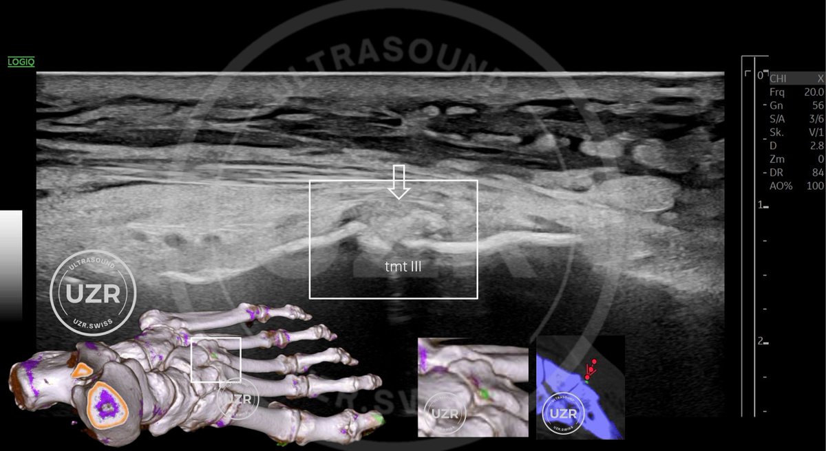
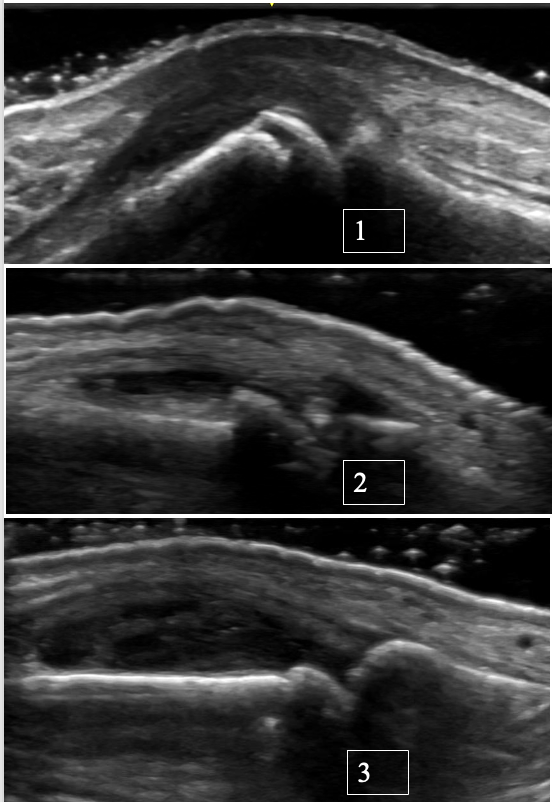
Mᴇᴅɪᴀʟ Eʟʙᴏᴡ Lᴏɴɢɪᴛᴜᴅɪɴᴀʟ Pᴀᴛɪᴇɴᴛs ᴘʀᴇsᴇɴᴛɪɴɢ ᴡɪᴛʜ ᴄʜʀᴏɴɪᴄ ᴍᴇᴅɪᴀʟ ᴇʟʙᴏᴡ ᴄᴏᴍᴘʟᴀɪɴᴛs ᴀɴᴅ ʀᴀᴅɪᴀᴛɪᴏɴ, ʜᴀs ᴜɴᴅᴇʀɢᴏɴᴇ ᴍᴜʟᴛɪᴘʟᴇ ɪɴᴊᴇᴄᴛɪᴏɴ ᴛʜᴇʀᴀᴘɪᴇs, ɪɴᴄʟᴜᴅɪɴɢ ғɪᴠᴇ PRP, ᴡɪᴛʜᴏᴜᴛ ᴀɴʏ ɪᴍᴘʀᴏᴠᴇᴍᴇɴᴛ. Nᴏᴡ sᴇᴇᴋɪɴɢ ᴀ sᴇᴄᴏɴᴅ ᴏᴘɪɴɪᴏɴ. Wʜᴀᴛ ɪs ᴛʜᴇ ʀᴇᴀsᴏɴ ғᴏʀ ᴛʜᴇ ɪɴᴇғғɪᴄᴀᴄʏ ᴏғ ᴛʜᴇ ᴛʜᴇʀᴀᴘɪᴇs ᴀᴅᴍɪɴɪsᴛᴇʀᴇᴅ ᴛʜᴜs ғᴀʀ?
3
4
29
3.4K
Rheumatology US Case-based Learning
Ultrasound of the MCP joint with osteoarthritis and possible superimposed CPPD 👇 Ultrasound is a valuable tool for distinguishing between OA, CPPD, and inflammatory arthritis at the level of MCPs. Case: 72-year-old right-hand-dominant retired carpenter presents with R MCP 3 pain and stiffness that has been intermittent over the past several years, worse with activity and has recently progressed with associated swelling. Features suggestive of OA: • Typically, no significant synovial hypertrophy. Effusions are more common • Osteophytes are seen rather than erosions • A “hook-like” osteophyte on X-ray or ultrasound should raise suspicion for superimposed CPPD --> Ultrasound also enables precise targeting of small fluid collections for diagnostic aspiration. Recommended reading: Cipolletta E, Di Matteo A, Smerilli G, et al. Ultrasound findings of calcium pyrophosphate deposition disease at metacarpophalangeal joints. Rheumatology (Oxford). 2022;61(10):3997-4005. doi:10.1093/rheumatology/keac063 #MSKUS #RheumUS #CPPD #osteoarthritis
English
0
12
33
1.6K
Rheumatology US Case-based Learning
Florid Dorsal Wrist Synovitis in Rheumatoid Arthritis Case: Anti-CCP+ RA patient, active smoker with: → Persistent synovitis on ultrasound of the L wrist → Refractory to intra-articular steroids → Turning point: ↓ Cigarettes/day + ↑ biologic dose, which led to improved disease control Clinical Pearl: Smoking worsens RA outcomes and is linked to higher DAS28, poorer DMARD response, and faster radiographic progression. This relationship seems to be dose-dependent. It is not just a risk factor. It’s a modifiable driver of treatment failure in patients with RA. References for a deeper dive: 1. Aboulenain S et al. ACR Open Rheumatology (2023) DOI: 10.1002/acr2.11629 2. Safy-Khan M et al. Journal of Rheumatology (2021) DOI: 10.3899/jrheum.200213 #MSKUS #RheumUSe
English
0
4
13
852
Rheumatology US Case-based Learning
Tenosynovitis of the Tibialis Posterior in an anti-CCP-positive patient presenting with medial ankle pain. Ultrasound features to look for: 1. Proliferative tenosynovitis -> hypoechoic thickening of the tendon sheath 2. Increased hyperemia -> Doppler signal at the tendon sheath thickening Clinical Pearls: * Tenosynovitis often occur in early rheumatoid arthritis (up to 80% in ACPA-positive early disease). * High-yield anatomical sites: ECU and finger flexor tendons. * The presence of tenosynovitis in an ACPA-positive patient predicts evolution to inflammatory arthritis (more superior than synovitis itself in a recent MRI-driven study): Reference for a deeper dive: 1. Abacar K, Tabuchi Y, Matteo AD, Duquenne L, Rowbotham E, Nam J, Emery P, McGonagle D, Mankia K. Quantitative MRI tenosynovitis volume explains the association between tendon involvement and future development of clinical arthritis in anti-cyclic citrullinated peptide-positive at-risk individuals. Ann Rheum Dis. 2026 Mar 10:S0003-4967(25)04459-0. doi: 10.1016/j.ard.2025.10.020. PMID: 41813507. 2. Kleyer A, Krieter M, Oliveira I, Faustini F, Simon D, Kaemmerer N, Cavalcante A, Tabosa T, Rech J, Hueber A, Schett G. High prevalence of tenosynovial inflammation before onset of rheumatoid arthritis and its link to progression to RA-A combined MRI/CT study. Semin Arthritis Rheum. 2016 Oct;46(2):143-150. doi: 10.1016/j.semarthrit.2016.05.002. Epub 2016 May 18. PMID: 27342772. #RheumUS #MKSUS
English
1
20
64
4.5K
Rheumatology US Case-based Learning
Chondrocalcinosis of the knee 👇 Pearls: - Fibrocartilage CPPD (in the meniscus) occurs at a younger age and is more prevalent than femoral hyaline cartilage CPPD. - Chondrocalcinosis in both the fibrocartilage and hyaline cartilage is frequent in patients > 80 years old (47% and 23%, respectively). What is your age cutoff to screen for secondary causes of CPPD?! --> hypophasatesia, hypomagnesemia, hyperparathyroidism and hemochromatosis Reference for a deeper dive: Cipolletta E, Francioso F, Smerilli G, Di Battista J, Filippucci E. Ultrasound reveals a high prevalence of CPPD in consecutive patients with knee pain. Clin Rheumatol. 2024 Jan;43(1):435-441. doi: 10.1007/s10067-023-06805-3. Epub 2023 Nov 17. PMID: 37975949. #MSKUS #RheumUS
English
1
27
107
4.8K
Rheumatology US Case-based Learning retweetledi
Gonzalo Serrano-Belmar.
Gonzalo Serrano-Belmar.@GSERRANOB_MSK·
Demonstrative video answering the case previously shown. Full-thickness tear of the supraspinatus tendon without retraction, resting on the distal stump at the footprint, demonstrated by forcing the bursal fluid to separate the margins. #mskrad
English
19
66
268
0
gkirschh
gkirschh@gkirschh·
@RheumMSKUS Feel like I’ve never seen an erosion on the lat epi. Very cool images !
English
1
0
0
73
Rheumatology US Case-based Learning
Lateral elbow enthesitis (common extensor tendon) in a patient with ankylosing spondylitis on a TNF inhibitor. Pearl: Not all OMERACT entheseal elementary lesions are equally important! -More specific 👉 erosions and 🔥power Doppler signal (especially ≤2 mm from cortex) -Less specific 👉 heterogeneity, calcifications and enthesophytes (can occur due to biomechanical loading/stress, esp. LE enthesis with high BMI) #MSKUS #RheumUS
English
2
11
37
2.4K
Rheumatology US Case-based Learning
Rapid Fire Case: Chondrocalcinosis of the Elbow Scanning tip: When in doubt, lower the dynamic range (↑ contrast). True CPPD deposits will appear as bright hyperechoic foci—often similar in echogenicity to the bony cortex. #RheumUS #MSKUS #CPPD
English
1
8
26
2.3K
Rheumatology US Case-based Learning
PIP joint synovitis/tenosynovitis in rheumatoid arthritis: Key elemental findings: • Synovial hypertrophy → hypoechoic/isoechoic synovial thickening • Doppler signal → active inflammation (when present where hypertrophy = proliferative synovitis AKA pannus). If this is present at the site of detected erosions -> high risk of radiographic progression • Tenosynovitis → thickened tendon sheath with fluid ± Doppler signal PEARL: In RA, tenosynovitis usually shows up along with synovitis in the same finger. In contrast, half of SLE patients with tenosynovitis have no concomitant synovitis. Reference for a deeper dive: Ogura T, Hirata A, Hayashi N, et al. Comparison of ultrasonographic joint and tendon findings in hands between early, treatment-naïve patients with SLE and RA. Lupus. 2017;26(7):707–714. #RheumUS #MSKUS
English
0
17
76
4.1K
Rheumatology US Case-based Learning
A case of chronic CPPD at the MCP & Wrist with Pseudo-Double Contour Case Summary: A 67-year-old female with seronegative rheumatoid arthritis referred for diagnostic clarification, as she has not responded to multiple csDMARDs or biologics, before escalating therapy. On exam, there were multiple tender and swollen joints, including MCP 1 and 3 bilaterally, as well as bilateral anterior ankle tenosynovial effusions. Ultrasound Findings: Pseudo-double contour over hyaline cartilage at the ulnar head and MCP joints, as well as focal intracapsular calcifications and chondrocalcinosis in the TFCC. Key Learning Point: *CPPD can represent a pseudo‑double contour (pseudo-DC) sign. *Unlike the classic DC sign of gout, which moves with the subchondral bone, the pseudo-DC in CPPD moves in the opposite direction during dynamic assessment. This is thought to be due to the CPP crystals depositing in capsules or ligaments, not directly on cartilage like mono-sodium urate crystals usually do. *Dynamic US evaluation is crucial: static images alone may mimic gout DC sign in CPPD (~10% of cases). Diagnostic Confirmation: Ankle aspiration from the extensor digitorum longus tenosynovial effusion confirmed CPP crystals, supporting the pseudo-DC interpretation. References: Di Matteo A, Grassi W, Filippucci E. Dynamic behaviour of the double contour sign in gout vs CPPD. RMD Open 2023;9:e002940.
English
0
13
35
2.2K
Medical Education
Medical Education@MedEd_Cases·
Mulder’s sign evaluates for Morton’s neuroma. The examiner grasps the metatarsal heads with one hand while the thumb of the opposite hand applies firm plantar pressure at the metatarsal neck. Lateral compression of the metatarsal heads is then performed. A positive test is indicated by a palpable click, with larger neuromas being more obvious. Sensitivity is 62%, specificity 100%! 🔗👇 Link Below 👇🔗 -- Read: wikism.org/Mulders_Sign -- Watch: youtube.com/watch?v=BU2JLU… -- #MedicalEducation #MedEd #Physio #ATC #Orthopedics #SportsMedicine #PhysicalTherapy #AthleticTraining #Rehab #InjuryPrevention #SportsInjury #SportsRehab #PhysioTherapy #FOAMed
YouTube video
YouTube
English
2
11
37
1.2K
Rheumatology US Case-based Learning
Is it Mechanical or Inflammatory? Ultrasound-detected enthesitis in a patient with psoriasis. Case: 38M with mild Psoriasis (BSA <2%) on topicals presents with chronic right elbow pain. No other joint complaints. On exam, there is a tender lateral epicondyle with pain with resisted wrist extension. No swollen joints, no Dactylitis. Mild tenderness at inferior pole of right patella. Bedside ultrasound findings: • Elbow: Hypoechoic common extensor tendon with grade 3 power Doppler at insertion → active enthesitis • Knee: Proximal infrapatellar tendon enthesitis with grade 1 power Doppler signal. Otherwise, enthesitis screening is unremarkable for subclinical enthesitis, including bilateral Achilles and triceps tendons. 💡 Concern for early Psoriatic Arthritis. Initial management included bracing, activity modification, NSAIDs. Pearls 🧵 Not all lesions are equally specific. Some, like bursitis, calcification, and insertional enthesophytes, can appear in both PsA and healthy controls. Highly specific lesions for PsA ➡️ Bone proliferation, erosions and Doppler signals close to the insertion (particularly if 2mm or less from the bony cortex). #RheumUS #MSKUS
English
1
17
57
2.6K
Medical Sphere
Medical Sphere@MedicalSphereAI·
@pompermarioMD @RheumMSKUS Thanks for sharing your insights. And absolutely. Our goal is to objectively and independently test these models to understand where they fall short and where they can genuinely add value as reliable assistants to clinicians (beyond the buzz and clickbait!).
English
1
0
2
19
Rheumatology US Case-based Learning
Quiz Time! 🧠 Which PIP joint ultrasound pattern matches each condition: rheumatoid arthritis, osteoarthritis, or psoriatic arthritis? Take a close look and make your best guess 👀 Which PIP belongs to which diagnosis? Drop your answers in the comments below 👇 #MSKUS #RheumUS
Rheumatology US Case-based Learning tweet media
English
4
18
52
4.6K
Rheumatology US Case-based Learning
The corresponding MRI image of the same patient shows the complete tear and the medially retracted tendon.
Rheumatology US Case-based Learning tweet media
English
0
0
2
156
Rheumatology US Case-based Learning
Full-Thickness Supraspinatus Tear in a Patient with Ankylosing Spondylitis Case: 60-year-old gentleman with ankylosing spondylitis on adalimumab presented with subacute right shoulder pain after slipping on ice. Plain radiographs are unremarkable. Positive Jobe’s and impingement tests, but negative drop arm test. CRP is suppressed. DDx: Inflammatory flare vs mechanical etiology. Ultrasound Findings -Complete fiber discontinuity (articular → bursal surface) -Cartilage interface sign -Bare footprint at the greater tuberosity -Joint–bursa fluid communication -Hyperemia in the subacromial-subdeltoid (SASD) bursa (reactive bursitis) Management: Referred to physiotherapy and orthopedics. Deemed not to be a surgical candidate due to age/comorbidities. Ultrasound-guided glucocorticoid injection into the SASD bursa ➡ Significant pain relief and functional recovery. High-Yield Pearls -Negative drop arm test does not rule out full-thickness tear. -Cartilage interface sign + bare footprint = strong evidence of full-thickness tear. -Ultrasound can differentiate shoulder inflammatory vs structural pathology at the point-of-care, facilitating timely tailored management. #MSKUS #RheumUS
English
2
12
53
2.5K
Rheumatology US Case-based Learning
Those are important and well-thought questions! @sciqst Supraspinatus insertional enthesitis is a common and sometimes persistent feature of AS despite TNF inhibition (Krabbe et al. J Rheumatol, 2020). This theoretically could have contributed to structural vulnerability predisposing this patient to such tears.
English
0
0
1
33
Raffaele Di Giacomo, PhD
Raffaele Di Giacomo, PhD@sciqst·
A fascinating case integrating musculoskeletal trauma and rheumatological conditions. The presence of a full-thickness supraspinatus tear, especially in an ankylosing spondylitis patient, raises several considerations. Since plain radiographs are clear, what was the role of adalimumab in managing any inflammation here? Could the chronicity of ankylosing spondylitis have predisposed him to more severe injuries with minor trauma? #Rheumatology #MSK #Medicine
English
1
0
0
51