Samar Aboulenain

54 posts

Samar Aboulenain

Samar Aboulenain

@Samar_Aboul

Rheumatologist @ UofT | Mom | RhMSUS. ~ views are my own~

Toronto, Ontario Katılım Nisan 2020
151 Takip Edilen74 Takipçiler
Samar Aboulenain retweetledi
Rheumatology US Case-based Learning
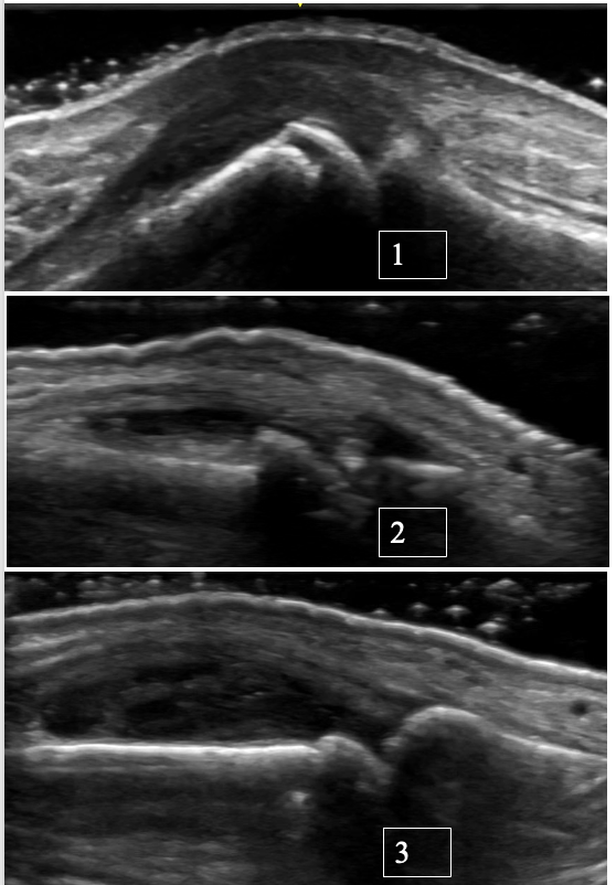
Ultrasound of the MCP joint with osteoarthritis and possible superimposed CPPD 👇 Ultrasound is a valuable tool for distinguishing between OA, CPPD, and inflammatory arthritis at the level of MCPs. Case: 72-year-old right-hand-dominant retired carpenter presents with R MCP 3 pain and stiffness that has been intermittent over the past several years, worse with activity and has recently progressed with associated swelling. Features suggestive of OA: • Typically, no significant synovial hypertrophy. Effusions are more common • Osteophytes are seen rather than erosions • A “hook-like” osteophyte on X-ray or ultrasound should raise suspicion for superimposed CPPD --> Ultrasound also enables precise targeting of small fluid collections for diagnostic aspiration. Recommended reading: Cipolletta E, Di Matteo A, Smerilli G, et al. Ultrasound findings of calcium pyrophosphate deposition disease at metacarpophalangeal joints. Rheumatology (Oxford). 2022;61(10):3997-4005. doi:10.1093/rheumatology/keac063 #MSKUS #RheumUS #CPPD #osteoarthritis
English
0
12
33
1.6K
Samar Aboulenain retweetledi
Rheumatology US Case-based Learning
Florid Dorsal Wrist Synovitis in Rheumatoid Arthritis Case: Anti-CCP+ RA patient, active smoker with: → Persistent synovitis on ultrasound of the L wrist → Refractory to intra-articular steroids → Turning point: ↓ Cigarettes/day + ↑ biologic dose, which led to improved disease control Clinical Pearl: Smoking worsens RA outcomes and is linked to higher DAS28, poorer DMARD response, and faster radiographic progression. This relationship seems to be dose-dependent. It is not just a risk factor. It’s a modifiable driver of treatment failure in patients with RA. References for a deeper dive: 1. Aboulenain S et al. ACR Open Rheumatology (2023) DOI: 10.1002/acr2.11629 2. Safy-Khan M et al. Journal of Rheumatology (2021) DOI: 10.3899/jrheum.200213 #MSKUS #RheumUSe
English
0
4
13
852
Samar Aboulenain retweetledi
Rheumatology US Case-based Learning
Tenosynovitis of the Tibialis Posterior in an anti-CCP-positive patient presenting with medial ankle pain. Ultrasound features to look for: 1. Proliferative tenosynovitis -> hypoechoic thickening of the tendon sheath 2. Increased hyperemia -> Doppler signal at the tendon sheath thickening Clinical Pearls: * Tenosynovitis often occur in early rheumatoid arthritis (up to 80% in ACPA-positive early disease). * High-yield anatomical sites: ECU and finger flexor tendons. * The presence of tenosynovitis in an ACPA-positive patient predicts evolution to inflammatory arthritis (more superior than synovitis itself in a recent MRI-driven study): Reference for a deeper dive: 1. Abacar K, Tabuchi Y, Matteo AD, Duquenne L, Rowbotham E, Nam J, Emery P, McGonagle D, Mankia K. Quantitative MRI tenosynovitis volume explains the association between tendon involvement and future development of clinical arthritis in anti-cyclic citrullinated peptide-positive at-risk individuals. Ann Rheum Dis. 2026 Mar 10:S0003-4967(25)04459-0. doi: 10.1016/j.ard.2025.10.020. PMID: 41813507. 2. Kleyer A, Krieter M, Oliveira I, Faustini F, Simon D, Kaemmerer N, Cavalcante A, Tabosa T, Rech J, Hueber A, Schett G. High prevalence of tenosynovial inflammation before onset of rheumatoid arthritis and its link to progression to RA-A combined MRI/CT study. Semin Arthritis Rheum. 2016 Oct;46(2):143-150. doi: 10.1016/j.semarthrit.2016.05.002. Epub 2016 May 18. PMID: 27342772. #RheumUS #MKSUS
English
1
20
64
4.5K
Samar Aboulenain retweetledi
CRUS-SURC
CRUS-SURC@CrusSurc·
🤩🏆 @crussurc 2025 research award winners announced - congratulations to Dr. Alan Zhou, Dr. Samar Aboulenain, Dr. Hsin Yen Liu and Dr. Jeanine McColl 👇👇👇crus-surc.ca/research/
CRUS-SURC tweet mediaCRUS-SURC tweet media
English
0
1
4
61
Samar Aboulenain retweetledi
Rheumatology US Case-based Learning
Chondrocalcinosis of the knee 👇 Pearls: - Fibrocartilage CPPD (in the meniscus) occurs at a younger age and is more prevalent than femoral hyaline cartilage CPPD. - Chondrocalcinosis in both the fibrocartilage and hyaline cartilage is frequent in patients > 80 years old (47% and 23%, respectively). What is your age cutoff to screen for secondary causes of CPPD?! --> hypophasatesia, hypomagnesemia, hyperparathyroidism and hemochromatosis Reference for a deeper dive: Cipolletta E, Francioso F, Smerilli G, Di Battista J, Filippucci E. Ultrasound reveals a high prevalence of CPPD in consecutive patients with knee pain. Clin Rheumatol. 2024 Jan;43(1):435-441. doi: 10.1007/s10067-023-06805-3. Epub 2023 Nov 17. PMID: 37975949. #MSKUS #RheumUS
English
1
27
107
4.8K
Samar Aboulenain retweetledi
Rheumatology US Case-based Learning
Lateral elbow enthesitis (common extensor tendon) in a patient with ankylosing spondylitis on a TNF inhibitor. Pearl: Not all OMERACT entheseal elementary lesions are equally important! -More specific 👉 erosions and 🔥power Doppler signal (especially ≤2 mm from cortex) -Less specific 👉 heterogeneity, calcifications and enthesophytes (can occur due to biomechanical loading/stress, esp. LE enthesis with high BMI) #MSKUS #RheumUS
English
2
11
37
2.4K
Samar Aboulenain retweetledi
CRUS-SURC
CRUS-SURC@CrusSurc·
✅ SAVE THE DATE: upcoming course crus 1) Basic course Sept 26-27, 2026 2) Martinoli masters in October 2026 3) Advanced injection course Nov 2026!!
CRUS-SURC tweet media
English
0
1
1
217
Samar Aboulenain retweetledi
Rheumatology US Case-based Learning
Rapid Fire Case: Chondrocalcinosis of the Elbow Scanning tip: When in doubt, lower the dynamic range (↑ contrast). True CPPD deposits will appear as bright hyperechoic foci—often similar in echogenicity to the bony cortex. #RheumUS #MSKUS #CPPD
English
1
8
26
2.3K
Samar Aboulenain retweetledi
Rheumatology US Case-based Learning
PIP joint synovitis/tenosynovitis in rheumatoid arthritis: Key elemental findings: • Synovial hypertrophy → hypoechoic/isoechoic synovial thickening • Doppler signal → active inflammation (when present where hypertrophy = proliferative synovitis AKA pannus). If this is present at the site of detected erosions -> high risk of radiographic progression • Tenosynovitis → thickened tendon sheath with fluid ± Doppler signal PEARL: In RA, tenosynovitis usually shows up along with synovitis in the same finger. In contrast, half of SLE patients with tenosynovitis have no concomitant synovitis. Reference for a deeper dive: Ogura T, Hirata A, Hayashi N, et al. Comparison of ultrasonographic joint and tendon findings in hands between early, treatment-naïve patients with SLE and RA. Lupus. 2017;26(7):707–714. #RheumUS #MSKUS
English
0
17
76
4.1K
Samar Aboulenain retweetledi
Rheumatology US Case-based Learning
A case of chronic CPPD at the MCP & Wrist with Pseudo-Double Contour Case Summary: A 67-year-old female with seronegative rheumatoid arthritis referred for diagnostic clarification, as she has not responded to multiple csDMARDs or biologics, before escalating therapy. On exam, there were multiple tender and swollen joints, including MCP 1 and 3 bilaterally, as well as bilateral anterior ankle tenosynovial effusions. Ultrasound Findings: Pseudo-double contour over hyaline cartilage at the ulnar head and MCP joints, as well as focal intracapsular calcifications and chondrocalcinosis in the TFCC. Key Learning Point: *CPPD can represent a pseudo‑double contour (pseudo-DC) sign. *Unlike the classic DC sign of gout, which moves with the subchondral bone, the pseudo-DC in CPPD moves in the opposite direction during dynamic assessment. This is thought to be due to the CPP crystals depositing in capsules or ligaments, not directly on cartilage like mono-sodium urate crystals usually do. *Dynamic US evaluation is crucial: static images alone may mimic gout DC sign in CPPD (~10% of cases). Diagnostic Confirmation: Ankle aspiration from the extensor digitorum longus tenosynovial effusion confirmed CPP crystals, supporting the pseudo-DC interpretation. References: Di Matteo A, Grassi W, Filippucci E. Dynamic behaviour of the double contour sign in gout vs CPPD. RMD Open 2023;9:e002940.
English
0
13
35
2.2K
Samar Aboulenain retweetledi
Rheumatology US Case-based Learning
Full-Thickness Supraspinatus Tear in a Patient with Ankylosing Spondylitis Case: 60-year-old gentleman with ankylosing spondylitis on adalimumab presented with subacute right shoulder pain after slipping on ice. Plain radiographs are unremarkable. Positive Jobe’s and impingement tests, but negative drop arm test. CRP is suppressed. DDx: Inflammatory flare vs mechanical etiology. Ultrasound Findings -Complete fiber discontinuity (articular → bursal surface) -Cartilage interface sign -Bare footprint at the greater tuberosity -Joint–bursa fluid communication -Hyperemia in the subacromial-subdeltoid (SASD) bursa (reactive bursitis) Management: Referred to physiotherapy and orthopedics. Deemed not to be a surgical candidate due to age/comorbidities. Ultrasound-guided glucocorticoid injection into the SASD bursa ➡ Significant pain relief and functional recovery. High-Yield Pearls -Negative drop arm test does not rule out full-thickness tear. -Cartilage interface sign + bare footprint = strong evidence of full-thickness tear. -Ultrasound can differentiate shoulder inflammatory vs structural pathology at the point-of-care, facilitating timely tailored management. #MSKUS #RheumUS
English
2
12
53
2.5K
Samar Aboulenain retweetledi
Rheumatology US Case-based Learning
Quiz Time! 🧠 Which PIP joint ultrasound pattern matches each condition: rheumatoid arthritis, osteoarthritis, or psoriatic arthritis? Take a close look and make your best guess 👀 Which PIP belongs to which diagnosis? Drop your answers in the comments below 👇 #MSKUS #RheumUS
Rheumatology US Case-based Learning tweet media
English
4
18
52
4.6K
Samar Aboulenain retweetledi
Rheumatology US Case-based Learning
❄️“Snowstorm” appearance of the 1st MTP tophaceous gout (plantar/volar views) Key Imaging Findings: - “Snowstorm” appearance -> extensive hyperechoic aggregates of monosodium urate crystal deposition. - Topheous deposits are seen within the flexor hallucis longus tendon as well as intra-capsular. - Overlying adventitial bursitis. 📌 The 1st MTP joint is the most commonly affected site in gout due to biomechanical stress and relatively lower temperature, favoring urate crystal precipitation. Recognizing MSU crystal deposition with POCUS in the form of aggregates, tophi, or double contour facilitates a timely diagnosis of gout and differentiates it from other DDx such as osteoarthritis or infection. #MSKUS #RheumUS
English
1
4
20
949
Samar Aboulenain retweetledi
Rheumatology US Case-based Learning
Deforming inflammatory arthritis with little-to-no pain: “Arthritis Robustus” Pattern Case: A physically active man in his 60s with chronic RF- and anti-CCP–positive polyarthritis reports no joint pain or functional limitation and therefore declined DMARD escalation. X-rays demonstrated severe symmetric erosive disease with carpal collapse. The patient was referred for assessment of subclinical synovitis to guide DMARD therapy. Ultrasound findings: See video 👇 - On B-mode, you see synovial hypertrophy (hypoechoic, non-compressible synovial thickening) in the radiocarpal, intercarpal and around the extensor compartment 4 (i.e., tenosynovitis). - Carpal crowding/collapse - also seen on X-rays - Active power Doppler, mostly seen in the intercarpal joint recess and thickened extensor compartment 4 tenosynovium. - Additionally, multiple MCPs revealed active synovitis with large marginal erosions (images not included). Key teaching points: 🔹 Ultrasound can detect subclinical synovitis in cases where symptoms do not correlate with physical exam, thereby informing decisions on DMARD therapy. 🔹 Subclinical active synovitis confirmed by power Doppler can predict radiographic progression, and in some cases, risk of future flares. 🔹“Arthritis robustus” is descriptive, not diagnostic This term has been used in case literature to describe rheumatoid-pattern synovitis in those with minimal symptoms and preserved hand function despite high inflammatory burden and radiographic progression. Reference for a deeper dive: Han J et al. Subclinical synovitis assessed by ultrasound predicts flare and progressive bone erosion in rheumatoid arthritis patients with clinical remission: a systematic review and meta analysis. J Rheumatol. 2016;43(11):2010–2018. #MSKUS #RheumUS
English
0
3
33
1.8K
Samar Aboulenain
Samar Aboulenain@Samar_Aboul·
5. Clinical Application Tips Lastly, integrate findings seen with PD within the clinical picture and physical exam.
English
0
0
0
51
Samar Aboulenain
Samar Aboulenain@Samar_Aboul·
“How do you optimize power Doppler sensitivity?” A key question for anyone learning rheumatology MSUS to detect low-flow signals in inflammatory arthritis Really enjoyed today’s lecture at the USSONAR Fundamental Hands-on MSK Course. Dr. VanDerVeer Key tips below 👇🏼 #RheumUS
Samar Aboulenain tweet media
English
5
5
6
699
Samar Aboulenain
Samar Aboulenain@Samar_Aboul·
4. Anatomy and Grading • Know Your Anatomy: Familiarize yourself with normal anatomical vessels • Use Standardized Grading: Apply a semiquantitative OMERACT scale (Grade 0–3) to assess the severity of flow.
English
0
0
0
58
Samar Aboulenain
Samar Aboulenain@Samar_Aboul·
3. Refine Scanning Technique • Minimize Probe Pressure: Use a "standoff view" • Limit Movement: Support your scanning arm and move the probe slowly or keep it still. • Scan Entirely: Evaluate the structure in orthogonal views (two planes).
English
0
0
0
48