Sabitlenmiş Tweet

🚨💡Excited to share our new publication @Histo_Journal
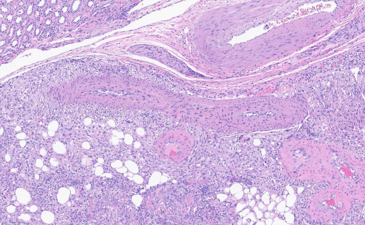
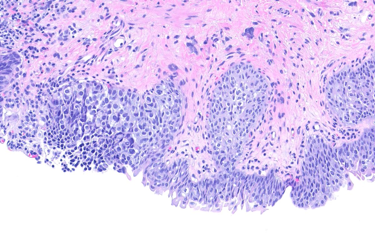
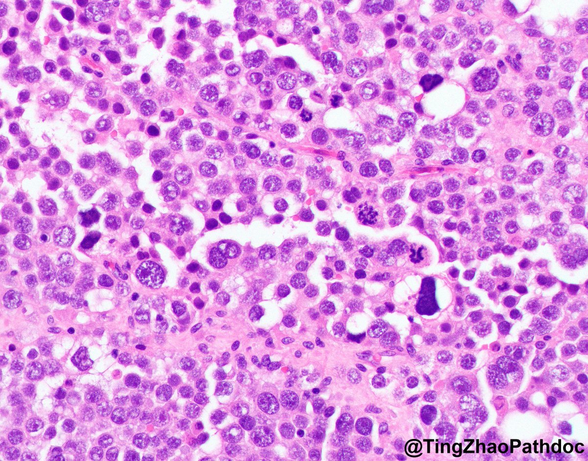
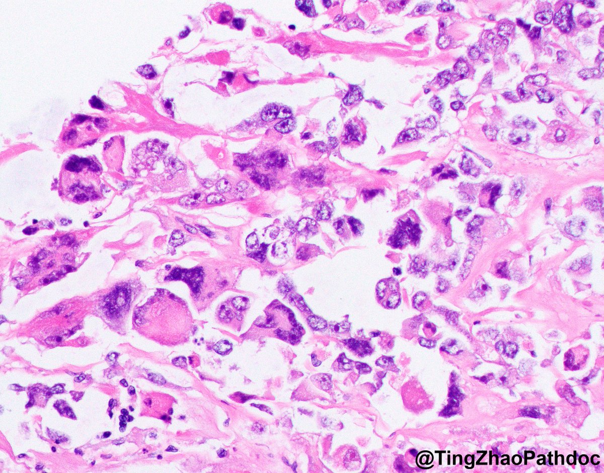
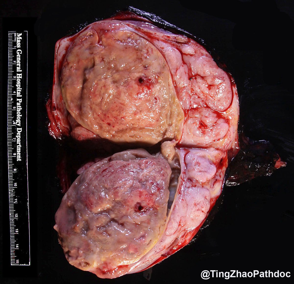
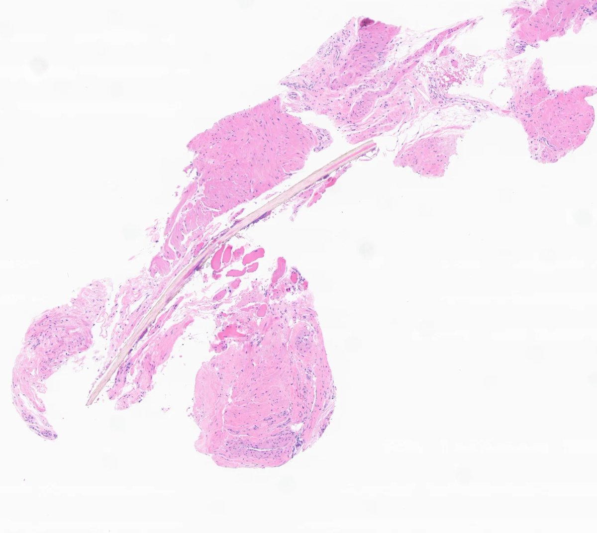
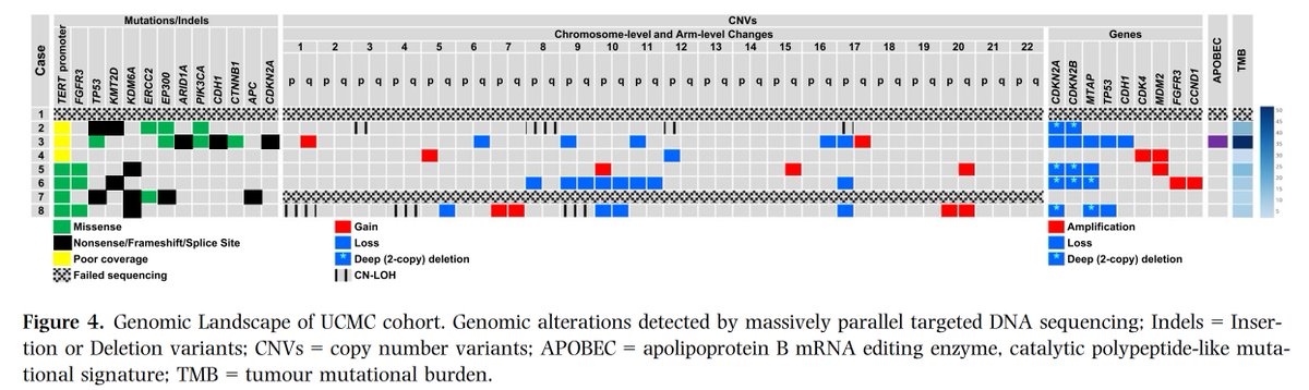
Clinicopathologic, immunohistochemical and genomic characterization of urothelial carcinoma with myxoid and chordoid features
@BWHPath @MGHPathology @MGBpathology
doi.org/10.1111/his.15…
#GUpath #Molpath 🧬


English