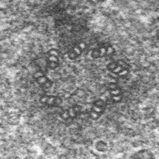
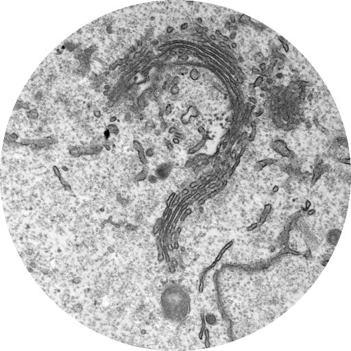
Sabitlenmiş Tweet
Kristina Micheva
190 posts

Kristina Micheva
@kdmicheva
Neuroscientist, array tomographist, learner of languages and martial arts
Katılım Mayıs 2016
134 Takip Edilen258 Takipçiler

@alon_douek I second @EM__Ian . Looks to me like they may be specifically within astrocytes, definitely not random and not lead. If you have serial sections I bet you could follow the same mysterious objects through the sections.
English

Any #TEM experts able to weigh in on something? We are seeing these small electron-dense things in brain. Not sure if it's real (samples are from a storage disease) or artifactual, but it looks worryingly like lead precipitate... #SciTwitter #ElectronMicroscopy


English
Kristina Micheva retweetledi

What a week at the #arraytomography workshop in Munich. Vivid exchange and great fun. Grateful for support from @czi, @SyNergy_Cluster and industry. Missed it? Stay tuned for Buenos Aires 2025: lnkd.in/dgSaXpTp
@kdmicheva @JemimaBurden @MSoizaReilly


English

Delicious & unique #arraytomography cake at the AT Workshop in Munich 2024! Thank you @MaSchifferer @JemimaBurden @MSoizaReilly @czi @DZNE_en

English
Kristina Micheva retweetledi

Great kick-off of our Array Tomography Workshop in Munich 2024🔬🎊 supported by @czi @DZNE_en with @MaSchifferer @kdmicheva @JemimaBurden and many other AT experts #arraytomography


English

@DaniBeckman Thank you for all your beautiful image and informative posts! You've created so many positive connections here, hopefully one day we bump into each other in real life! Take care of yourself 🌷🌻🪻
English

5 days of hands-on learning, discussions and talks + gorgeous electron microscopy and immunofluorescence ➡️ apply now!
Martina Schifferer@MaSchifferer
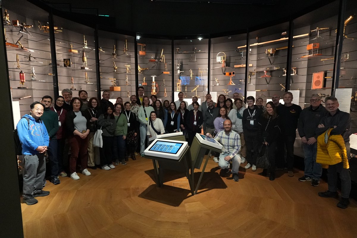
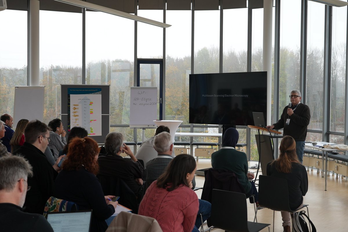
For all Array Tomography enthusiasts: Apply now for our AT workshop in Munich, Germany: arraytomography.org/workshop-2024.… #volumeEM #CZI #arraytomography #CLEM #electronmicroscopy
English

Just look at this: axon is blue, astrocyte is in red, myelin loops (oligodendrocyte) are green. Axon initial segment remodeling in action! Congratulations to Miki Furusho and Mark Terasaki @aminiata for this important work!

English

Beautiful #volumeEM detective work: oligodendrocytes, astrocytes and neurons are definitely up to something at the first myelin segment!
Journal of Comparative Neurology | Systems Neuroscience Journal | Wiley Online Library …rary-wiley-com.laneproxy.stanford.edu/doi/10.1002/cn…
English

10 women in STEM pick it up and keep it moving! @JoJoBuchanan1 @HaniehFalahati @ahmari_lab @AdemaRibic @ClaudiaLopez_EM @Meng2Fu @doctheagrif @MocaerK @Sci_Wanderlust @S_Nowotarski
Jemima Burden@JemimaBurden
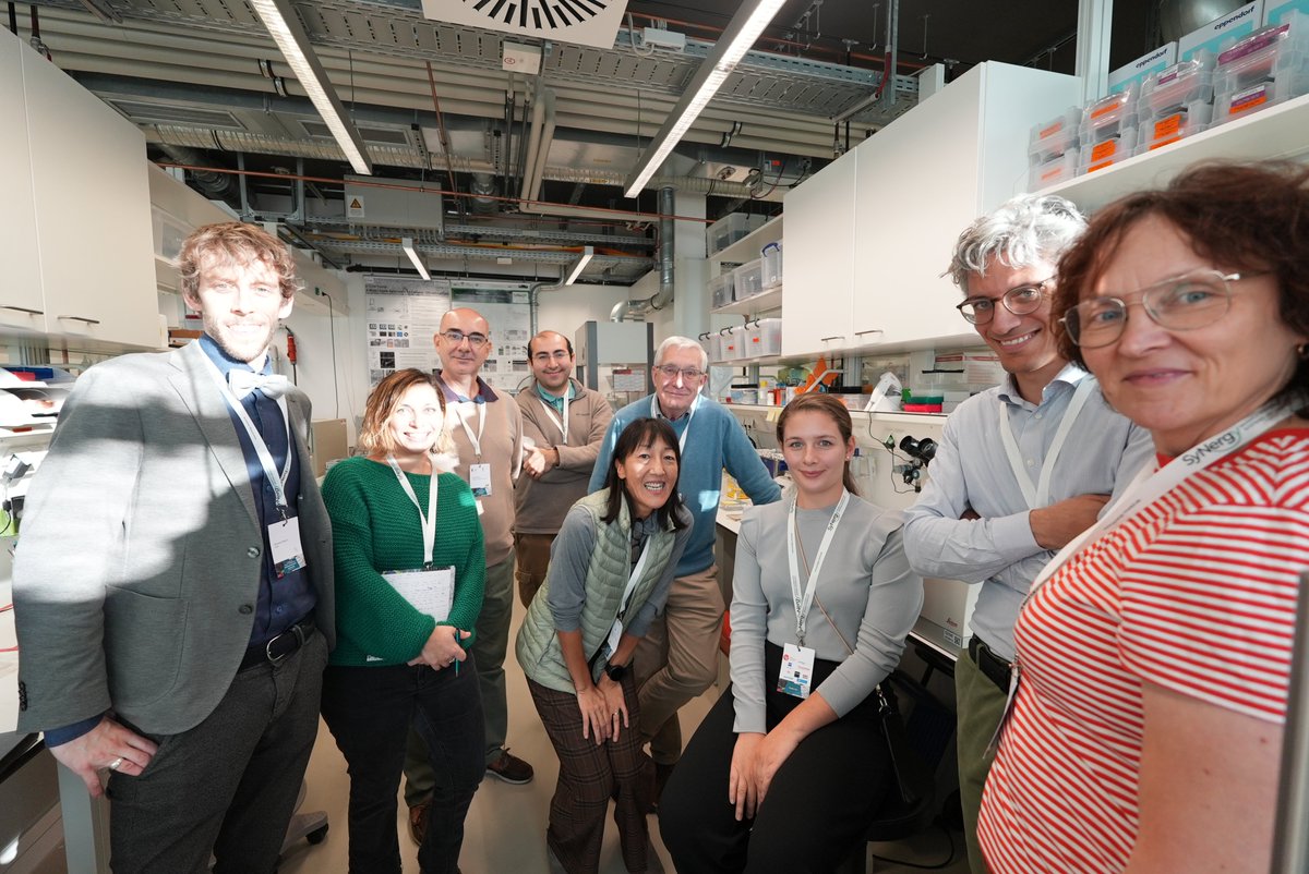
10 women in STEM pick it up and keep it moving! @EM_STP @MaSchifferer @kdmicheva @SuperResoluSian @VirginieUhlmann @AnwenBullen @giulia_casal @AlisonBeckett11 @edenlab_ucl @triciagoggin
English
Kristina Micheva retweetledi

In this #ImagingTheFuture Week we are launching our @cziscience funded community website on #ArrayTomography for AT enthusiasts and those who would like to know more!
arraytomography.org
#volumeEM @volumeEM1
@kdmicheva @JemimaBurden @aminiata @MSoizaReilly

English

@DaniBeckman Looking at plaques in the brain always brings this contradiction of how can something so nasty look so beautiful!
English

Happy #MicroscopyMonday! Going back to my roots with some amyloid in this 3D image 🤓🔬! It shows two different markers for amyloid pathology:
🔴Anti-Amyloid Fibrils OC;🟢Thioflaving S: a Dye for β sheet-rich deposits found in misfolding protein diseases. Surrounding this amyloid plaque are some very activated astrocytes (🔵Gfap). You can see some delineating a blood vessel nearby, passing in the middle of the amyloid plaque. @zeiss_micro

English

@MKirkcaldie @aniai_commune I kept this for my weekend read, and so glad I did - thank you for this engaging piece! It is a great reminder to look beyond the "cellular scale", across that gulf to the human/animal mind.
English

@MattRGR None of these 😄 It's postembedding immunofluorescence on ultrathin sections of the brain embedded in Lowicryl, then heavy metal staining and SEM (conjugate IF-SEM array tomography).
English


This is gorgeous! What blows my mind however is that you managed to find the same tiny cilium, 10 years later, and take an even better image of it!


JoAnn Buchanan@JoJoBuchanan1
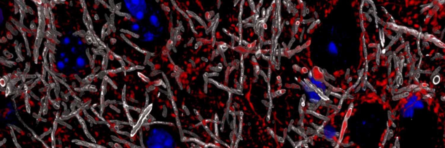
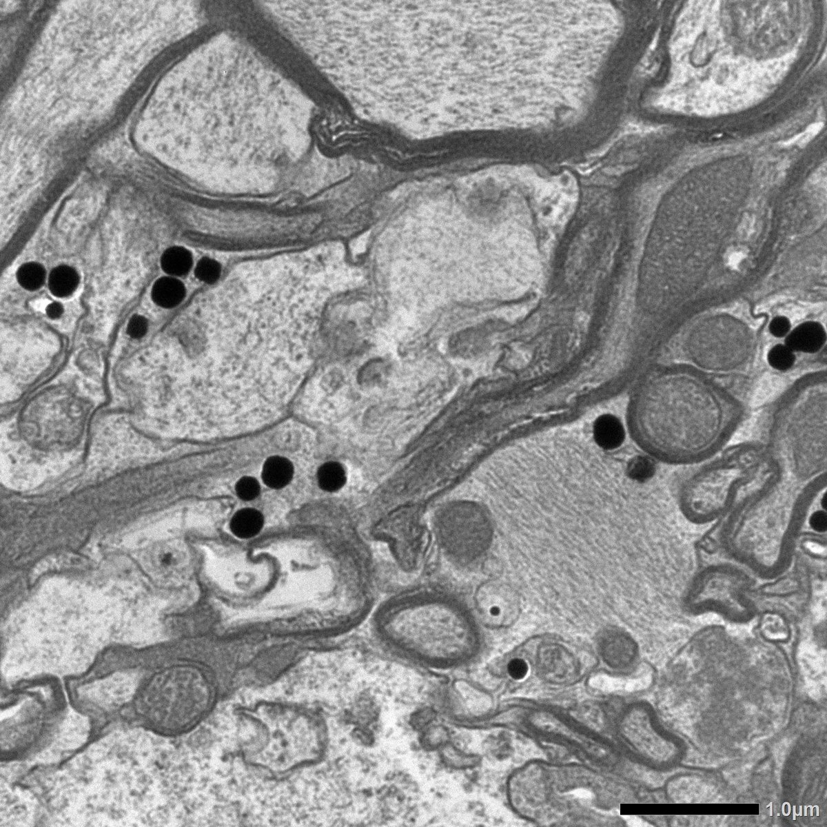
So excited to share this beautiful preprint ! Our collaboration with @JLS and @Microns Volume EM data made it possible 👏🏻🔬🧠🎊
English


