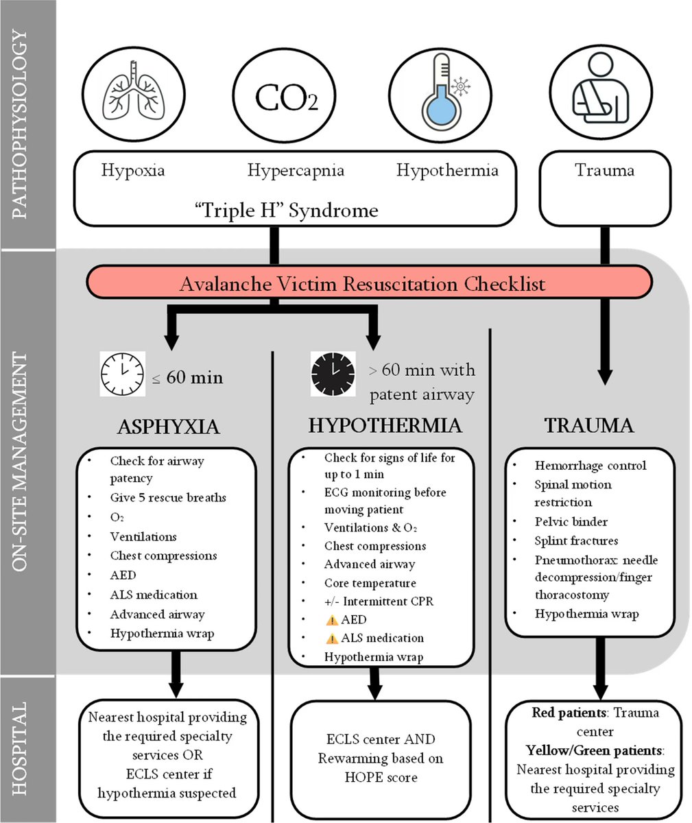
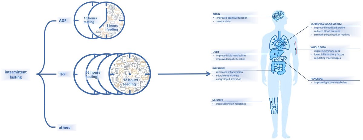
M-Mode Echocardiography Explained
This figure demonstrates M-mode measurements from the parasternal long-axis (PLAX) view, crucial for assessing valvular & ventricular function:
🔹 Aortic Valve Motion (Top Panel)
- The right coronary cusp (RCC) and non-coronary cusp (NCC) move apart in systole (valve opens) and come together in diastole (valve closes).
🔹 Mitral Valve Motion (Middle Panel)
- The anterior mitral leaflet (AML) and posterior mitral leaflet (PML) exhibit characteristic movements:
- Diastole: AML moves towards the septum (E wave) due to rapid LV filling, then slightly back (A wave) due to atrial contraction.
- Systole: Leaflets close tightly, preventing regurgitation.
🔹 Left Ventricular Dimensions (Bottom Panel)
- End-diastolic diameter (EDD): Largest LV dimension measured before systole.
- End-systolic diameter (ESD): Smallest LV dimension after contraction.
- These values help calculate LV function (e.g., fractional shortening & ejection fraction).
📌 Courtesy of Bernard E. Bulwer, MD, FASE
#Echocardiography#Cardiology#Mmode
😵💫 Does the differential for dizziness make your head spin? Need help finding your balance in the world of vertigo?
Let's break down how to approach the confusing consults for “dizziness” 👇
Se acaban de actualizar las guías de Fibrilación Auricular (FA) de la European Society of Cardiology (ESC), que reemplazan a las del 2020. Vamos a sintetizar lo más importante de las más de 100 páginas. 👇
Por emanaciones de gases está inhabilitado el centro regulador del @Samumetropolita. Se está operando por contingencia en otro lado inadecuado. No operando con normalidad. Los funcionarios empezaron movilización.
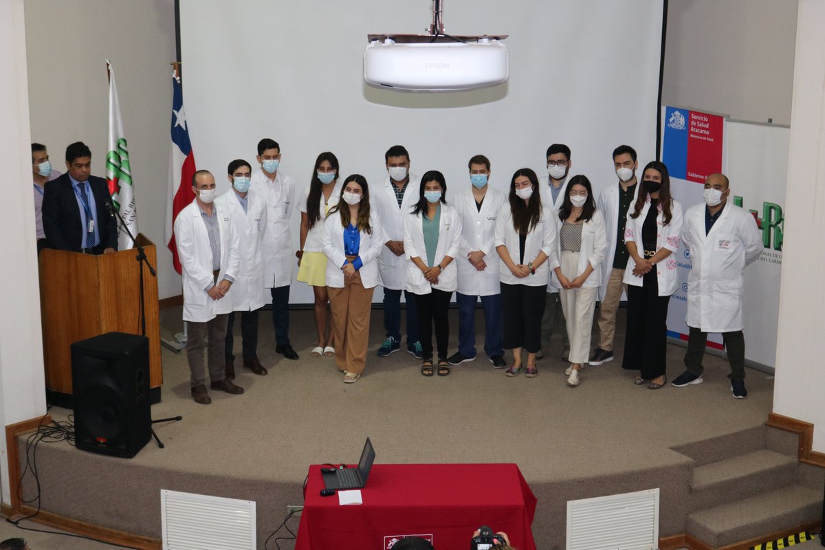
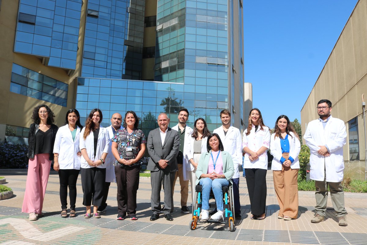
Hoy se realizó la bienvenida a nuevos especialistas que se suman a nuestro equipo de médicos del @hospitalcopiapo.
Médicos ginecólogos, de urgencia, anestesiología, neurología, cirugía pediátrica, otorrinolaringólogo, entre otros