
Fatima Shamsuddin
430 posts

Fatima Shamsuddin
@FatimaShamsPath
MD, FRCPath, FIAC
Kuwait เข้าร่วม Haziran 2023
115 กำลังติดตาม1.1K ผู้ติดตาม
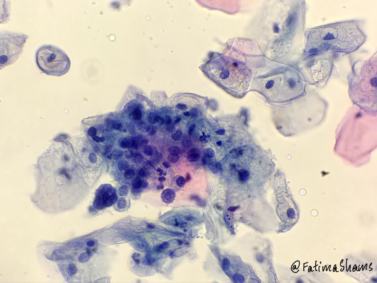
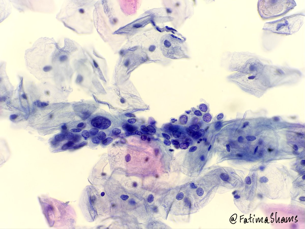
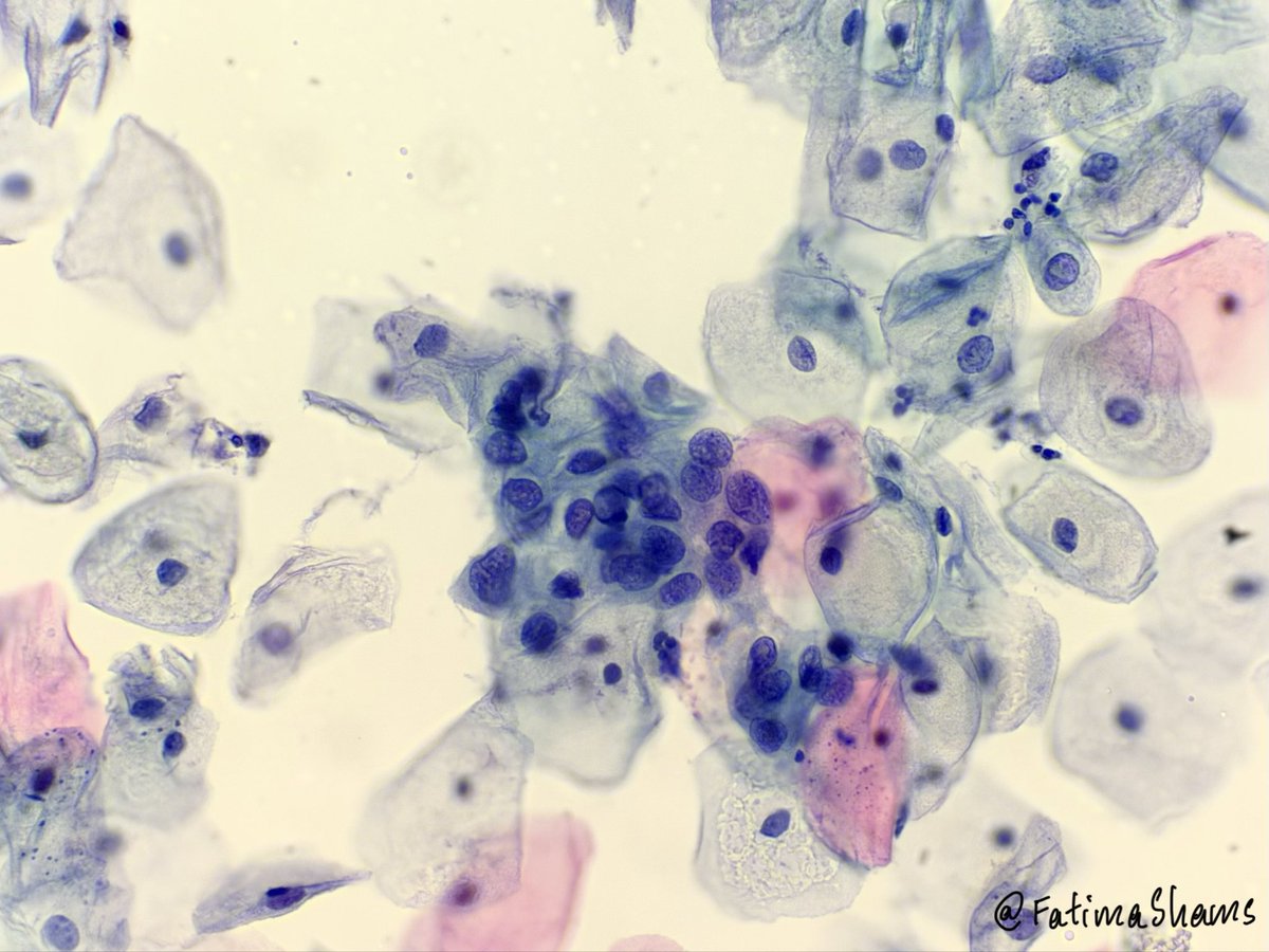
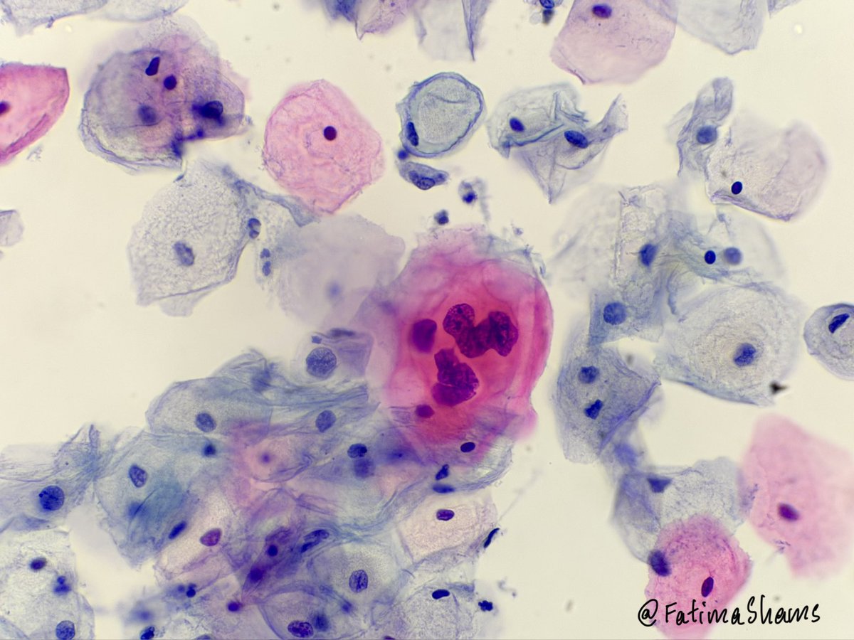
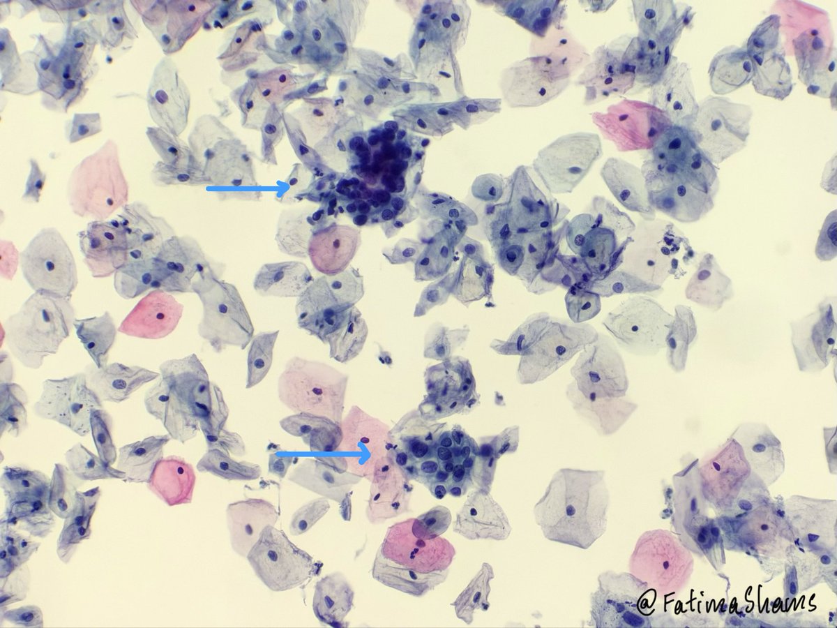
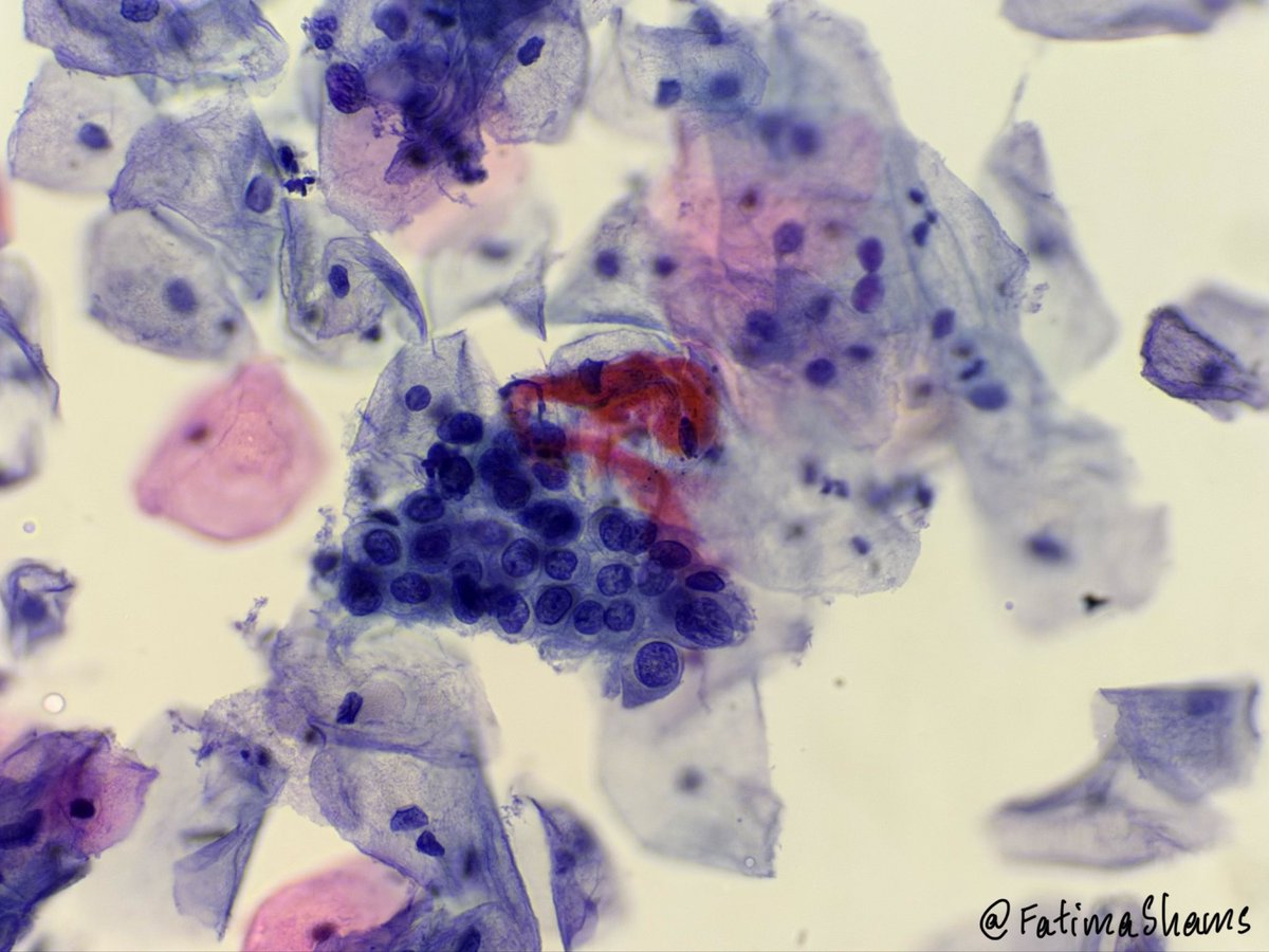
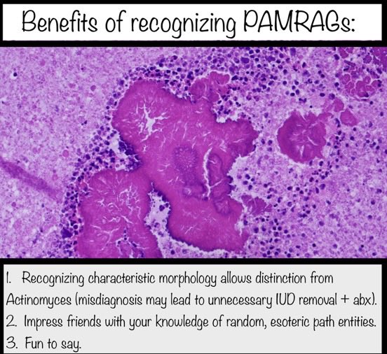
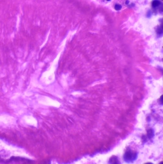
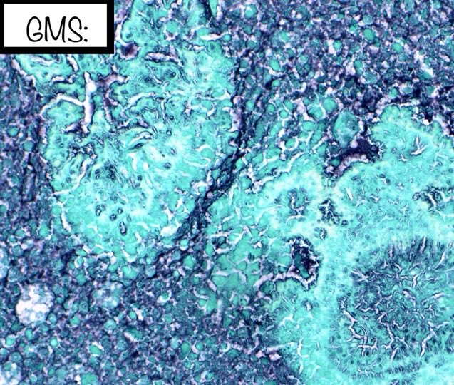
👉 42YO female - pap smear.
#PapSmear #Pathology #PathTwitter #MedTwitter #FOAMed #MedEd #Cytology #Screening #HPV
HT


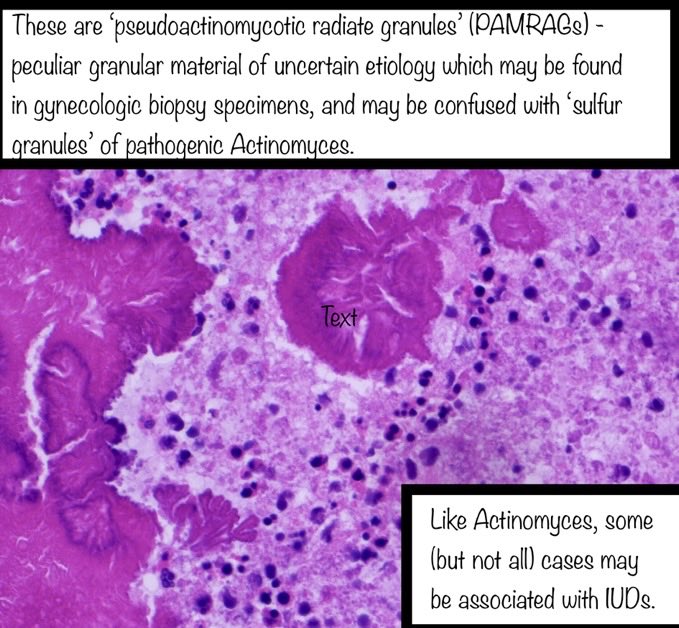
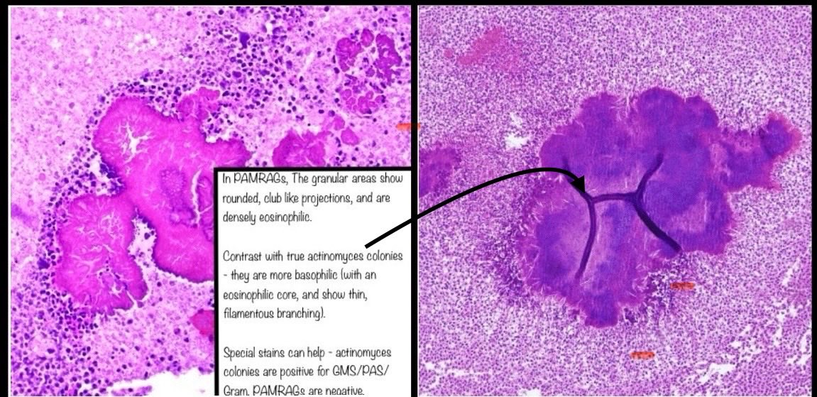
For more information on PAMRAGs, here is the seminal paper by Pritt et al: pmc.ncbi.nlm.nih.gov/articles/PMC18…
Actinomyces image source: PMID 25896158

English
#Gynpath #surgpath #pathresidents
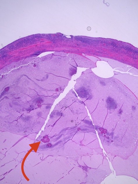
A 37 female was found to have a cervical polyp during IUD removal. The polyp was excised.
Poll in reply.

English
Answer
Fatima Shamsuddin@FatimaShamsPath
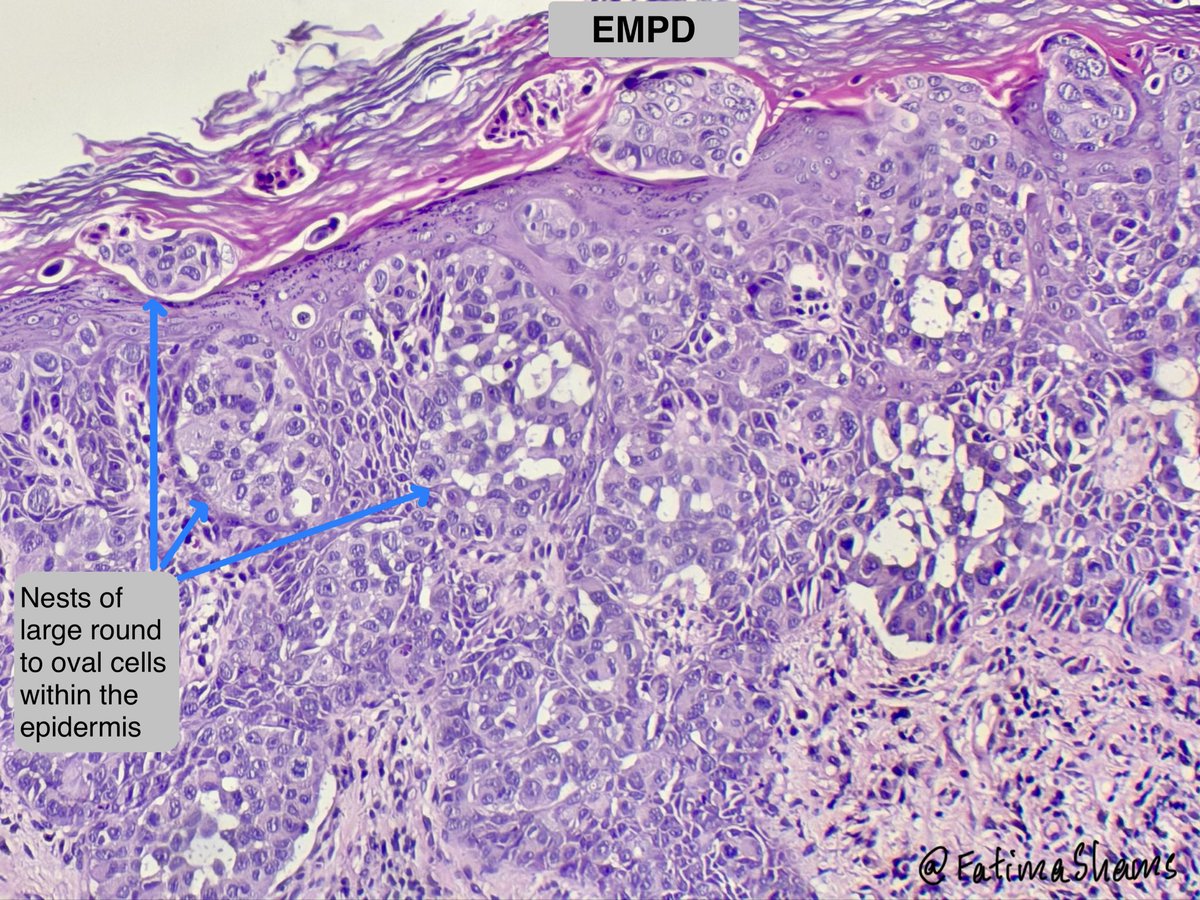
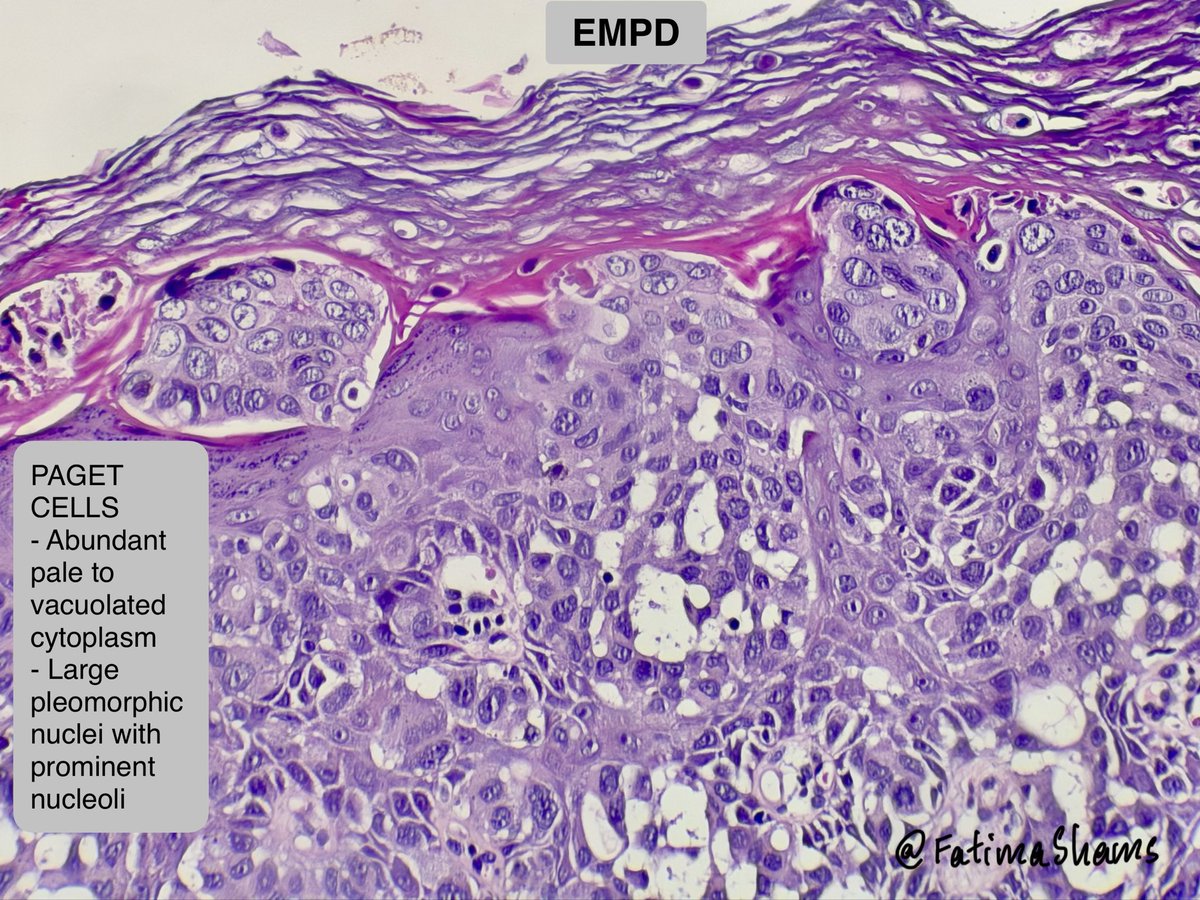
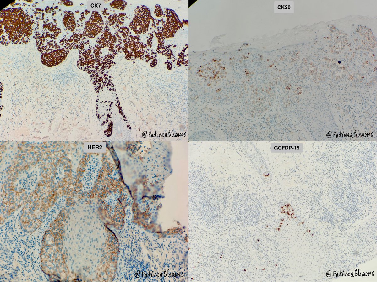
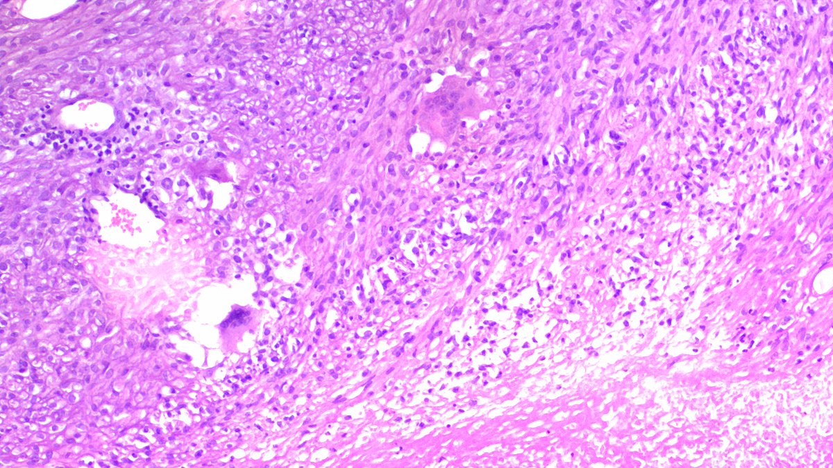
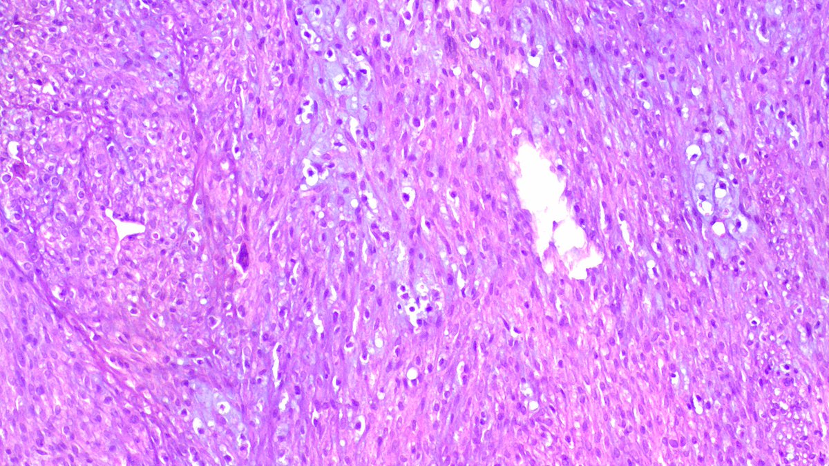
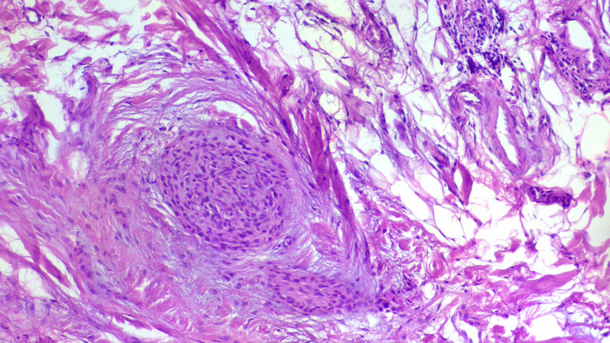
💎 Extramammary Paget Disease (EMPD) •Rare intraepithelial adenocarcinoma of apocrine gland–bearing skin, most commonly involving the vulva, perianal region, scrotum, and axilla. •It typically affects elderly patients and clinically presents as a persistent erythematous, pruritic eczematous plaque, often leading to delayed diagnosis. 🔬 Histology The hallmark is pagetoid spread of malignant glandular cells within the epidermis. Key features: - see histology pictures Typical immunoprofile: Primary EMPD: CK7⁺ / EMA⁺ / CEA⁺ / GCDFP-15⁺ / CK20⁻ Secondary EMPD: CK7⁺ / EMA⁺ / CEA⁺ / CK20⁺ / GCDFP-15⁻ (suggests epidermotropic spread from colorectal or urothelial carcinoma) 🔎 Prognosis & Treatment •Most cases are intraepidermal with relatively good prognosis, though local recurrence is common. Prognosis worsens if dermal invasion or associated malignancy is present. •Treatment usually involves: - Wide local excision or Mohs surgery - Selected cases may receive topical therapy, radiotherapy, or HER2-targeted therapy. ⚓️ Differential Diagnosis (Pagetoid Epidermal Lesions) Melanoma in situ • Pagetoid melanocytes with atypia and pigment • S100, SOX10, HMB45 positive and CK7 negative Pagetoid Bowen disease (SCC in situ) • Atypical keratinocytes with epidermal dysplasia • p63/p40 and HMWCK positive and CK7 negative Langerhans cell histiocytosis • Cells with grooved nuclei and eosinophilic infiltrate • CD1a, langerin, S100 positive and Epithelial markers negative #Dermpath #ExtramammaryPaget #Pathology #GynePath #Histopathology #PathTwitter #TeachingCase
English
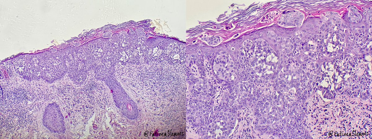
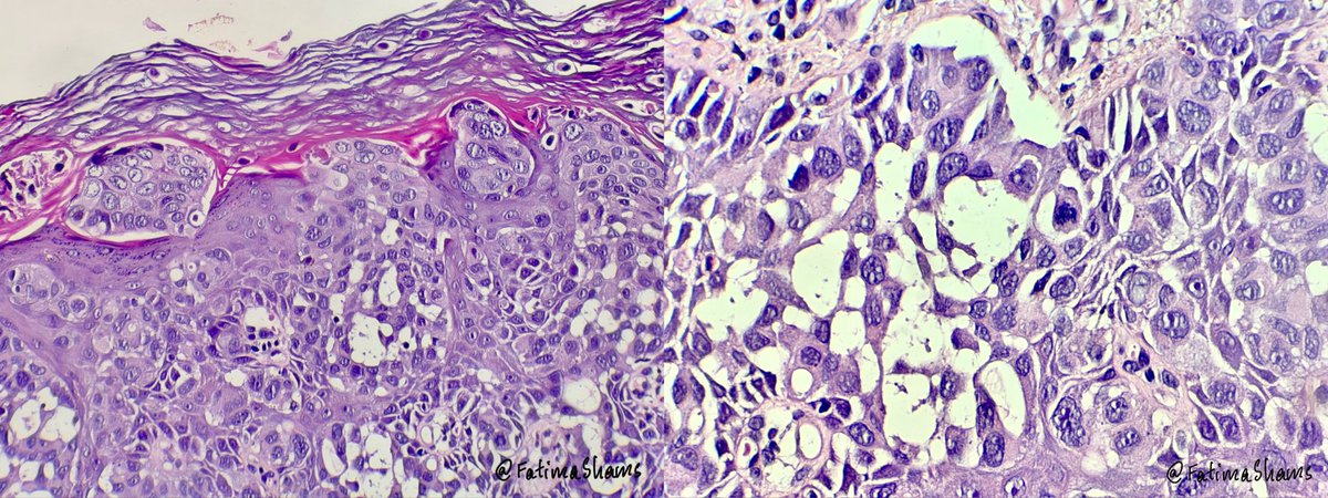
👉 68 YO postmenopausal woman presents with a persistent erythematous plaque on the right labia majora.
⁉️ What is the most likely diagnosis?
#Pathology #Dermatopathology #GynPath #PathTwitter
#MedTwitter #HistoPath #PathPearls


English
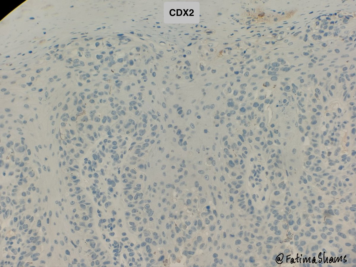

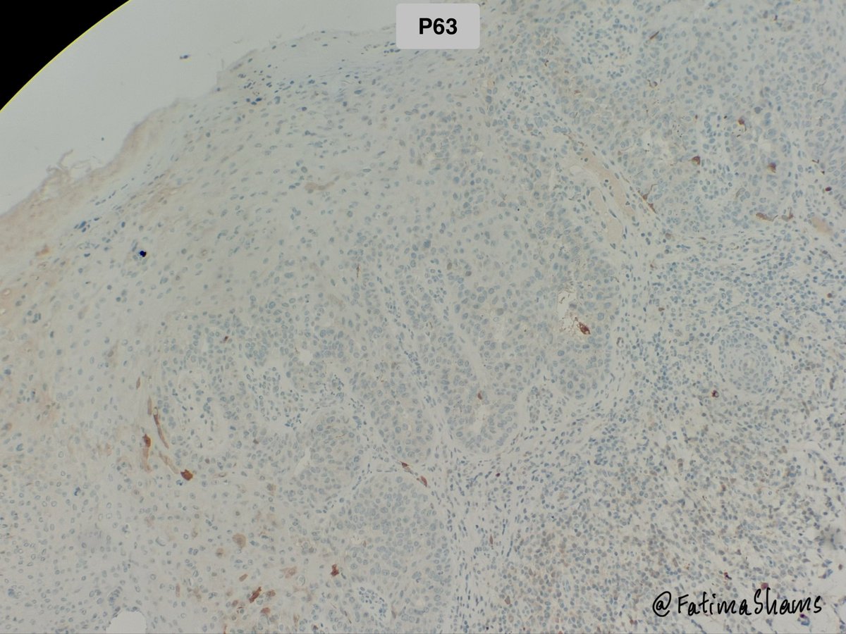
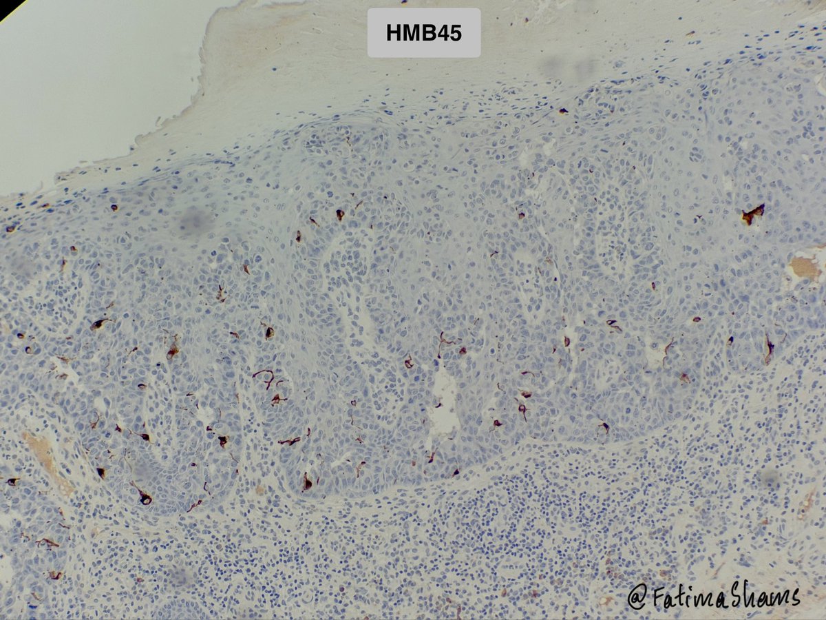
💎 Extramammary Paget Disease (EMPD)
•Rare intraepithelial adenocarcinoma of apocrine gland–bearing skin, most commonly involving the vulva, perianal region, scrotum, and axilla.
•It typically affects elderly patients and clinically presents as a persistent erythematous, pruritic eczematous plaque, often leading to delayed diagnosis.
🔬 Histology
The hallmark is pagetoid spread of malignant glandular cells within the epidermis.
Key features: - see histology pictures
Typical immunoprofile:
Primary EMPD: CK7⁺ / EMA⁺ / CEA⁺ / GCDFP-15⁺ / CK20⁻
Secondary EMPD: CK7⁺ / EMA⁺ / CEA⁺ / CK20⁺ / GCDFP-15⁻ (suggests epidermotropic spread from colorectal or urothelial carcinoma)
🔎 Prognosis & Treatment
•Most cases are intraepidermal with relatively good prognosis, though local recurrence is common. Prognosis worsens if dermal invasion or associated malignancy is present.
•Treatment usually involves:
- Wide local excision or Mohs surgery
- Selected cases may receive topical therapy, radiotherapy, or HER2-targeted therapy.
⚓️ Differential Diagnosis (Pagetoid Epidermal Lesions)
Melanoma in situ
• Pagetoid melanocytes with atypia and pigment
• S100, SOX10, HMB45 positive and CK7 negative
Pagetoid Bowen disease (SCC in situ)
• Atypical keratinocytes with epidermal dysplasia
• p63/p40 and HMWCK positive and CK7 negative
Langerhans cell histiocytosis
• Cells with grooved nuclei and eosinophilic infiltrate
• CD1a, langerin, S100 positive and Epithelial markers negative
#Dermpath #ExtramammaryPaget #Pathology #GynePath #Histopathology #PathTwitter #TeachingCase

English
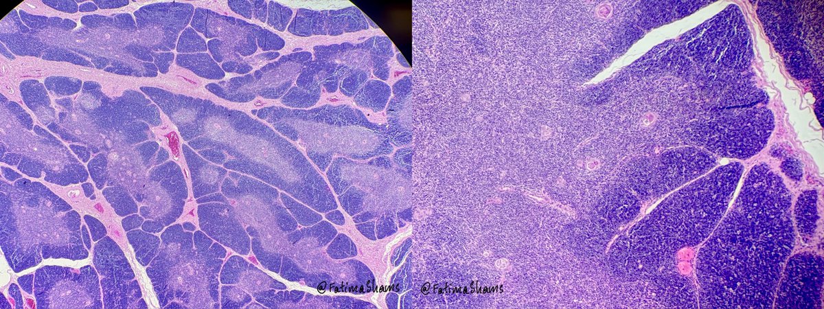
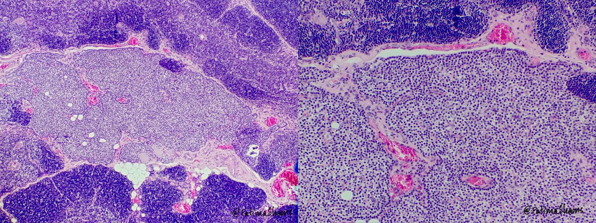
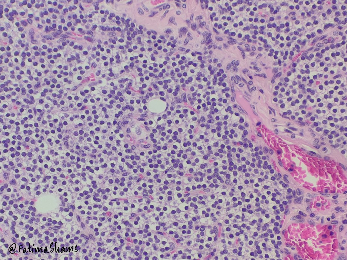
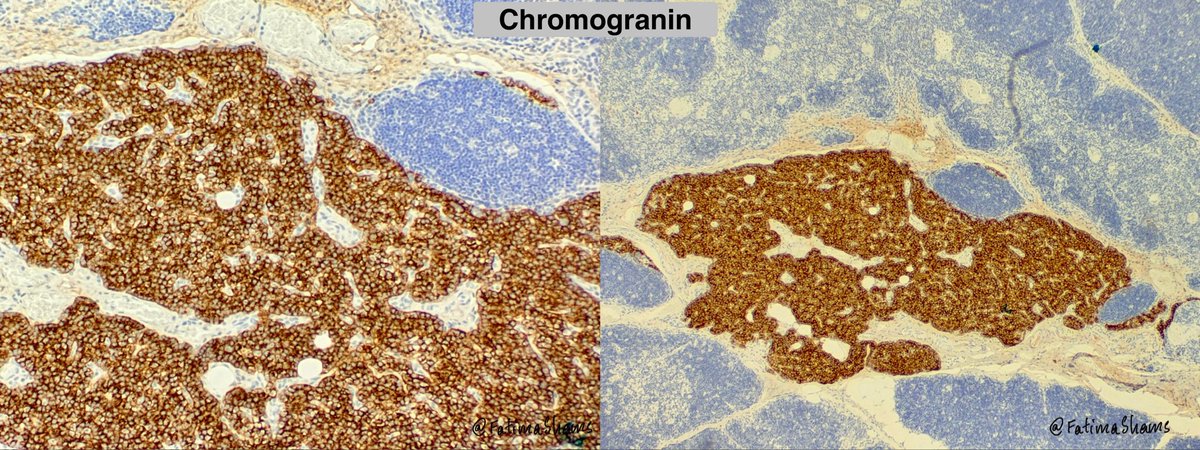
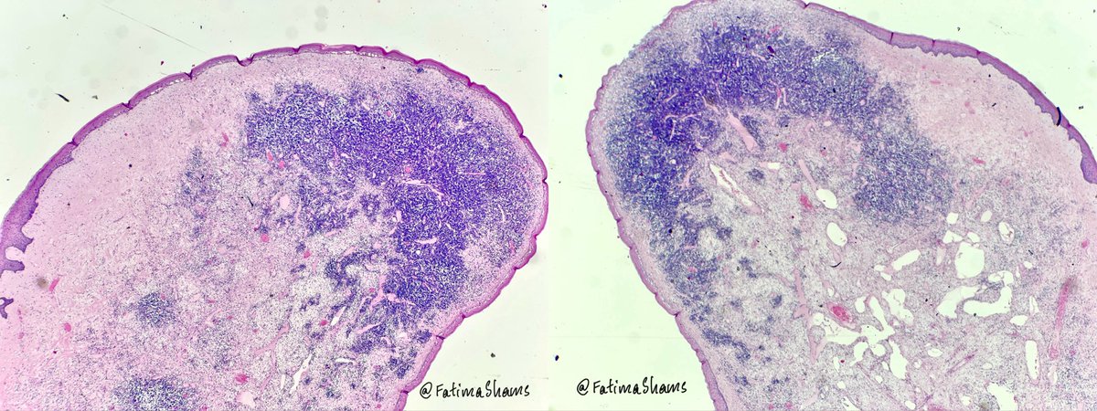
Diagnosis: Ectopic thymus with associated ectopic parathyroid tissue.
A classic example of third pharyngeal pouch derivatives migrating together — a key embryologic concept reflected in surgical pathology.
English
💎 Ectopic Thymus with an Interesting Companion – A Pediatric Neck Mass !
2-year-old boy with right-sided neck swelling.
Histology shows beautifully lobulated thymic tissue with cortex–medulla differentiation and Hassall corpuscles.
But what is this small adjacent nest of uniform cells highlighted on IHC? 👀
🔎 Clue: Think embryologic migration.
#Pathology #PediatricPathology #HeadAndNeckPathology #Embryology #PathTwitter #MedTwitter
English

@DrGeeONE @_Wondweson64 @DanGrahamMD @RFutoran @Immanuel_Paul30 @DrDanok @teddy_wub @MirunaPopescu13 Agree with these. These will be up in my differentials too.
Other lesions with somewhat similar morphology - dermatofibroma, neurothekeoma and other neural tumors, LGFMS - less likely
English

@_Wondweson64 @DanGrahamMD @RFutoran @Immanuel_Paul30 @DrDanok @teddy_wub @MirunaPopescu13 @FatimaShamsPath Differentials:
Plexiform fibrohistiocytic tumour
Giant cell tumour of soft tissue
Nodular fasciitis
English
A 22 Yr 👱♀️
📑Recurrent scalp soft tissue mass, 2x2cm!
💡What do you think X-fam?
👇More in the comments
#pathx #pathtweeter #softtissue

English
Answer provided here
Fatima Shamsuddin@FatimaShamsPath
💎 Tonsillar lymphangiomatous polyp Case history: 26 YO female presented with a left tonsillar polyp. 🔬 Histology Highlights •Polypoid lesion lined by stratified squamous epithelium •Subepithelial proliferation of numerous dilated lymphatic channels containing proteinaceous fluid •Prominent lymphoid aggregates with germinal centers •No cytologic atypia or infiltrative growth IHC: D2-40 highlights lymphatic endothelial lining → confirms lymphatic nature 🎓 Key Teaching Points •Rare benign hamartomatous lesion •Can clinically mimic papilloma or neoplasm •Composed of: ◦Dilated lymphatic channels ◦Fibrous stroma ◦Lymphoid tissue •Complete excision is curative •No malignant potential #Pathology #ENTpathology #HeadAndNeckPathology #Lymphangiomatouspolyp #Histopathology #PathTwitter
English
👉 26 YO female.
Site of lesion - Left tonsil
Diagnosis?
#PathTwitter #TonsilPathology #HeadAndNeckPathology
#Histopathology #SurgicalPathology


English
💎 Tonsillar lymphangiomatous polyp
Case history: 26 YO female presented with a left tonsillar polyp.
🔬 Histology Highlights
•Polypoid lesion lined by stratified squamous epithelium
•Subepithelial proliferation of numerous dilated lymphatic channels containing proteinaceous fluid
•Prominent lymphoid aggregates with germinal centers
•No cytologic atypia or infiltrative growth
IHC: D2-40 highlights lymphatic endothelial lining → confirms lymphatic nature
🎓 Key Teaching Points
•Rare benign hamartomatous lesion
•Can clinically mimic papilloma or neoplasm
•Composed of:
◦Dilated lymphatic channels
◦Fibrous stroma
◦Lymphoid tissue
•Complete excision is curative
•No malignant potential
#Pathology #ENTpathology #HeadAndNeckPathology
#Lymphangiomatouspolyp #Histopathology #PathTwitter


English
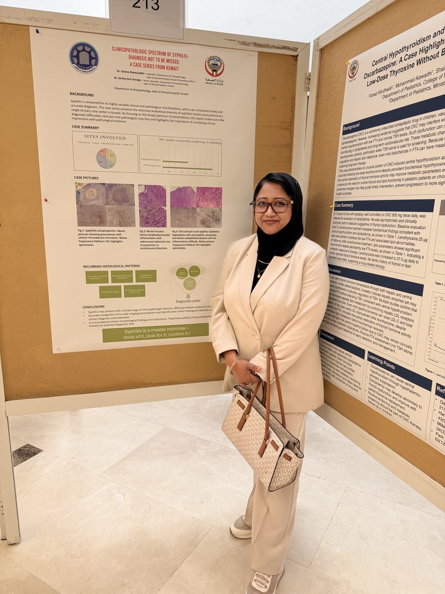
Presented our poster “Clinicopathologic Spectrum of Syphilis – A Diagnosis Not to Be Missed” at the 30th HSC Scientific Conference (11 Feb).
A reminder that syphilis remains a true master mimicker — clinicopathologic correlation is key.
Courtesy @annsmiley78
#Pathology #Syphilis #HSCConference #MedicalResearch #Kuwait
English
Answer provided here
Fatima Shamsuddin@FatimaShamsPath
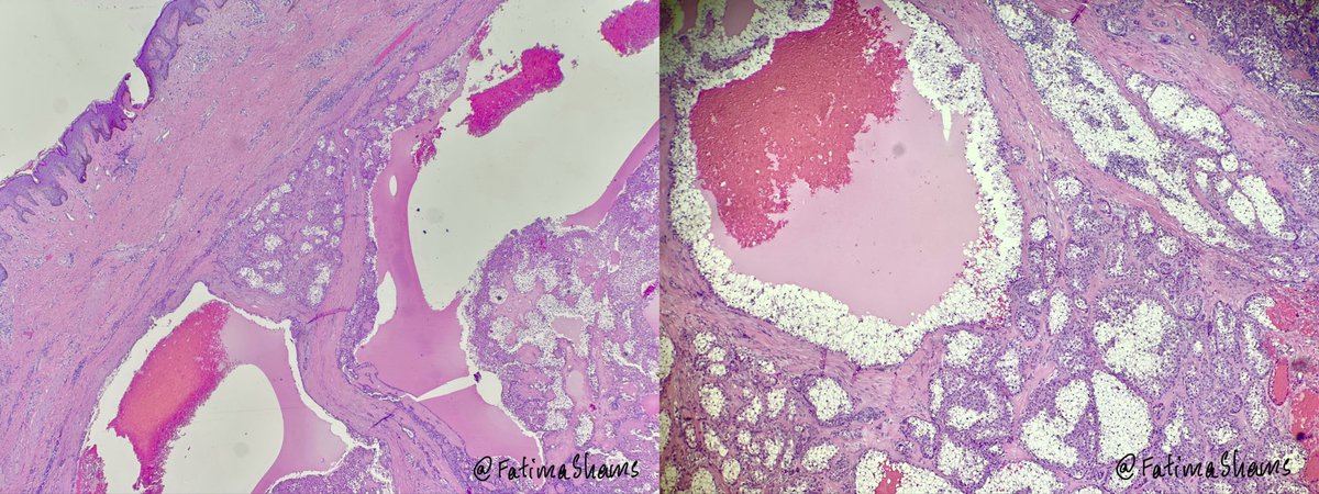
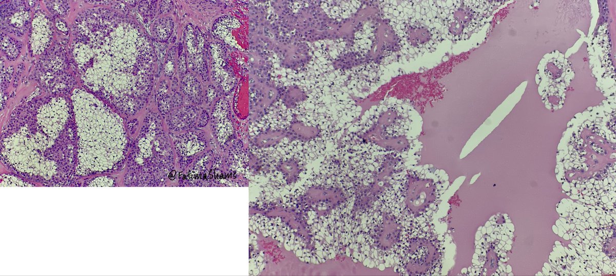
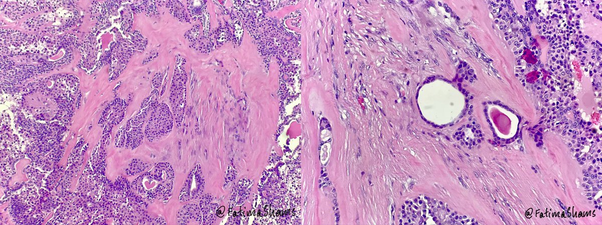
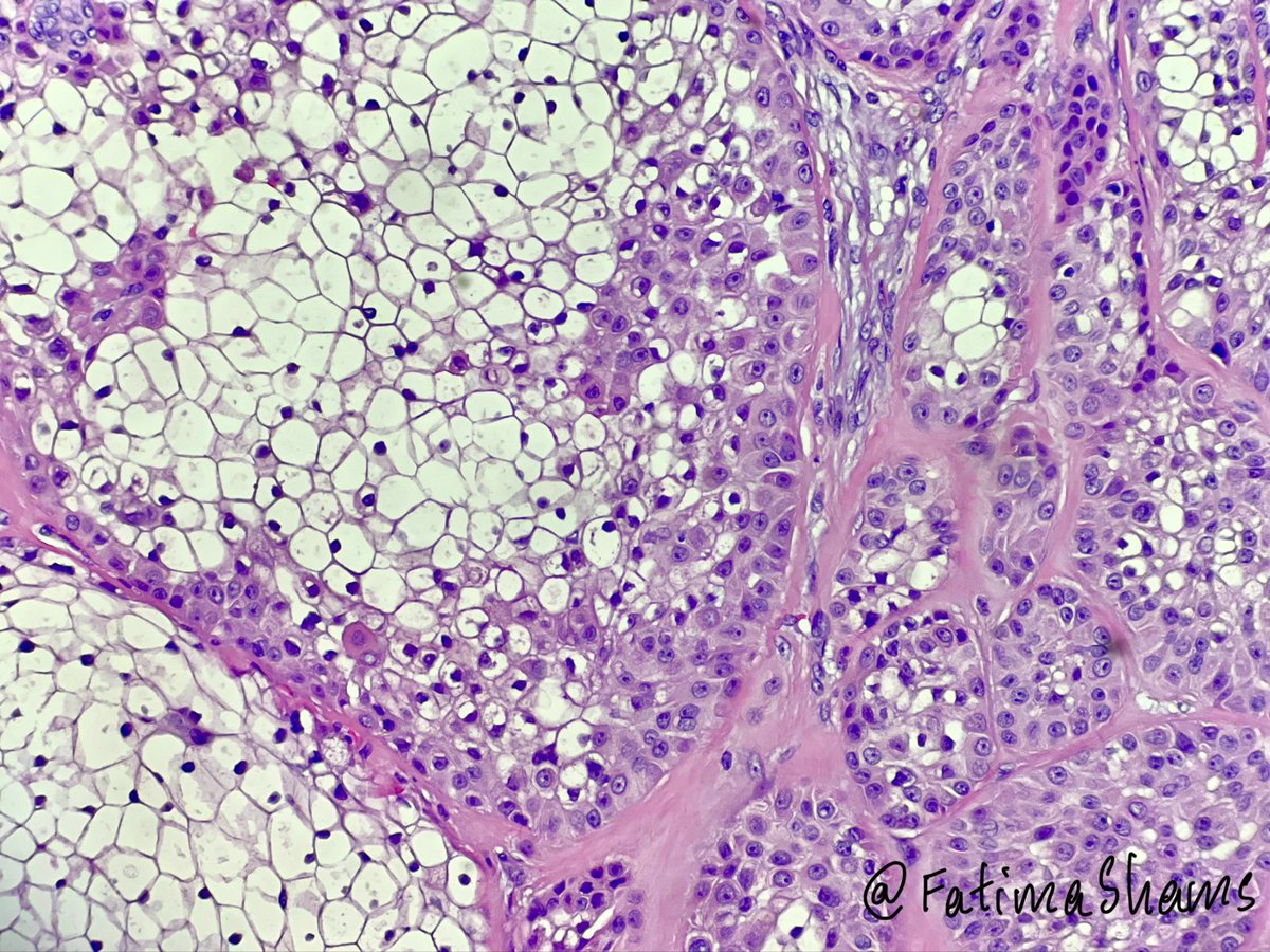
🎯 Clear cell hidradenoma 👉 64-year-old male with right arm soft tissue swelling 🔬Histology highlights:- •Well-circumscribed dermal tumor •Solid and cystic architecture •Lobules of epithelial cells in hyalinized stroma •Prominent clear cells with glycogen-rich cytoplasm •Evidence of ductal differentiation PanCK and EMA positivity shows epithelial and highlights ductal differentiation 🔎 Key differentials:- •Metastatic renal cell carcinoma •Poroma / poroid hidradenoma •Hidradenocarcinoma •Clear cell squamous cell carcinoma 💎 How to distinguish •Lack of infiltrative growth, necrosis, and significant atypia •Presence of ductal structures and biphasic cell population •Clinicopathologic correlation is essential ⚓️ Prognosis •Benign tumor •Excellent prognosis after complete excision •Local recurrence possible if incompletely excised #PathTwitter #Dermatopathology #SkinAdnexalTumor #Hidradenoma #PathologyCase
English
👉 64 YO male presents with a slowly growing soft tissue swelling over the right arm.
Excisional biopsy shows the following histomorphology.
What is your diagnosis?
#Pathology #Dermatopathology #Histopathology #PathTwitter #PathologyCase
English
@Markie27070028 Since clear cells are prominent here, it would be a clear cell hidradenoma.
English
