echobasics

1.1K posts

echobasics banner
echobasics

echobasics

@echobasics

echobasics | introduction to echocardiography | Derliz Mereles, MD | Heidelberg, Germany

Heidelberg Beigetreten Eylül 2010
3.6K Folgt3.5K Follower
Angehefteter Tweet
echobasics
echobasics@echobasics·
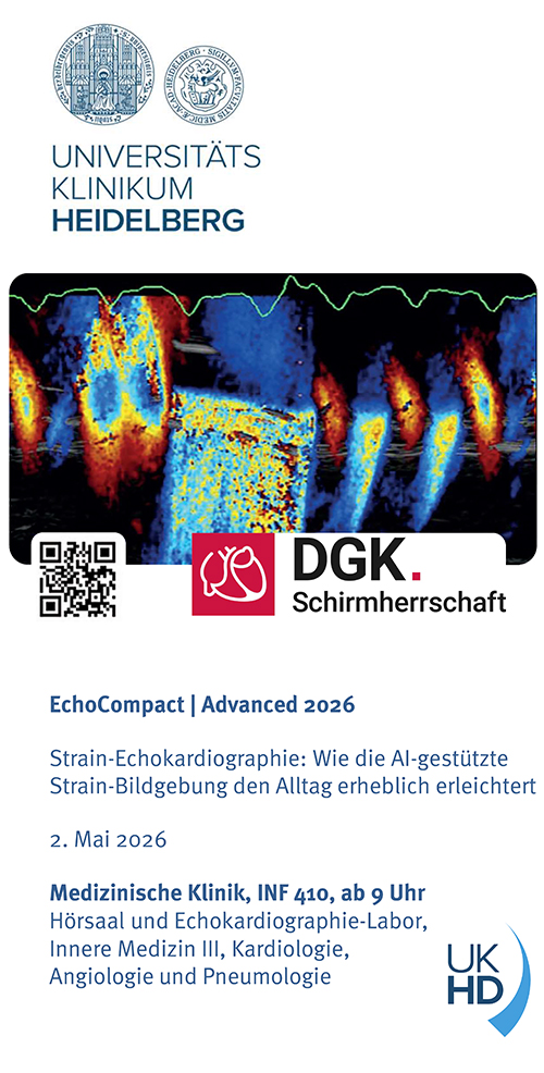
EchoCompact | Advanced am 2. Mai 2026 
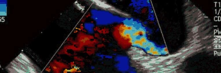
Strain-Echokardiographie: Wie die AI-gestützte Strain-Bildgebung den Alltag erheblich erleichtert echocompact.de
echobasics tweet media
Deutsch
0
0
1
462
echobasics retweetet
Carlos El-Tallawi, MD, FACC, FASE
Intra- and extra- cardiac shunt can coexist. Differentiate mainly via flow pattern not cycle timing. •PFO: transient/intermittent bolus which can be late if RAP>LAP occurs late. •Transpulmonary: continuous trail of bubbles; can occur early for example in liver cirrhosis patients with large stroke volumes.
English
3
31
152
9.6K
echobasics retweetet
Corrado Fiore
Corrado Fiore@fiore_corrado·
💡 Take a look to all the features of rheumatic mitral disease in an young female. 2D/3D TEE with Vivid Pioneer ✅ Hockey stick of AML 🏒 ✅ Extensive fusion of anterolateral commissure ✅ Chordae thickening ✅ Severe MS & mild to moderate MR What should we do? Surgical repair, replacement or Inoue Balloon?
English
6
18
90
4.9K
echobasics retweetet
CardiovascularCorner
CardiovascularCorner@TrackYourHeart·
Diagnosis? A. Cardiac thrombus B. Myxoma C. Vegetation D. Artifact
English
59
70
462
155.5K
echobasics retweetet
Irina Akhmedova
Irina Akhmedova@Irina67790690·
And one more BAV.
English
1
8
60
2.3K
echobasics retweetet
Ross Prager
Ross Prager@ross_prager·
What is your preferred approach to draining an effusion like this causing tamponade? How many mL would you drain in this case.. all or do you stop after XmL to avoid RV failure?
English
10
24
123
19.4K
echobasics retweetet
Nicolas Merke
Nicolas Merke@NMerke·
Quantification of MR especially primary MR using #echofirst requires attention PISA EROA is often the problem 👉 due to many pitfalls 👀 here review shorturl.at/DZsNs But there is a Solution 👊shorturl.at/xVAfb Using volumetric method
Nicolas Merke tweet mediaNicolas Merke tweet media
English
5
33
97
7.8K
echobasics retweetet
gracielly barros
gracielly barros@gracybarros·
Mitral valve with localized degenerative disease (P2 flail), preserved and symmetric annular geometry, showing a primary mechanism of mitral regurgitation (Carpentier type II). Volumetric reconstruction with precise anatomical mapping for valve repair planning.#echofirst #philips
English
1
13
56
4.1K
echobasics retweetet
Ahmed Ata
Ahmed Ata@Ahmedata7777·
Septal Bounce in Constrictive Pericarditis: 🫀 What is Septal Bounce? Definition: A paradoxical early diastolic movement of the interventricular septum toward the left ventricle. Timing: Occurs during early diastole, often visible on M-mode, 2D, or with real-time imaging. Cause: Results from exaggerated ventricular interdependence due to a stiff, non-compliant pericardium. In constrictive pericarditis, the thickened pericardium limits normal diastolic filling, causing pressure discordance between ventricles. 🔍 Echocardiographic Findings: ▪︎2D echo: “Bounce” or abrupt septal shift with respiration (usually toward the left during inspiration). ▪︎M-mode: Shows a “notch” or abrupt early diastolic motion. ▪︎Doppler: May show respiratory variation in mitral and tricuspid inflow velocities. 📋 Differential Diagnoses ▪︎Constrictive pericarditis (most common cause). Occasionally seen in: ▪︎Severe right ventricular volume overload. ▪︎Post-pericardiectomy. ▪︎Effusive-constrictive pericarditis. 🎯 Clinical Relevance Helps differentiate constrictive pericarditis from restrictive cardiomyopathy: Septal bounce = Constrictive pericarditis.
Ahmed Ata tweet media
English
2
76
313
14.6K
echobasics retweetet
Irina Akhmedova
Irina Akhmedova@Irina67790690·
Double Orifice of the Mitral Valve. 17 years old As a child, he underwent surgery for coarctation of the aorta and VSD.
English
4
40
156
8.6K