Carlos El-Tallawi, MD, FACC, FASE
1.5K posts

Carlos El-Tallawi, MD, FACC, FASE
@HeartToProve
Conscious lump of atoms posting edutaining tweets | AUBMC cardiology | Houston Methodist Multimodality CV Imaging | Echo + CMR | Valvular heart disease — MVP
Houston Methodist Hospital Katılım Nisan 2017
86 Takip Edilen12.5K Takipçiler

@bwoody58 @ahmad_70x @PWesslyMD @LucySafi @PriyaPanday27 @NadeenFaza @ASE360 @JournalASEcho The initial E1 deflection represents the emptied prolapse volume, and E2 is the atrial volume that typically follows.
English

@ahmad_70x @HeartToProve @PWesslyMD @LucySafi @PriyaPanday27 @NadeenFaza @ASE360 @JournalASEcho I have this ? also. My guess is relaxation of the curled segment out of phase with rest of LV 🤔
English

#ASEchoJC Q 10 What are curling and the Picklehaube sign and how are these related to papillary muscle traction? @LucySafi @PriyaPanday27 @NadeenFaza @HeartToProve
#ASEchoJC #EchoFirst #CardioTwitter #EPeeps #MVP @ASE360 @JournalASEcho

English
Carlos El-Tallawi, MD, FACC, FASE retweetledi

🔍 What exactly is prolapse volume?
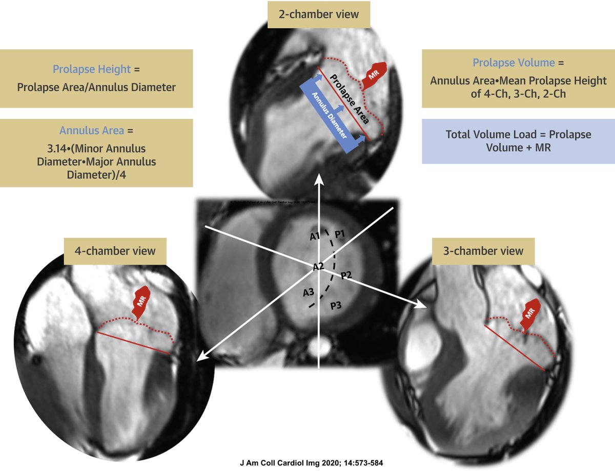
Prolapse volume is the "hidden" volume trapped between the prolapsing mitral leaflets and the annulus at end-systole.
This volume:
➡️ Is not forward stroke volume
➡️ Is not transvalvular MR
➡️ Returns to the LV during diastole
🫀 Total LV volume load = MR volume + prolapse volume
💎 A paradigm-shifting concept introduced by @HeartToProve @DipanJShah @WilliamZoghbi and colleagues that may help explain disproportionate LV enlargement and remodeling in Barlow MVP.
#ASEchoJC #EchoFirst #MVP @ASE360 #CardioX #MVP @JournalASEcho

English
Carlos El-Tallawi, MD, FACC, FASE retweetledi

💎 Why does the LV sometimes appear "too enlarged" for the degree of MR in MVP?
🫀 Because MR is not the entire volume load.
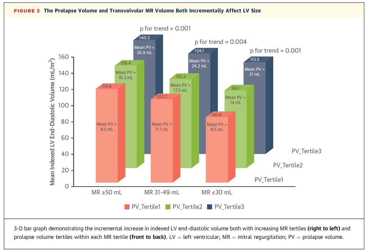
In the landmark work by @HeartToProve and colleagues, adding prolapse volume to MR volume strengthened the relationship between volume load and LV enlargement.
🎯 Total volume load = MR volume + prolapse volume
A powerful concept that may explain disproportionate LV remodeling in Barlow MVP.
#ASEchoJC #EchoFirst #MVP @ASE360 @JournalASEcho #CardioX

English
Carlos El-Tallawi, MD, FACC, FASE retweetledi

Such an elegant observation, @HeartToProve 👏
💎 The bifid-E wave may be a novel Doppler marker of advanced MVP.
Associated with:
➡️ Bileaflet MVP (33% vs 6% in single leaflet prolapse)
➡️ Larger prolapse volume
➡️ Disproportionate LV enlargement despite mild MR
➡️ LV replacement fibrosis
🔑 On multivariable analysis, the bifid-E wave was the only echocardiographic parameter independently associated with MVP-related fibrosis.
🔍 Sometimes the most important clues are hiding in plain sight on the mitral inflow Doppler.
#ASEchoJC #EchoFirst @ASE360 @JournalASEcho #MVP #CardioX
Carlos El-Tallawi, MD, FACC, FASE@HeartToProve
@PWesslyMD @LucySafi @PriyaPanday27 @NadeenFaza @ASE360 @JournalASEcho Our group also identified a novel Doppler sign —Bifid-E wave— that could represent an additional echocardiographic marker of advanced MVP and myocardial fibrosis. Keep an eye out for this next time you're reading a Barlow Echo. #ASEchoJC
English
Carlos El-Tallawi, MD, FACC, FASE retweetledi

Such an important point, @HeartToProve 💎
As highlighted in your editorial 👇, not all flails are created equal.
🎯 A flail leaflet should raise suspicion for severe MR—but it is not synonymous with severe MR. An integrated assessment remains essential.
#ASEchoJC #EchoFirst @ASE360 @JournalASEcho #MVP #WhyCMR

Carlos El-Tallawi, MD, FACC, FASE@HeartToProve
@PWesslyMD @ASE360 @JournalASEcho Very important -- flail =/= severe MR! In our data a flail leaflet had 81% specificity for severe MR as defined on CMR >60 mL/50% #ASEchoJC
English
Carlos El-Tallawi, MD, FACC, FASE retweetledi

✨ Huge thank you to everyone who joined tonight's #ASEchoJC!
What an incredible discussion on MVP, MR, MAD, ventricular arrhythmias, and the evolving concept of arrhythmic MVP. 🫀⚡️
Grateful to our outstanding guest authors Dr. Jeffrey Silbiger @LucySafi, @priyaPanday27, and for sharing their expertise and insights, and to my wonderful co-moderators @NadeenFaza and @HeartToProve. A special thank you to our #EchoFirst enthusiasts and engaged audience for contributing to such a lively, thoughtful, and high-yield discussion.
Proud to be part of this amazing global #EchoFirst community! 💙🌍 @ASE360 @JournalASEcho

English

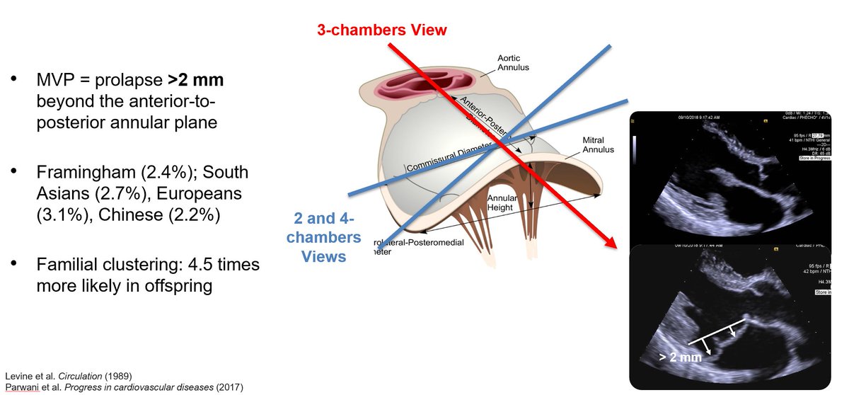
#ASEchoJC Q 1 What are the echocardiographic definition, prevalence, and clinical importance of mitral valve prolapse (MVP)?
@LucySafi @PriyaPanday27 @NadeenFaza @HeartToProve @ASE360 @JournalASEcho #Echofirst #MVP #CardioX

English

@PWesslyMD @LucySafi @PriyaPanday27 @NadeenFaza @ASE360 @JournalASEcho Prolapsing valves pull on chords/papillary muscles, stretching the LV wall inward. This causes "curling" (LV crest rotates posteriorly) & the Pickelhaube sign -- a sharp late-systolic S’ wave >16 cm/s on TDI. Both are markers of increased arrhythmia risk! #ASEchoJC

English

@PWesslyMD @ASE360 @JournalASEcho Very important -- flail =/= severe MR! In our data a flail leaflet had 81% specificity for severe MR as defined on CMR >60 mL/50% #ASEchoJC

English

⚠️ Yet not all flails are created equal—not every flail leaflet results in severe MR.
🔑 MR severity depends on the extent of coaptation loss, not simply the presence of a flail leaflet. 👇
#ASEchoJC #EchoFirst #CardioTwitter #MVP @ASE360 @JournalASEcho


English

ASEchoJC Q 4 What mechanisms contribute to mitral regurgitation in MVP?
@LucySafi @PriyaPanday27 @NadeenFaza @HeartToProve @ASE360 @JournalASEcho

Română

@PWesslyMD @LucySafi @PriyaPanday27 @NadeenFaza @ASE360 @JournalASEcho @DavidWienerMD @iamritu @purviparwani @argulian @boegel_kelly @SIwa23288585 @EACVIPresident We have shown that for the same amount of transvalvular MR, the LV volume increases with increasing prolapse volume. Be careful with using PISA in Barlow MR -- use volumetric methods instead. #ASEchoJC

English

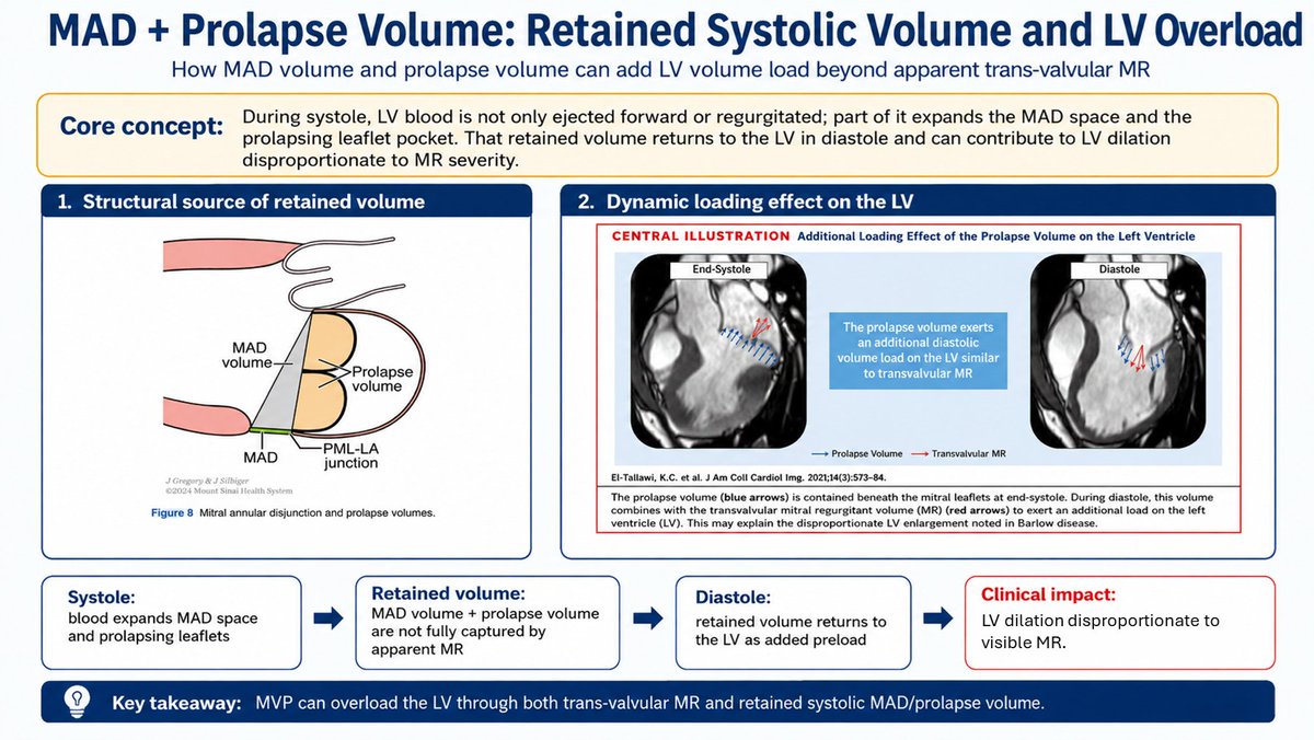
#ASEchoJC Q 8 How do MAD and prolapse volume contribute to LV overload and MVP-related cardiomyopathy?
@LucySafi @PriyaPanday27 @NadeenFaza @HeartToProve
#ASEchoJC #EchoFirst #CardioX #MVP @ASE360 @JournalASEcho

English

@PWesslyMD @LucySafi @PriyaPanday27 @NadeenFaza @ASE360 @JournalASEcho @DavidWienerMD @iamritu @purviparwani @argulian @boegel_kelly @SIwa23288585 @EACVIPresident #ASEchoJC Blood isn't just regurgitated; it also expands the prolapsing leaflets (prolapse volume) during systole. This "retained" volume drives LV volume overload independently of MR, leading to excess LV dilation that is disproportionate to the perceived trans-valvular MR.

English

@PWesslyMD @LucySafi @PriyaPanday27 @NadeenFaza @ASE360 @JournalASEcho @DavidWienerMD @iamritu @purviparwani @SIwa23288585 @boegel_kelly MAD is an epiphenomenon for arrhythmias, as opposed to a true trigger. True MAD reflects advanced myxomatous degeneration and MVP. MAD is a reflection of the prolapse severity/height and extent of pulling on the pap muscles, which eventually contributes to arrhythmias. #ASEchoJC
English

@PWesslyMD @LucySafi @PriyaPanday27 @NadeenFaza @ASE360 @JournalASEcho @DavidWienerMD @iamritu @purviparwani @SIwa23288585 @boegel_kelly MAD is a true separation between the LV crest & posterior mitral leaflet/LA junction. Pseudo-MAD mimics this during systole when a voluminous leaflet presses against the LA wall, but it vanishes in diastole. Distinguishing them is key! #ASEchoJC

English

#ASEchoJC Q 6 What is mitral annular disjunction (MAD), how is it distinguished from pseudo-MAD, and why is it important?
@LucySafi @PriyaPanday27 @NadeenFaza @HeartToProve @ASE360 @JournalASEcho

English

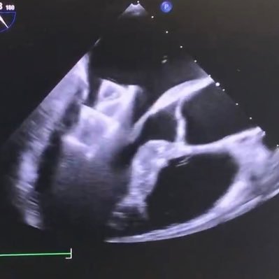
@PWesslyMD @LucySafi @PriyaPanday27 @NadeenFaza @ASE360 @DavidWienerMD @iamritu @purviparwani @SIwa23288585 @boegel_kelly @JournalASEcho Mitral commissural flails are the trickiest to diagnose on echo because of their very eccentric MR jets. Demo of medial commissure (MC) flail and expected jet trajectory:
#ASEchoJC
English

#ASEchoJC Q 5 What are the characteristic MR jet patterns in MVP, and what are the pitfalls in MR quantification?
@LucySafi @PriyaPanday27 @NadeenFaza @HeartToProve

English

@PWesslyMD Very educational—an important reminder that MVP should be defined in the PLAX view, given the saddle-shaped mitral annulus. Thank you for sharing🥹
English

#ASEchoJC Q 2 How do Barlow's disease and fibroelastic deficiency differ in morphology, genetics, and clinical presentation? @LucySafi @PriyaPanday27 @NadeenFaza @HeartToProve @ASE360

English

@NadeenFaza @PWesslyMD @LucySafi @PriyaPanday27 @ASE360 @JournalASEcho @purviparwani @iamritu @DavidWienerMD @SIwa23288585 @EACVIPresident If you happen to find this heart abnormality intersting -- follow this #ASEchoJC
English

📍 Definition: MVP is defined by systolic displacement of one or both mitral valve leaflets ≥2 mm above the mitral annular plane in the parasternal or apical long-axis view. 📈
👥 Prevalence:MVP affects ~2.4–2.5% of the population, making it one of the most common valvular heart diseases. 🌎
⚠️ Why does it matter?
🔹Most common cause of primary mitral regurgitation (MR)
🔹Can lead to progressive valve degeneration and chordal rupture
🔹Associated with ventricular arrhythmias and sudden cardiac death in selected patients
🔹Early recognition helps guide surveillance, timing of intervention, and arrhythmic risk assessment
💡 MVP is much more than a "benign click"—it represents a spectrum ranging from mild leaflet prolapse to severe MR and arrhythmic MVP. #ASEchoJC
English

@mderdem3 @WilliamZoghbi @DeBakeyCVedu @Sadeer_AlKindi @SachinGoelMD @SLittleMD @NadeenFaza Star ⭐️ Well deserved Saliha and colleagues for this important research effort.
English

Post call , after hours and some sleepless nights, this is how much this project took 🙂 I am grateful for this recognition by Dr @WilliamZoghbi , Dr @HeartToProve and @DeBakeyCVedu . This would not have been possible without supervision of Dr @Sadeer_AlKindi , thank you 🙏🏻


English