Kevin Cheng retweetledi

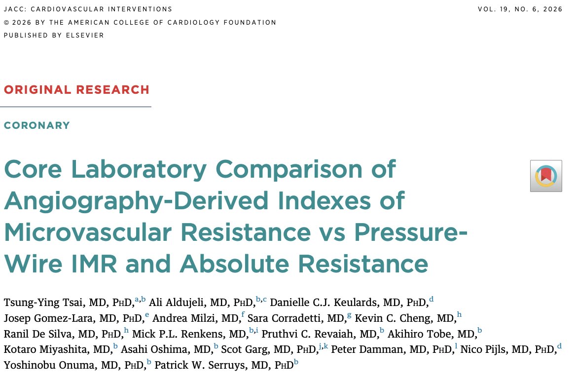
Tsai et al. just published a very elegant core lab comparison of 5 angio-IMR methods against pressure wire IMR and continuous thermodilution Rmicro in JACC Interventions (@JACCJournals). The results were perhaps a bit disappointing: all 5 methods performed poorly — AUCs of 0.53–0.58 against PW-IMR ≥25. Essentially coin-flip territory.
I guess everyone’s natural reaction is to blame angio-IMR. But maybe the real story here goes a bit deeper. So, before we condemn the index test, I wanted to look into the ruler.
Tsai et al. compared 5 angio-IMR formulas from 3 software platforms (QAngio XA 3D, FlashAngio, AngioPlus Core) against PW-IMR in 274 vessels and Rmicro in 109 vessels, all analyzed by an independent core lab (CORRIB, Galway) with rigorous blinding. Three independent analysts, anonymized software, a dedicated matcher investigator selecting frames. Methodologically, this is as clean as it gets.
The results well, were not good: correlations between angio-IMR and PW-IMR ranged from r = 0.04 to 0.12. Three methods massively overestimated IMR (mean bias –20 to –26 units). None discriminated PW-IMR ≥25 better than chance. Against Rmicro, three methods did somewhat better (AUC 0.68–0.70), but still clinically inadequate.
IMR was introduced by Fearon et al. in 2003, validated in a porcine model where microspheres obliterated the microcirculation. The logic is Ohm's law: resistance = pressure / flow. Since flow ∝1/mean transit time, IMR = distal pressure × transit time during hyperemia.
It was then tested in STEMI patients, where it correlated with infarct size (biomarkers, PET, CMR) and predicted mortality. In a registry of 253 patients, IMR >40 post-primary PCI was the only independent predictor of death or heart failure readmission at 1 year.
From there, IMR became a widespread measurement method for microvascular resistance. But notice the leap: it was validated in extreme microvascular destruction. But what about other populations, including ANOCA where the measured differences might be more subtle?
Gallinoro et al. (EuroIntervention 2023) performed duplicate bolus thermodilution measurements in 102 ANOCA patients. The test-retest variability of IMR was 24.2 ± 19.3%.
What does that mean clinically? If a patient's true IMR is 22 (just below the threshold of 25), the 95% range on a repeat measurement spans roughly 17–27. That patient has about a 30% chance of being classified as "abnormal" on any given measurement. Conversely, a true IMR of 28 spans ~21 to 35 — roughly 25–30% chance of being called "normal."
In STEMI, this noise actually doesn't matter as much. I mean, if your true IMR is 70, the 95% range is ~53–87. So the chance of this case be misclassified is very low. The signal (IMR values of 60–100 vs. 10–20) overwhelms the noise, with a very strong signal-to-noise ratio (3:1 to 10:1).
In ANOCA, the population clusters right on the threshold. Median PW-IMR in Tsai et al. was 19.02, IQR 12.79–28.08. The "abnormal" ANOCA patients sit at ~28–40 and the "normal" ones at ~12–22. The between-group gap is 10–20 units. Apply 24% noise and the signal-to-noise ratio drops to ~1:1. Your discriminatory capacity shrinks considerably.
When continuous thermodilution came along — validated against ¹⁵O-H₂O PET (the closest thing we have to a true gold standard for myocardial flow) — it provided absolute volumetric flow and resistance. It is in fact more reproducible: variability of ~12% vs. ~24% for bolus thermodilution.
Now, here’s the thing: IMR does not correlate with absolute hyperemic microvascular resistance (Rμ-hyper) derived from continuous thermodilution. r = 0.06, P = 0.425 (Gallinoro et al., JACC Intv 2023). From another Gallinoro paper, published in EuroIntervention in 2023, reproducibility paper (n=102, ANOCA), in which they explicitly report reclassification rates for CFR, the percentage of disagreement between CFRcont and CFRbolus was 35.3%. So at the standard CFR ≤ 2.5 cutoff, the two methods disagree on classification in over a third of patients. For IMR vs. MRR specifically, the correlation was essentially zero (r = 0.1, P = 0.305), meaning binary agreement can't be much better. An article by Fawaz et al. (IJC Heart Vasc 2024, 96 patients, 116 vessels post-revascularization) confirmed this in a different population: when assessed at CFR cut-off values of 2.0 and 2.5, the methods disagreed in 35% and 39% of cases, respectively. An article by Jansen et al., published in the JAHA in 2023 (n=246 ANOCA) is explicit about the overall picture, finding a relatively high amount of classification disagreement between bolus and continuous thermodilution measurements, of which the pathophysiological meaning is unknown.
The Jansen et al. paper adds something else. CFR and IMR from bolus thermodilution did not correlate with anginal symptoms, while continuous thermodilution-derived parameters did. The Stanford transplant study (Circ Cardiovasc Interv 2025) added even more confusion, since in 20 transplant patients, IMR had excellent reproducibility (ICC 0.95) — far better than in ANOCA. So it appears that IMR behaves differently depending on the population.
Now, I wanted to take a moment to think about the validation of IMR. To me, there’s some circular reasoning here, because the validation chain went something like this:
1. Tested in large microvascular destruction (animal models, acute STEMI) → IMR was able to differentiate injured from intact microvascular circulation in these very extreme scenarios.
2. Therefore, we assumed that IMR measures microvascular resistance accurately.
3. So, we assumed we can correctly measure & classify patients in other clinical settings.
4. So, we used it to measure other tools against because IMR works in all scenarios because it worked in extreme ones.
I mean, to me it seems that we might be saying something like: I have this thermometer that can correctly tell ice from boiling water (it is). But I can’t say it's accurate at distinguishing 36.8°C from 37.2°C.
Now let’s place angio-IMR in this context. It compounds multiple noisy inputs: contrast flow velocity from TIMI frame counts (which correlates poorly with true hyperemic velocity: r = 0.17–0.26 in Tsai et al.), angiographic contour detection (with known inaccuracies that get squared when converting diameter to area), estimated hyperemic pressure, and angio-FFR itself (which had only modest correlation with PW-FFR in this study: r = 0.33–0.38). Each error source multiplies through the formula. It's not surprising that the output is noisy. Maybe we’re measuring noise vs noise.
The theoretical maximum AUC that any index test (even a perfect one) could achieve against PW-IMR ≥25, given PW-IMR's own ~25% misclassification rate around the threshold, is probably 0.80–0.85. The observed 0.53–0.58 is still below that ceiling (yes), so it appears that angio-IMR as a whole has real problems beyond reference noise. But the gap to "perfection" is much narrower than it looks at first glance.
Now, someone might say: "But CorMicA proved IMR-guided therapy works in ANOCA." True, so true — randomized, stratified treatment based on IMR/CFR improved angina & quality of life at 1 year. But to me it seems that IMR probably works as a crude triage tool, not as a precise diagnostic test. You don't need a calibrated thermometer to know someone with a fever should take antipyretics — a hand on the forehead often suffices. CorMicA proved the hand-on-the-forehead approach to CMD management is better than guessing. It didn't prove the thermometer reads accurately.
Now, where do we stand?
Regardless of all of that, what Tsai et al. proves:
→ Current angio-IMR methods cannot replace invasive PW-IMR at the individual patient level.
→ The compounding of errors in frame-count-based formulas degrades signal below clinical utility.
What Tsai et al. does NOT prove:
→ That angio-IMR is clinically useless. Prognostic value ≠ diagnostic accuracy. A noisy estimate that correlates with outcomes may still have clinical value as a screening or stratification tool, even if it can't match the absolute number.
What I think is reasonable:
→ PW-IMR is not the stable, precise reference standard we treat it as. Its validation was built in high-signal environments and extrapolated without verification to low-signal ones.
→ Continuous thermodilution (Rmicro, MRR) is more reproducible and correlates with symptoms, but isn't widely available and has its own limitations.
→ We may be trying to reduce a complex, multidimensional pathophysiology (structural vs. functional vs. vasospastic CMD) to a single number.
What should come next:
→ Validate angio-IMR (and any future wire-free tool) against Rmicro as the primary reference, not PW-IMR.
→ Prospective studies in pure ANOCA cohorts with full endotyping (including acetylcholine) and sufficient power.
→ Move from single-frame CFD equations to complete cine-run analysis with machine learning — the data is there in every angiogram, we're just not extracting it yet.
→ Accept that the microcirculation may require multiple measurements, not a single index.
The story of angio-IMR isn't a story of failure. The same goes for IMR. It's a story of a field trying to measure something very subtle, very fragile, very complex. And to me this disposition to distill the complex interplay of cardiac disease and willingness to go deep into problems is something we should be proud of.
#CardiologyX #InterventionalCardiology #IMR #JACC #angioIMR
English