
Leidysabel Celis
1.5K posts
Leidysabel Celis
@LeidysabelC
Medico Cirujano UC. Especialista en Radiodiagnóstico UCV. HMPC. PCM. CMM
Caracas Katılım Şubat 2014
899 Takip Edilen267 Takipçiler

Leidysabel Celis retweetledi


Leidysabel Celis retweetledi

"El secreto del demagogo es hacerse tan estúpido como su audiencia, de tal modo que crean que son tan inteligentes como él"
Karl Krauss

Español
Leidysabel Celis retweetledi

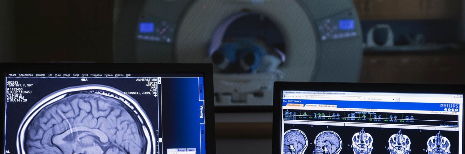
l've got your back — literally!
A solid grasp of lumbar spine anatomy is essential to be able to find pathology and be accurate in reporting.
You need to know where the nerve roots lie in the central canal and neural foramina to know when they are compressed.
And you need to know your disc nomenclature to accurately describe the pathology
This video shows you the anatomy and nomenclature you NEED to know!
Because when your anatomy knowledge is weak, your reports can really be a pain in the back!
English

Me informan que María Corina Machado está herida en una pierna al caer y en un brazo…
Español

hahahahaha me recuerdo de esto🤣🤣🤣
Fader SHK@faderg2_
Shakira: A la orden, ahhh no tú no estás a la orden @lelepons. 😂💋
Português
@shakira Imposible comprar las boletas en Colombia 😭
Ven a Venezuela por favor 🥹🙏
Español

Uno de los vídeos más divertidos de mi vida! Espero lo disfruten! One of the most fun and funniest videos of my life! I hope you enjoy it 💋#Soltera youtu.be/oBofuVYDoG4

YouTube


Español
Leidysabel Celis retweetledi

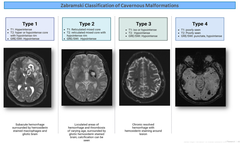
Zabramski classification of cavernous malformations
A type 1 cavernous malformation is one with acute or subacute hemorrhage characterized radiographically by hyperintense T1 signal and hyper- or hypointense T2 core (1st panel, axial T2 brain MRI). Pathologically, these lesions display subacute hemorrhage surrounded by hemosiderin-laden macrophages and gliotic brain.
A type 2 cavernous malformation is the classic-appearing "popcorn"-like lesion characterized radiographically by a mixed reticulated core on T1 and T2 images (2nd panel, axial T2 brain MRI). There is often a "ring" of hemosiderin (hypointense T2). This correlates pathologically with loculated, intracavernous hemorrhage and thrombosis, surrounded by gliotic hemosiderin-stained brain.
A type 3 cavernous malformation is hypointense on both T1 and T2. Pathologically, type 3 cavernous malformations display chronic hemorrhage with hemosiderin.
Type 4 cavernous malformations are not visible on standard MRI sequences. They can only be visualized on hemosiderin-sensitive sequences (gradient echo or susceptibility-weighted imaging [SWI])
Reference: ncbi.nlm.nih.gov/books/NBK1293/…
English


Leidysabel Celis retweetledi
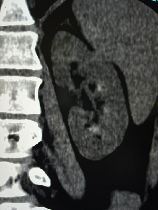
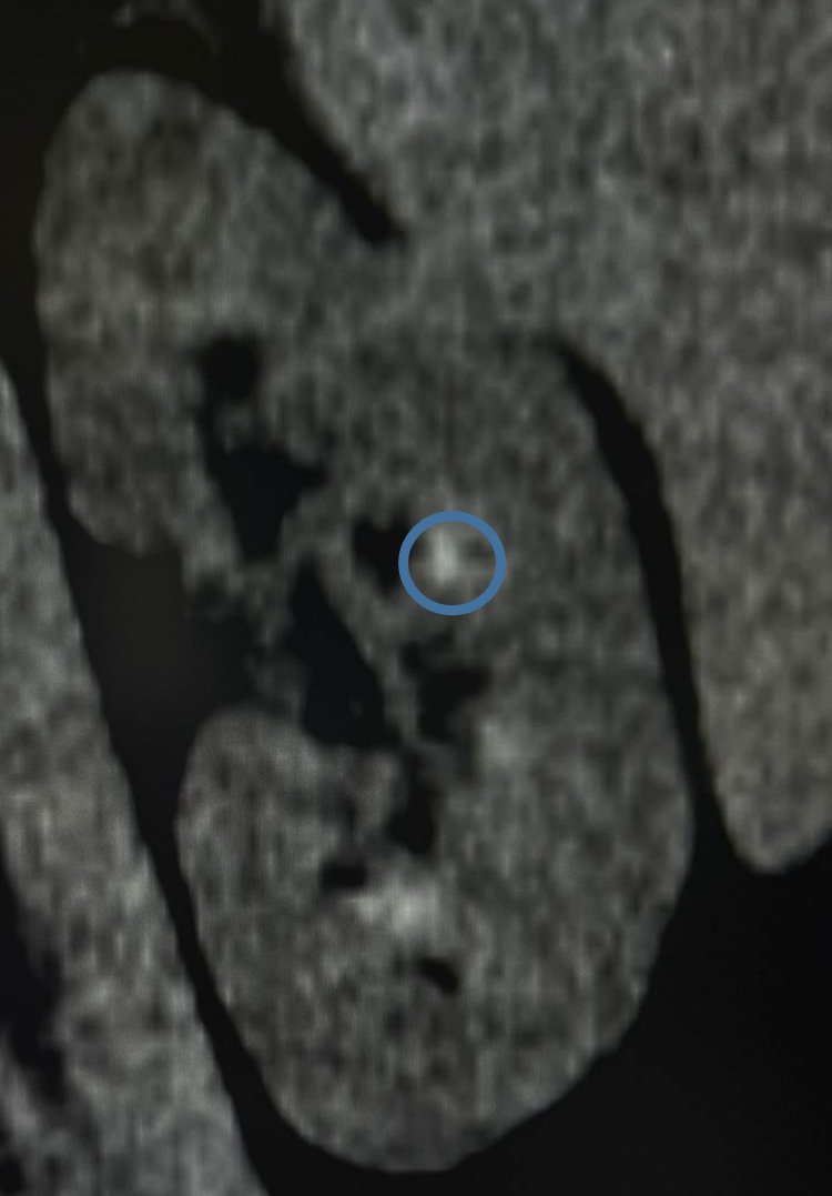
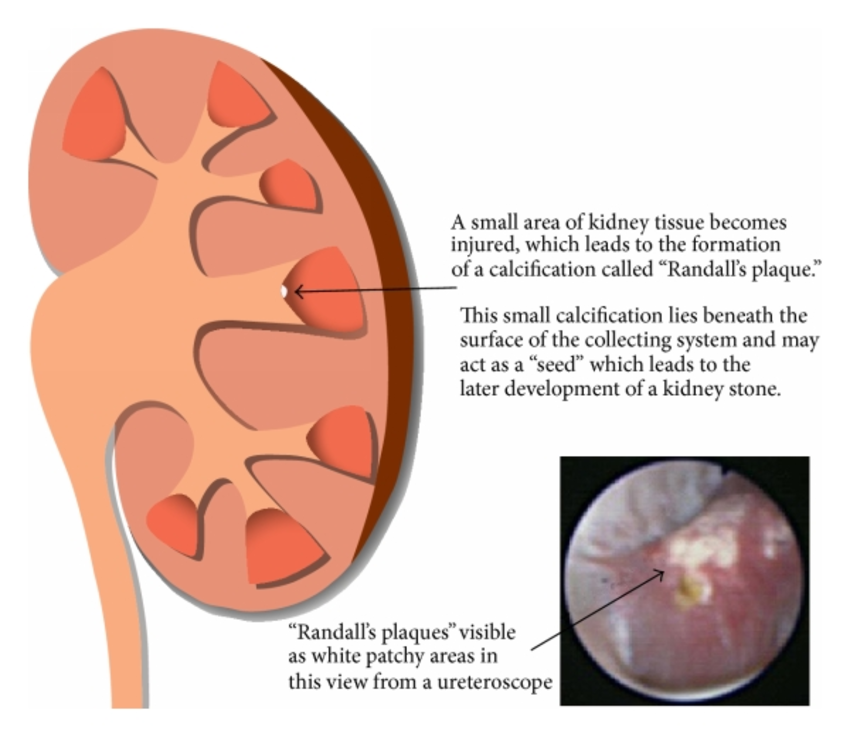
Randall plaques are microcalcifications of the tip of the renal papilla, which is <2 mm (papillary microlithasis): precursor/predisposing factor for renal stone formation.
--Today's ER cases
English


Un experto cocinero chino prepara sopa Yin Yang…
twitter.com/visualfeastwan…
Español


@ManavendraUpad1 Was it diagnosticated just by ultrasound?
English

Peri hilar cholangiocarcinoma ( peri ductal infiltrating type, Bismuth- Corlette Type IIIa) with liver metastasis.
Română

@maps_black La gran Colombia sacando la cara por América.
Español


Leidysabel Celis retweetledi


Leidysabel Celis retweetledi


Leidysabel Celis retweetledi

El cielo de Pampatar en la tarde de hoy. De sal y fuego.

Español