Optical Nanoscopy

5.7K posts

Optical Nanoscopy banner
Optical Nanoscopy

Optical Nanoscopy

@OptNanoscopy

* Thoughts on #imaging, #microscopy and optical #nanoscopy / #superresolution microscopy! * Tweets by @Ulrike_Boehm - Enjoy!

Oberkochen, Deutschland Katılım Mart 2014
711 Takip Edilen3.2K Takipçiler
Optical Nanoscopy retweetledi
Ulrike Boehm 🔬👩🏻‍💻
🔬 Conducting experiments in a vibration-free environment is crucial, especially for optical systems that require precise alignment. Learn more about vibration sources & how to mitigate them 😀👉 ulrikeboehm.org/sources-of-vib…
English
0
11
34
2.6K
Optical Nanoscopy retweetledi
biohub
biohub@biohub·
Imaging scientist @juliafrodri is part of a collaborative team developing + implementing novel sample preparation workflows, imaging acquisition & correlated software for multimodal imaging across scales
biohub tweet media
English
1
5
39
3.1K
Optical Nanoscopy retweetledi
ZEISS Group
ZEISS Group@ZEISS_Group·
Say hi to Carl, the very first mini version of our founding father! 👋 Born on this day 208 years ago, Carl Zeiss spent most of his life in #Jena, defining the history of optics. Now, the limited edition #PLAYMOBIL figure can keep his spirit alive. Happy birthday, Carl Zeiss! 🎉
ZEISS Group tweet media
English
0
13
41
2.9K
Optical Nanoscopy retweetledi
Dylan Burnette
Dylan Burnette@MAG2ART·
A cell photographed through a microscope. DNA in the nucleus, the Golgi apparatus, and actin filaments are shown. #CellBiology
GIF
English
8
109
517
46.3K
Optical Nanoscopy retweetledi
Stefan W. Hell
Stefan W. Hell@Stefan_W_Hell·
Just scratching the surface of what #Minflux can really do in tissue…:
Stefan Hell Labs@StefanHellLabs

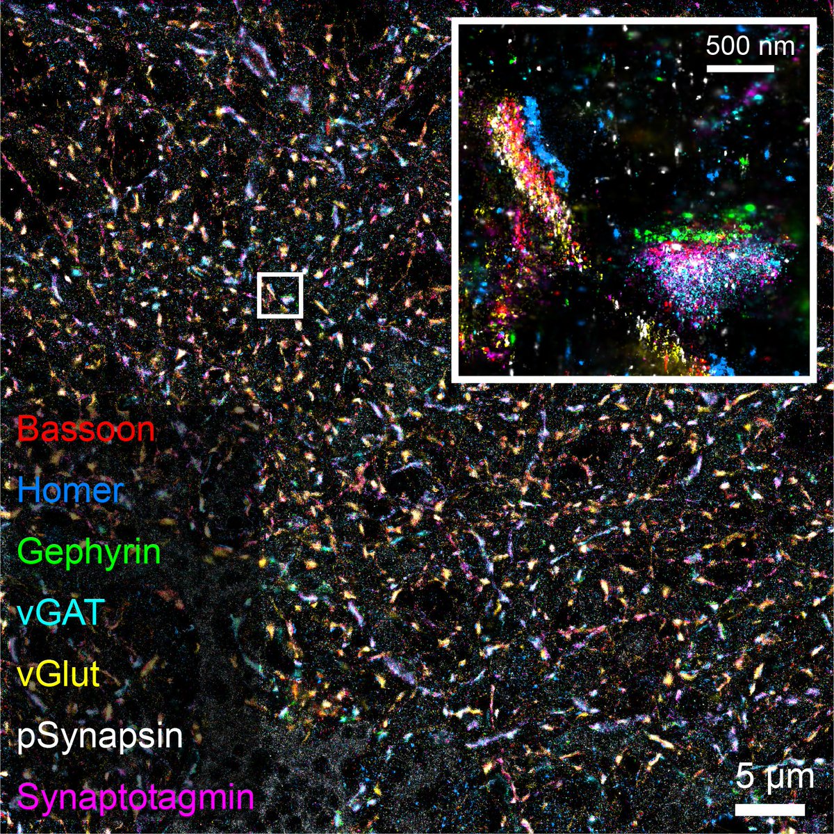
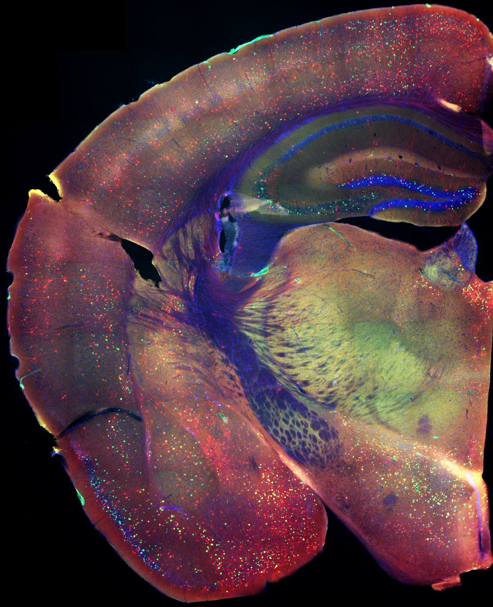
#MINFLUX fluorescence nanoscopy in biological tissue New preprint by T. Moosmayer, K.A. Kiszka et al. including PSD95 relative to the spine morphology (example below), presynaptic VGlut clustering and AMPAR clustering at the post-synapse. Check it out at biorxiv.org/content/10.110…

English
0
7
53
6.7K
Optical Nanoscopy retweetledi
Ulrike Boehm 🔬👩🏻‍💻
Ulrike Boehm 🔬👩🏻‍💻@Ulrike_Boehm·
Optics, such as lenses & mirrors, play a vital role in various fields, including #microscopy, & laser systems. To ensure optimal performance of these optical components, it is crucial to handle and clean them correctly. Here are some recommendations: 😀👉 ulrikeboehm.org/cleaning-optic…
Ulrike Boehm 🔬👩🏻‍💻 tweet media
English
1
12
53
5.3K
Optical Nanoscopy retweetledi
Ilaria Testa
Ilaria Testa@IlariaTesta4·
Fun chat with Peter O’Toole @YorkBioimaging happy to be part of the #Microscopists podcast serie
Peter O'Toole@YorkBioimaging

Chill out with Ilaria Testa (@karolinskainst) whilst she chats about: - The challenges and triumphs of grant writing - Balancing family life with groundbreaking science - Her love for Magritte's paintings because - Her lab's award-winning cake shaped like a neuron! See/hear: bit.ly/microscopists8… #TheMicroscopists

English
0
4
35
2.5K
Optical Nanoscopy retweetledi
Eduard Unterauer
Eduard Unterauer@EduardUnterauer·
How many synapses can you detect in a single Field of View? The answer is many😊. Here we have a 7plex image with 450 Synapses imaged with SUM-PAINT @Jungmannlab, that I am excited to share with you. Happy #FluorescenceFriday
Eduard Unterauer tweet media
English
3
54
351
28.3K
Optical Nanoscopy retweetledi
Justin Botterill
Justin Botterill@jjb930·
#FluorescenceFriday Also a good time to mention my lab is still recruiting graduate students!
Justin Botterill tweet media
English
7
47
453
46.4K
Optical Nanoscopy retweetledi
Adam Cohen
Adam Cohen@AdamEzraCohen·
Here are some neural fireworks for 4th of July! A 3D reconstruction of a pair of optogenetically triggered spikes in a CA1 pyramidal cell, acute slice. Stim to blue branch. With Liam Paninski's lab, supported by @cziscience. biorxiv.org/content/10.110…
English
6
58
271
33.7K
Optical Nanoscopy retweetledi
Eric Betzig
Eric Betzig@Eric_Betzig·
#FluorescenceFriday #cellbiology #bioart A neutrophilic HL-60 cell exhibits both sheet- and rosette-like dynamic pseudopods as it migrates in 3D through a collagen mesh. Here, the cell center is marked in yellow, and each pseudopod is marked with an orange ball of size proportional to its volume. As seen by lattice light sheet microscopy.
English
3
39
189
22.2K